
A English
Structure and function of heart Questions in English
Class 11 Biology · Body Fluids and Circulations · Structure and function of heart
410+
Questions
English
Language
100%
With Solutions
Showing 50 of 410 questions in English
351
EasyMCQ
Blood circulation was discovered by
A
William Harvey
B
Hippocrates
C
Karl Landsteiner
D
Paul Ehrlich
Solution
(A) The circulation of blood in the human body was discovered by William Harvey in the year $1628$. He demonstrated that blood is pumped by the heart and circulates through the body in a closed system.
0 likes
View Solution352
EasyMCQ
Give the location of the heart.
A
In the abdominal cavity
B
In the thoracic cavity,between the two lungs
C
In the cranial cavity
D
Below the diaphragm
Solution
(B) The heart is a mesodermally derived organ.
It is situated in the thoracic cavity,in the mediastinum space between the two lungs,and is slightly tilted to the left.
It is situated in the thoracic cavity,in the mediastinum space between the two lungs,and is slightly tilted to the left.
0 likes
View Solution353
Easy
Give the location of the tricuspid valve.
Solution
(N/A) The tricuspid valve is located at the opening between the right atrium and the right ventricle of the heart. It consists of three flaps (cusps) that prevent the backflow of blood from the right ventricle into the right atrium during ventricular systole.
0 likes
View Solution354
EasyMCQ
What is the Mitral valve?
A
$A$ valve between the right atrium and right ventricle
B
$A$ valve between the left atrium and left ventricle
C
$A$ valve at the opening of the aorta
D
$A$ valve at the opening of the pulmonary artery
Solution
(B) The valve that guards the opening between the left atrium and the left ventricle is called the Mitral valve.
It is also known as the bicuspid valve because it consists of two cusps or flaps.
This valve prevents the backflow of blood from the left ventricle into the left atrium during ventricular systole.
It is also known as the bicuspid valve because it consists of two cusps or flaps.
This valve prevents the backflow of blood from the left ventricle into the left atrium during ventricular systole.
0 likes
View Solution355
EasyMCQ
What is the function of valves in the heart?
A
To pump blood to the lungs.
B
To prevent the backward flow of blood.
C
To increase the heart rate.
D
To store oxygenated blood.
Solution
(B) The valves in the heart allow the flow of blood only in one direction,i.e.,from the atria to the ventricles and from the ventricles to the pulmonary artery or aorta.
These valves prevent any backward flow of blood,ensuring efficient circulation throughout the body.
These valves prevent any backward flow of blood,ensuring efficient circulation throughout the body.
0 likes
View Solution356
EasyMCQ
How many times does the heart beat in one minute?
A
$60-75$ times
B
$70-75$ times
C
$80-85$ times
D
$90-95$ times
Solution
(B) Our heart normally beats $70-75$ times in a minute (average $72$ beats $min^{-1}$).
0 likes
View Solution357
Easy
What is meant by cardiac output?
Solution
(N/A) Cardiac output is defined as the volume of blood pumped by each ventricle per minute. It is calculated by multiplying the stroke volume (the amount of blood pumped by each ventricle per beat) by the heart rate (the number of beats per minute). The formula is: $\text{Cardiac Output} = \text{Stroke Volume} \times \text{Heart Rate}$.
0 likes
View Solution358
EasyMCQ
Why is the heart called myogenic?
A
Because it is made of muscles.
B
Because its heartbeat originates from specialized muscle cells.
C
Because it is controlled by the brain.
D
Because it is controlled by hormones.
Solution
(B) The heart is called myogenic because its rhythmic contractions are initiated and regulated by specialized cardiac muscle cells known as nodal tissue (e.g.,$SA$ node).
Unlike neurogenic hearts,which require nerve impulses from the central nervous system to contract,the myogenic heart generates its own electrical impulses intrinsically.
This nodal tissue possesses properties of both muscle and nerve cells,allowing the heart to maintain its beat even when disconnected from the nervous system.
0 likes
View Solution359
Easy
Define the following terms and give their location:
$(a)$ Purkinje fibres
$(b)$ Bundle of His
$(a)$ Purkinje fibres
$(b)$ Bundle of His
Solution
(N/A) Purkinje fibres: These are specialized cardiac muscle fibres that conduct electrical impulses from the $AV$ node to the ventricular myocardium. They are located in the subendocardial surface of the ventricular walls.
$(b)$ Bundle of His: This is a collection of heart muscle cells specialized for electrical conduction that transmits the electrical impulses from the $AV$ node to the point of the apex of the fascicular branches. It is located in the interventricular septum of the heart.
$(b)$ Bundle of His: This is a collection of heart muscle cells specialized for electrical conduction that transmits the electrical impulses from the $AV$ node to the point of the apex of the fascicular branches. It is located in the interventricular septum of the heart.
0 likes
View Solution360
Easy
What is the significance of the time gap in the passage of the action potential from the sino-atrial node to the ventricle?
Solution
(N/A) The time gap allows the ventricles to remain relaxed while the action potential travels from the sino-atrial $(SA)$ node to the ventricles. This delay ensures that the atria complete their contraction (atrial systole) and empty their blood into the ventricles before the ventricles begin to contract. This maximizes ventricular filling,thereby increasing cardiac output.
0 likes
View Solution361
MediumMCQ
Which of the following organs contains all four types of tissues?
A
Brain
B
Lungs
C
Kidney
D
Heart
Solution
(D) The four primary types of tissues in the human body are epithelial,connective,muscular,and nervous tissues.
An organ is a structure composed of two or more types of tissues working together to perform a specific function.
The heart is a complex organ that contains all four types of tissues:
$1$. Epithelial tissue: Lines the inner chambers (endocardium) and outer surface (epicardium) of the heart.
$2$. Connective tissue: Forms the valves,fibrous skeleton,and blood vessels within the heart.
$3$. Muscular tissue: The cardiac muscle (myocardium) is responsible for the pumping action.
$4$. Nervous tissue: The nodal tissue ($SA$ node,$AV$ node) and nerve fibers regulate the heart rate and conduction system.
0 likes
View Solution362
MediumMCQ
The human circulatory system consists of $.....$
A
Heart $+$ Blood
B
Heart $+$ Network of branched blood vessels $+$ Blood
C
Heart $+$ Network of branched blood vessels $+$ Blood $+$ Lungs
D
Heart $+$ Network of branched blood vessels
Solution
(B) The human circulatory system,also known as the blood vascular system,consists of a muscular chambered heart,a network of closed branching blood vessels,and blood,which is the fluid that is circulated.
Therefore,the correct components are the heart,the network of branched blood vessels,and the blood.
Therefore,the correct components are the heart,the network of branched blood vessels,and the blood.
0 likes
View Solution363
MediumMCQ
Select the incorrect statement regarding the heart.
A
It is mesodermal in origin.
B
It is situated in the abdominal cavity.
C
It is situated in between the two lungs,slightly tilted to the left.
D
It has the size of a clenched fist.
Solution
(B) The heart is a mesodermal organ,which means it originates from the mesoderm layer during embryonic development.
It is located in the thoracic cavity,specifically in the mediastinum,between the two lungs.
It is slightly tilted towards the left side.
The size of the human heart is approximately that of a clenched fist.
Therefore,the statement that the heart is situated in the abdominal cavity is incorrect,as it is located in the thoracic cavity.
0 likes
View Solution364
MediumMCQ
Which of the following statements regarding the size of heart chambers is correct?
A
The size of atria and ventricles is equal.
B
Ventricles are smaller than atria.
C
Ventricles are larger than atria.
D
None of the above.
Solution
(C) The human heart is a four-chambered organ consisting of two atria and two ventricles.
Anatomically,the atria are the upper receiving chambers,while the ventricles are the lower pumping chambers.
Because the ventricles are responsible for pumping blood to the entire body (left ventricle) and the lungs (right ventricle),they possess much thicker muscular walls and a larger overall volume capacity compared to the atria.
Therefore,the ventricles are larger than the atria.
0 likes
View Solution365
EasyMCQ
The protective membrane surrounding the heart is called .........
A
Peritoneum
B
Pericardium
C
Pleura
D
Perimetrium
Solution
(B) The heart is enclosed in a double-walled sac called the $Pericardium$.
This membrane protects the heart,anchors it within the chest cavity,and prevents overfilling with blood.
$Peritoneum$ is the lining of the abdominal cavity.
$Pleura$ is the membrane surrounding the lungs.
$Perimetrium$ is the outer layer of the uterus.
This membrane protects the heart,anchors it within the chest cavity,and prevents overfilling with blood.
$Peritoneum$ is the lining of the abdominal cavity.
$Pleura$ is the membrane surrounding the lungs.
$Perimetrium$ is the outer layer of the uterus.
0 likes
View Solution366
MediumMCQ
Match the following columns:
| Column-$I$ | Column-$II$ |
|---|---|
| $P$. Interatrial septum | $I$. Thick fibrous tissue |
| $Q$. Interventricular septum | $II$. Thin wall |
| $R$. Atrioventricular septum | $III$. Thick wall |
A
$(P-I), (Q-II), (R-III)$
B
$(P-II), (Q-III), (R-I)$
C
$(P-II), (Q-I), (R-III)$
D
$(P-III), (Q-II), (R-I)$
Solution
(B) The heart structure consists of various septa that separate its chambers:
$1$. The $P$. Interatrial septum is a thin muscular wall that separates the right and left atria.
$2$. The $Q$. Interventricular septum is a thick wall that separates the right and left ventricles to withstand higher pressure.
$3$. The $R$. Atrioventricular septum is a thick fibrous tissue that separates the atria from the ventricles,containing the valves.
Therefore,the correct matching is $P-II, Q-III, R-I$.
0 likes
View Solution367
MediumMCQ
Identify the correct location of the valves in the human heart.
A
Tricuspid valve: Left atrium and left ventricle; Bicuspid valve: Right atrium and right ventricle; Aortic semilunar valve: Aorta and right ventricle; Pulmonary semilunar valve: Pulmonary artery and left ventricle.
B
Tricuspid valve: Right atrium and right ventricle; Bicuspid valve: Left atrium and left ventricle; Aortic semilunar valve: Aorta and right ventricle; Pulmonary semilunar valve: Pulmonary artery and left ventricle.
C
Tricuspid valve: Left atrium and left ventricle; Bicuspid valve: Right atrium and right ventricle; Aortic semilunar valve: Aorta and left ventricle; Pulmonary semilunar valve: Pulmonary artery and right ventricle.
D
Tricuspid valve: Right atrium and right ventricle; Bicuspid valve: Left atrium and left ventricle; Aortic semilunar valve: Aorta and left ventricle; Pulmonary semilunar valve: Pulmonary artery and right ventricle.
Solution
(D) The human heart contains four main valves that ensure unidirectional blood flow:
$1$. Tricuspid valve: Located between the right atrium and the right ventricle.
$2$. Bicuspid (Mitral) valve: Located between the left atrium and the left ventricle.
$3$. Aortic semilunar valve: Located at the base of the aorta,separating it from the left ventricle.
$4$. Pulmonary semilunar valve: Located at the base of the pulmonary artery,separating it from the right ventricle.
Comparing these with the options,option $D$ correctly identifies all these locations.
0 likes
View Solution368
MediumMCQ
In the given diagram of the vertical section of the human heart,which label represents the papillary muscles?
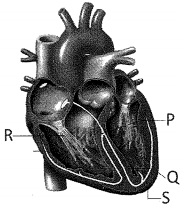
A
$P$
B
$Q$
C
$R$
D
$S$
Solution
(A) In the provided diagram of the human heart:
- Label $P$ points to the papillary muscles,which are muscular projections in the ventricles that attach to the chordae tendineae.
- Label $Q$ points to the myocardium (cardiac muscle layer).
- Label $R$ points to the chordae tendineae.
- Label $S$ points to the endocardium or the inner wall of the ventricle.
Therefore,the correct label for papillary muscles is $P$.
0 likes
View Solution369
EasyMCQ
Select the correct pathway for the transmission of impulses in the heart.
A
$SA$ node $\rightarrow$ $AV$ node $\rightarrow$ Purkinje fibers $\rightarrow$ $AV$ bundle
B
$AV$ node $\rightarrow$ $SA$ node $\rightarrow$ Purkinje fibers $\rightarrow$ $AV$ bundle
C
$SA$ node $\rightarrow$ $AV$ node $\rightarrow$ $AV$ bundle $\rightarrow$ Purkinje fibers
D
$AV$ node $\rightarrow$ $SA$ node $\rightarrow$ $AV$ bundle $\rightarrow$ Purkinje fibers
Solution
(C) The cardiac impulse originates in the $SA$ (Sinoatrial) node,which acts as the natural pacemaker of the heart.
From the $SA$ node,the impulse travels to the $AV$ (Atrioventricular) node.
From the $AV$ node,the impulse is transmitted to the $AV$ bundle (Bundle of His).
Finally,the impulse spreads through the Purkinje fibers to the ventricular walls,causing ventricular contraction.
Therefore,the correct sequence is: $SA$ node $\rightarrow$ $AV$ node $\rightarrow$ $AV$ bundle $\rightarrow$ Purkinje fibers.
0 likes
View Solution370
DifficultMCQ
State the location of the $SA$ node and the $AV$ node.
A
$SA$ node - Lower right corner of the right atrium
$AV$ node - Upper left corner of the right atrium
$AV$ node - Upper left corner of the right atrium
B
$SA$ node - Upper left corner of the right atrium
$AV$ node - Lower right corner of the right atrium
$AV$ node - Lower right corner of the right atrium
C
$SA$ node - Lower left corner of the right atrium
$AV$ node - Upper right corner of the right atrium
$AV$ node - Upper right corner of the right atrium
D
$SA$ node - Upper right corner of the right atrium
$AV$ node - Lower left corner of the right atrium
$AV$ node - Lower left corner of the right atrium
Solution
(D) The $SA$ node (Sino-atrial node) is located in the upper right corner of the right atrium.
It acts as the natural pacemaker of the heart.
The $AV$ node (Atrio-ventricular node) is located in the lower left corner of the right atrium,close to the atrio-ventricular septum.
Therefore,the correct option is $D$.
It acts as the natural pacemaker of the heart.
The $AV$ node (Atrio-ventricular node) is located in the lower left corner of the right atrium,close to the atrio-ventricular septum.
Therefore,the correct option is $D$.
0 likes
View Solution371
EasyMCQ
Which structure generates an action potential without any external stimulus?
A
$SA$ node
B
$AV$ node
C
$AV$ bundle
D
Purkinje fibers
Solution
(A) The $SA$ node (Sinoatrial node) is known as the pacemaker of the heart. It is a specialized patch of cardiac muscle fibers located in the right atrium that possesses the property of self-excitation. It generates an action potential spontaneously without any external neural or hormonal stimulus,which initiates the heartbeat.
0 likes
View Solution372
EasyMCQ
Which of the following is known as the pacemaker of the heart?
A
$SA$ node
B
$AV$ node
C
$AV$ bundle
D
Purkinje fibers
Solution
(A) The $SA$ node (Sinoatrial node) is a specialized patch of tissue present in the right upper corner of the right atrium. It is responsible for initiating and maintaining the rhythmic contractile activity of the heart. Because it generates the maximum number of action potentials per minute ($70-75$ per minute) and controls the heart rate,it is known as the natural pacemaker of the heart.
0 likes
View Solution373
MediumMCQ
Which structure is known as the 'heart of the heart'?
A
$SA$ node
B
$AV$ node
C
$AV$ bundle
D
Purkinje fibers
Solution
(A) The $SA$ node (Sinoatrial node) is known as the 'pacemaker' of the heart. It is often referred to as the 'heart of the heart' because it initiates the rhythmic electrical impulses that cause the heart to contract,thereby setting the pace for the entire cardiac cycle.
0 likes
View Solution374
MediumMCQ
Our heart normally beats ....... times in a minute.
A
$60-65$
B
$65-70$
C
$70-75$
D
$90-95$
Solution
(C) The human heart is a myogenic organ that initiates its own rhythmic contractions.
In a healthy adult,the normal heart rate at rest is typically between $70$ and $75$ beats per minute,with an average value often cited as $72$ beats per minute.
Therefore,the correct range is $70-75$ beats per minute.
In a healthy adult,the normal heart rate at rest is typically between $70$ and $75$ beats per minute,with an average value often cited as $72$ beats per minute.
Therefore,the correct range is $70-75$ beats per minute.
0 likes
View Solution375
MediumMCQ
Cardiac output $= ......$
A
Volume of blood pumped out by the heart per minute
B
Volume of blood pumped out by the heart per cardiac cycle
C
Volume of blood entering the heart per minute
D
Volume of blood entering the heart per cardiac cycle
Solution
(A) Cardiac output is defined as the volume of blood pumped out by each ventricle per minute.
It is calculated by multiplying the stroke volume (the volume of blood pumped by each ventricle per cardiac cycle, which is approximately $70 \ mL$) by the heart rate (the number of heartbeats per minute, which is approximately $72$ beats per minute).
Therefore, $\text{Cardiac Output} = \text{Stroke Volume} \times \text{Heart Rate} \approx 70 \ mL \times 72 \ \text{beats/min} \approx 5040 \ mL$ or $5 \ L$ per minute.
0 likes
View Solution376
MediumMCQ
What are the values of stroke volume and cardiac output under normal conditions?
Stroke Volume $\quad$ Cardiac Output
Stroke Volume $\quad$ Cardiac Output
A
$70 \text{ mL} \quad 25 \text{ L}$
B
$25 \text{ L} \quad 70 \text{ mL}$
C
$70 \text{ mL} \quad 5040 \text{ mL}$
D
$5040 \text{ mL} \quad 70 \text{ mL}$
Solution
(C) The stroke volume is the volume of blood pumped by each ventricle per beat,which is approximately $70 \text{ mL}$.
Cardiac output is the volume of blood pumped by each ventricle per minute.
It is calculated as: $\text{Cardiac Output} = \text{Stroke Volume} \times \text{Heart Rate}$.
Assuming an average heart rate of $72 \text{ beats/min}$,$\text{Cardiac Output} = 70 \text{ mL} \times 72 = 5040 \text{ mL}$ (or approximately $5 \text{ L}$).
Therefore,the correct values are $70 \text{ mL}$ for stroke volume and $5040 \text{ mL}$ for cardiac output.
0 likes
View Solution377
MediumMCQ
Heart sounds can be heard using a:
A
Sphygmomanometer
B
Spirometer
C
Stethoscope
D
All of the above
Solution
(C) The heart sounds,specifically the '$lub$' and '$dub$' sounds,are produced by the closure of the heart valves during the cardiac cycle.
These sounds are low-frequency vibrations that can be heard by a physician using a medical instrument called a stethoscope.
$A$ sphygmomanometer is used to measure blood pressure,and a spirometer is used to measure lung capacity.
Therefore,the correct instrument for listening to heart sounds is the stethoscope.
0 likes
View Solution378
EasyMCQ
When do the 'lub' and 'dub' sounds occur during the cardiac cycle?
Lub $\quad$ $\quad$ $\quad$ Dub
Lub $\quad$ $\quad$ $\quad$ Dub
A
Closure of $AV$ valves $\quad$ Closure of semilunar valves
B
Closure of semilunar valves $\quad$ Closure of $AV$ valves
C
Opening of $AV$ valves $\quad$ Opening of semilunar valves
D
Opening of semilunar valves $\quad$ Opening of $AV$ valves
Solution
(A) The cardiac cycle involves two primary heart sounds:
$1$. The first heart sound,'$lub$',is produced by the closure of the atrioventricular $(AV)$ valves (tricuspid and bicuspid valves) at the beginning of ventricular systole.
$2$. The second heart sound,'$dub$',is produced by the closure of the semilunar valves at the beginning of ventricular diastole.
Therefore,the correct sequence is: $Lub$ = Closure of $AV$ valves,$Dub$ = Closure of semilunar valves.
0 likes
View Solution379
EasyMCQ
The heart sound can be heard by using this instrument.
A
Stethoscope
B
Sphygmomanometer
C
Spirometer
D
Barometer
Solution
(A) The $Stethoscope$ is a medical instrument used for auscultation, or listening to the internal sounds of an animal or human body, such as the heart and lungs.
$Sphygmomanometer$ is used to measure blood pressure.
$Spirometer$ is used to measure the volume of air inspired and expired by the lungs.
$Barometer$ is used to measure atmospheric pressure.
$Sphygmomanometer$ is used to measure blood pressure.
$Spirometer$ is used to measure the volume of air inspired and expired by the lungs.
$Barometer$ is used to measure atmospheric pressure.
0 likes
View Solution380
EasyMCQ
Who discovered the circulation of blood?
A
Robert May
B
William Harvey
C
Robert Hooke
D
Wilkinson
Solution
(B) William Harvey is credited with the discovery of the systemic circulation of blood in the human body. In his landmark work,'Exercitatio Anatomica de Motu Cordis et Sanguinis in Animalibus' published in $1628$,he demonstrated that blood is pumped throughout the body by the heart.
0 likes
View Solution381
MediumMCQ
Following are the stages of the pathway for the conduction of an action potential through the heart:
$A.$ $AV$ bundle
$B.$ Purkinje fibres
$C.$ $AV$ node
$D.$ Bundle branches
$E.$ $SA$ node
Choose the correct sequence of the pathway from the options given below:
$A.$ $AV$ bundle
$B.$ Purkinje fibres
$C.$ $AV$ node
$D.$ Bundle branches
$E.$ $SA$ node
Choose the correct sequence of the pathway from the options given below:
A
$A-E-C-B-D$
B
$B-D-E-C-A$
C
$E-A-D-B-C$
D
$E-C-A-D-B$
Solution
(D) The correct answer is option $D$ $(E-C-A-D-B)$.
The conduction system of the heart follows a specific pathway to ensure coordinated contraction:
$1$. The action potential originates at the $SA$ node $(E)$.
$2$. It travels to the $AV$ node $(C)$.
$3$. From the $AV$ node,it passes to the $AV$ bundle $(A)$.
$4$. The $AV$ bundle divides into right and left bundle branches $(D)$.
$5$. Finally,the impulse spreads through the Purkinje fibres $(B)$ to the ventricular myocardium.
0 likes
View Solution382
EasyMCQ
The first heart sound is $:-$
A
'Dub' sound produced at the beginning of ventricular systole
B
'Lubb' sound produced at the beginning of ventricular systole
C
'Dub' sound produced at the end of ventricular systole
D
'Lubb' sound produced at the end of ventricular systole
Solution
(B) The heart produces two main sounds during each cardiac cycle, which can be heard through a stethoscope.
$1$. The first heart sound is known as '$Lubb$'. It is produced by the closure of the atrioventricular $(AV)$ valves (tricuspid and bicuspid valves) at the beginning of ventricular systole.
$2$. The second heart sound is known as '$Dub$'. It is produced by the closure of the semilunar valves at the end of ventricular systole.
Therefore, the first heart sound is the '$Lubb$' sound produced at the beginning of ventricular systole.
0 likes
View Solution383
EasyMCQ
Cardiac output is defined as the volume of blood:
A
received by the heart per minute
B
pumped by the ventricles per second
C
pumped by the left ventricle per minute
D
pumped by the left ventricle per hour
Solution
(C) Cardiac output is defined as the volume of blood pumped by the left ventricle into the aorta per minute.
It is calculated by multiplying the stroke volume (the amount of blood pumped per beat,approximately $70 \ mL$) by the heart rate (number of beats per minute,approximately $72 \ beats/min$).
Thus,the average cardiac output is approximately $5000 \ mL$ or $5 \ L$ per minute.
It is calculated by multiplying the stroke volume (the amount of blood pumped per beat,approximately $70 \ mL$) by the heart rate (number of beats per minute,approximately $72 \ beats/min$).
Thus,the average cardiac output is approximately $5000 \ mL$ or $5 \ L$ per minute.
0 likes
View Solution384
MediumMCQ
Choose the incorrect statement about the human heart $:-$
A
The entire heart is made up of cardiac muscles.
B
The nodal tissue in the right upper corner of the right atrium is the pacemaker.
C
The nodal tissue in the lower left corner of the right atrium can generate $70-75 \text{ min}^{-1}$ action potentials.
D
The nodal tissue is auto-excitable.
Solution
(C) The human heart is composed of cardiac muscle,but it also contains connective tissue (valves,chordae tendineae,fibrous skeleton) and specialized nodal tissue. Thus,statement $A$ is incorrect as the heart is not made entirely of cardiac muscle.
Statement $B$ refers to the Sino-Atrial Node $(SAN)$,which is located in the right upper corner of the right atrium and acts as the pacemaker.
Statement $C$ is incorrect because the Atrio-Ventricular Node $(AVN)$ is located in the lower left corner of the right atrium,but it generates action potentials at a rate of $40-60 \text{ min}^{-1}$,not $70-75 \text{ min}^{-1}$.
Statement $D$ is correct as nodal tissue is auto-excitable.
0 likes
View Solution385
DifficultMCQ
The $SA$ node is located in
A
Upper lateral wall of the left atrium
B
Lower lateral wall of the left atrium
C
Lower lateral wall of the right atrium
D
Upper lateral wall of the right atrium
Solution
(D) The $SA$ node (Sinoatrial node) is a specialized bundle of cardiac muscle fibers located in the wall of the right atrium.
Specifically,it is situated in the upper lateral wall of the right atrium,near the opening of the superior vena cava.
It acts as the natural pacemaker of the heart,initiating the electrical impulses that lead to cardiac contraction.
Specifically,it is situated in the upper lateral wall of the right atrium,near the opening of the superior vena cava.
It acts as the natural pacemaker of the heart,initiating the electrical impulses that lead to cardiac contraction.
0 likes
View Solution386
MediumMCQ
Ventricular systole increases the ventricular pressure causing the
A
Closure of semilunar valves
B
Closure of tricuspid and bicuspid valves
C
Opening of $AV$ valves
D
Opening of tricuspid and bicuspid valves
Solution
(B) During the cardiac cycle,ventricular systole refers to the contraction of the ventricles.
As the ventricles contract,the ventricular pressure rises significantly.
This increase in pressure forces the $AV$ (tricuspid and bicuspid) valves to close to prevent the backflow of blood into the atria.
Consequently,the correct answer is the closure of tricuspid and bicuspid valves.
As the ventricles contract,the ventricular pressure rises significantly.
This increase in pressure forces the $AV$ (tricuspid and bicuspid) valves to close to prevent the backflow of blood into the atria.
Consequently,the correct answer is the closure of tricuspid and bicuspid valves.
0 likes
View Solution387
EasyMCQ
Given below are two statements.
Statement $I$ - The heart sound $Dub$ is heard during ventricular diastole.
Statement $II$ - The heart sound $Lubb$ is heard during atrial systole.
In the light of above statements,select the correct option given below:
Statement $I$ - The heart sound $Dub$ is heard during ventricular diastole.
Statement $II$ - The heart sound $Lubb$ is heard during atrial systole.
In the light of above statements,select the correct option given below:
A
Both statement $I$ and statement $II$ are correct.
B
Both statement $I$ and statement $II$ are incorrect.
C
Statement $I$ is correct but statement $II$ is incorrect.
D
Statement $I$ is incorrect but statement $II$ is correct.
Solution
(B) Statement $I$ is incorrect because the heart sound $Dub$ (or $S2$) is produced by the closure of the semilunar valves at the beginning of ventricular diastole.
Statement $II$ is incorrect because the heart sound $Lubb$ (or $S1$) is produced by the closure of the atrioventricular valves at the beginning of ventricular systole,not during atrial systole.
Therefore,both statements are incorrect.
Statement $II$ is incorrect because the heart sound $Lubb$ (or $S1$) is produced by the closure of the atrioventricular valves at the beginning of ventricular systole,not during atrial systole.
Therefore,both statements are incorrect.
0 likes
View Solution388
EasyMCQ
Match the valves of the human heart in column $I$ with the respective opening they guard in column $II$ and select the correct option.
| Column $I$ | Column $II$ |
| $i$. Eustachian valve | $a$. Opening of pulmonary aorta |
| $ii$. Thebesian valve | $b$. Left atrioventricular aperture |
| $iii$. Mitral valve | $c$. Opening of inferior vena cava |
| $iv$. Semilunar valve | $d$. Opening of coronary sinus |
A
$i-c, ii-d, iii-b, iv-a$
B
$i-c, ii-d, iii-a, iv-b$
C
$i-d, ii-c, iii-b, iv-a$
D
$i-b, ii-a, iii-c, iv-d$
Solution
(A) The correct matching is as follows:
$1$. Eustachian valve $(i)$ guards the opening of the inferior vena cava $(c)$.
$2$. Thebesian valve $(ii)$ guards the opening of the coronary sinus $(d)$.
$3$. Mitral valve $(iii)$ is located at the left atrioventricular aperture $(b)$.
$4$. Semilunar valve $(iv)$ guards the opening of the pulmonary aorta and the aorta $(a)$.
Therefore,the correct sequence is $i-c, ii-d, iii-b, iv-a$.
0 likes
View Solution389
EasyMCQ
Select the mismatched pair from the following.
A
Pericardial fluid - shock absorber
B
Epicardium - Protection
C
Myocardium - Systole and diastole
D
Thebesian valve - guards the opening of postcaval
Solution
(D) The $Thebesian$ valve (also known as the valve of the coronary sinus) guards the opening of the coronary sinus into the right atrium. The opening of the postcaval vein (inferior vena cava) is guarded by the $Eustachian$ valve. Therefore,option $D$ is the mismatched pair.
0 likes
View Solution390
EasyMCQ
Bicuspid and tricuspid valves are attached with inelastic fibres to the papillary muscles in the lumen of the ventricles. These fibres are . . . . . . .
A
Chordae tendinae
B
Purkinje fibres
C
Ciliary body
D
Sino-atrial node
Solution
(A) The bicuspid (mitral) and tricuspid valves are atrioventricular valves that prevent the backflow of blood into the atria during ventricular systole.
These valves are anchored to the papillary muscles of the ventricular walls by tough,inelastic,collagenous cords known as $Chordae \ tendinae$.
These structures ensure that the valves do not invert into the atria when the ventricles contract under high pressure.
These valves are anchored to the papillary muscles of the ventricular walls by tough,inelastic,collagenous cords known as $Chordae \ tendinae$.
These structures ensure that the valves do not invert into the atria when the ventricles contract under high pressure.
0 likes
View Solution391
EasyMCQ
Given below are two statements.
Statement $I$: Visceral layer of serous pericardium forms the outer wall of human heart.
Statement $II$: Epicardium is formed of a thick layer of cardiac muscles.
In the light of the above two statements,choose the correct answer from the options given below.
Statement $I$: Visceral layer of serous pericardium forms the outer wall of human heart.
Statement $II$: Epicardium is formed of a thick layer of cardiac muscles.
In the light of the above two statements,choose the correct answer from the options given below.
A
Both statement $I$ and statement $II$ are correct.
B
Both statement $I$ and statement $II$ are incorrect.
C
Statement $I$ is correct but statement $II$ is incorrect.
D
Statement $I$ is incorrect but statement $II$ is correct.
Solution
(C) $i$. The visceral layer of the serous pericardium,also known as the epicardium,forms the outermost layer of the heart wall. Thus,Statement $I$ is correct.
$ii$. The epicardium is a thin layer composed of simple squamous epithelium and connective tissue,not a thick layer of cardiac muscles. The thick layer of cardiac muscles is called the myocardium. Thus,Statement $II$ is incorrect.
$ii$. The epicardium is a thin layer composed of simple squamous epithelium and connective tissue,not a thick layer of cardiac muscles. The thick layer of cardiac muscles is called the myocardium. Thus,Statement $II$ is incorrect.
0 likes
View Solution392
EasyMCQ
Which one of the following acts as a pace setter of the human heart?
A
$AV$ Node
B
$SA$ Node
C
Bundle of His
D
Node of Ranvier
Solution
(B) The $SA$ Node (Sinoatrial node) is known as the natural pacemaker of the human heart because it initiates the rhythmic electrical impulses that determine the heart rate.
The $AV$ Node (Atrioventricular node) acts as a pace setter or relay station that delays the impulse slightly to allow for ventricular filling.
Therefore,the $SA$ Node sets the pace,and the $AV$ Node regulates the transmission of that pace.
The $AV$ Node (Atrioventricular node) acts as a pace setter or relay station that delays the impulse slightly to allow for ventricular filling.
Therefore,the $SA$ Node sets the pace,and the $AV$ Node regulates the transmission of that pace.
0 likes
View Solution393
EasyMCQ
Pericardial fluid is present between . . . . . . of heart.
A
Parietal layer and fibrous pericardium
B
Fibrous pericardium and visceral layer of serous pericardium
C
Fibrous and serous pericardium
D
Parietal and visceral layers of serous pericardium
Solution
(D) The heart is enclosed in a double-walled membranous sac called the pericardium.
The pericardium consists of two main layers: the outer fibrous pericardium and the inner serous pericardium.
The serous pericardium is further divided into two layers: the parietal layer (outer) and the visceral layer (inner),which is also known as the epicardium.
The pericardial cavity is the space between the parietal and visceral layers of the serous pericardium.
This cavity is filled with pericardial fluid,which reduces friction between the heart walls during contraction and relaxation.
0 likes
View Solution394
EasyMCQ
Select the $INCORRECT$ statement with respect to heart sounds.
A
Dub sound is heard due to closure of semilunar valves.
B
Lub sound is heard due to closure of Thebesian valve.
C
Sounds heard during measurement of blood pressure are called Korotkoff sounds.
D
During measurement of $B.P.$ by sphygmomanometer, when the first pulsatile sound is heard, the pressure indicated is systolic pressure.
Solution
(B) The first heart sound, '$Lub$', is produced by the closure of the atrioventricular $(AV)$ valves (tricuspid and bicuspid valves) at the beginning of ventricular systole.
Option '$B$' is incorrect because the '$Lub$' sound is not caused by the closure of the Thebesian valve (which is a valve of the coronary sinus).
Option '$A$' is correct as the second heart sound, '$Dub$', is caused by the closure of the semilunar valves.
Option '$C$' and '$D$' are correct statements regarding the clinical measurement of blood pressure using a sphygmomanometer.
0 likes
View Solution395
EasyMCQ
The human heart lies in the space present between the two lungs,which is called . . . . . . .
A
Rathke's pouch
B
Mediastinum
C
Median eminence
D
Sella turcica
Solution
(B) The $Mediastinum$ is the central compartment of the thoracic cavity,located between the two lungs. The heart is situated within this space.
$Rathke's$ pouch is an embryonic precursor to the anterior pituitary gland.
$Median$ eminence is a part of the hypothalamus.
$Sella$ turcica is a bony depression in the sphenoid bone that houses the pituitary gland.
$Rathke's$ pouch is an embryonic precursor to the anterior pituitary gland.
$Median$ eminence is a part of the hypothalamus.
$Sella$ turcica is a bony depression in the sphenoid bone that houses the pituitary gland.
0 likes
View Solution396
EasyMCQ
Which one of the following blood vessels does $NOT$ possess any valve at its point of connection with the heart?
A
Aorta
B
Pulmonary vein
C
Inferior vena cava
D
Pulmonary artery
Solution
(B) The heart has specific valves to ensure unidirectional blood flow.
$1$. The $Aorta$ and $Pulmonary \text{ artery}$ (Pulmonary aorta) have semilunar valves at their base to prevent backflow of blood into the ventricles.
$2$. The $Inferior \text{ vena cava}$ and $Superior \text{ vena cava}$ enter the right atrium without any valves.
$3$. The $Pulmonary \text{ veins}$ enter the left atrium without any valves.
$4$. Among the given options, the $Pulmonary \text{ vein}$ is the correct answer as it lacks a valve at its junction with the left atrium.
0 likes
View Solution397
EasyMCQ
The remnant of the embryonic aperture on the inter-auricular septum is called . . . . . . .
A
foramen ovale
B
foramen of Monro
C
foramen of Luschka
D
foramen of Magendie
Solution
(A) During fetal development,the $foramen \ ovale$ is an opening in the inter-atrial (inter-auricular) septum that allows blood to bypass the non-functional fetal lungs.
After birth,this opening closes and becomes a depression known as the $fossa \ ovalis$.
The term $foramen \ ovale$ refers to the embryonic aperture itself,while the remnant is technically the $fossa \ ovalis$. Given the options provided,$foramen \ ovale$ (often misspelled as $foramen \ ovalis$ in some contexts) is the structure being referred to as the embryonic aperture.
0 likes
View Solution398
EasyMCQ
Purkinje fibers are located in the following structure/s:
A
Cuspid valve
B
Coronary sulcus
C
Atria
D
Ventricles
Solution
(D) Purkinje fibers are specialized conducting cells located in the subendocardial layer of the heart's ventricles.
They are part of the cardiac conduction system,which transmits electrical impulses from the atrioventricular $(AV)$ bundle to the ventricular myocardium.
This conduction ensures the synchronized contraction of the ventricles,which is essential for the rhythmic and efficient pumping of blood throughout the body.
0 likes
View Solution399
EasyMCQ
Right atrium: Coronary sinus:: Left atrium: . . . . . . .
A
Coronary artery
B
Inferior vena cava
C
Pulmonary artery
D
Pulmonary vein
Solution
(D) The coronary sinus is a collection of veins joined together to form a large vessel that collects blood from the heart muscle and opens into the Right atrium.
Similarly,the pulmonary veins are responsible for carrying oxygenated blood from the lungs and opening into the Left atrium.
Similarly,the pulmonary veins are responsible for carrying oxygenated blood from the lungs and opening into the Left atrium.
0 likes
View Solution400
EasyMCQ
Neurogenic heart receives stimulus for contraction from . . . . . . fibres.
A
elastin
B
collagen
C
nerve
D
muscle
Solution
(C) neurogenic heart is a type of heart where the heartbeat is initiated by nerve impulses generated by a specialized group of nerve cells (ganglia) located near the heart.
In contrast,a myogenic heart (found in humans) initiates its own heartbeat through specialized muscle cells.
Since the stimulus for contraction in a neurogenic heart originates from nerve cells,it is transmitted via nerve fibres.
Therefore,the correct option is $C$.
In contrast,a myogenic heart (found in humans) initiates its own heartbeat through specialized muscle cells.
Since the stimulus for contraction in a neurogenic heart originates from nerve cells,it is transmitted via nerve fibres.
Therefore,the correct option is $C$.
0 likes
View SolutionBody Fluids and Circulations — Structure and function of heart · Frequently Asked Questions
1Are these Body Fluids and Circulations questions useful for JEE and NEET?
Yes. All questions in this section are mapped to JEE Main and NEET exam patterns. Previous year questions from JEE Main, NEET, GUJCET and state-level exams are included with full solutions.
2Can I switch to Hindi or Gujarati for these questions?
Yes. Use the language tabs in the hero section or the sidebar to view the same questions and solutions in English, Hindi or Gujarati.
3How do I generate a question paper from this subtopic?
Use the Vedclass Exam Paper Generator — select the chapter and subtopic, set difficulty, and generate Sets A, B, C, D automatically. First 3 chapters of every subject are free.
Vedclass Products
For Students
Vedclass Test Series
Mock tests in real JEE/NEET style with performance analysis. 5-day free trial.
Start Free TrialFor Teachers
Exam Paper Generator
Generate Set A/B/C/D papers from this chapter in 2 minutes. 3 chapters free.
Try FreeFor Institutes
Online Exam Module
Live online exams with unlimited students, 360° analytics & white-label branding.
See DemoFor Teachers & Institutes
Generate a Body Fluids and Circulations Exam Paper in 2 Minutes
Select subtopic & difficulty — Sets A, B, C, D auto-generated with No Repeat logic.
First 3 chapters of every subject are free — no payment required.