
A English
Structure and function of heart Questions in English
Class 11 Biology · Body Fluids and Circulations · Structure and function of heart
410+
Questions
English
Language
100%
With Solutions
Showing 49 of 410 questions in English
251
EasyMCQ
The correct pathway of the impulse of heartbeat through the heart is ...........
A
$AV$ node $\rightarrow$ Bundle of His $\rightarrow$ $SA$ node $\rightarrow$ Purkinje fibers $\rightarrow$ Heart muscles
B
$AV$ node $\rightarrow$ $SA$ node $\rightarrow$ Purkinje fibers $\rightarrow$ Bundle of His $\rightarrow$ Heart muscles
C
$SA$ node $\rightarrow$ Purkinje fibers $\rightarrow$ Bundle of His $\rightarrow$ $AV$ node $\rightarrow$ Heart muscles
D
$SA$ node $\rightarrow$ $AV$ node $\rightarrow$ Bundle of His $\rightarrow$ Purkinje fibers $\rightarrow$ Heart muscles
Solution
(D) The heartbeat is initiated by the $SA$ (Sinoatrial) node,which acts as the natural pacemaker of the heart.
From the $SA$ node,the impulse travels to the $AV$ (Atrioventricular) node.
From the $AV$ node,it is conducted through the Bundle of His.
Finally,the impulse spreads through the Purkinje fibers to the ventricular heart muscles,causing contraction.
Therefore,the correct sequence is: $SA$ node $\rightarrow$ $AV$ node $\rightarrow$ Bundle of His $\rightarrow$ Purkinje fibers $\rightarrow$ Heart muscles.
0 likes
View Solution252
MediumMCQ
The His bundle is a network of .........
A
Nerve fibers distributed throughout the heart.
B
Muscle fibers distributed throughout the heart wall.
C
Muscle fibers distributed only in the ventricular wall.
D
Nerve fibers distributed in the ventricles.
Solution
(C) The Bundle of His (or His bundle) is a collection of specialized cardiac muscle fibers (modified cardiac muscle cells) that conduct electrical impulses from the atrioventricular $(AV)$ node to the ventricles.
These fibers are specialized for rapid conduction of the cardiac impulse,ensuring that the ventricles contract in a coordinated manner.
They are located within the ventricular walls (specifically the interventricular septum).
Therefore,the Bundle of His is a network of muscle fibers distributed in the ventricular wall.
0 likes
View Solution253
MediumMCQ
The systemic heart refers to:
A
The two ventricles in humans acting together.
B
The heart contracting under the stimulation of the nervous system.
C
The left auricle and left ventricle in higher vertebrates.
D
The entire heart in lower vertebrates.
Solution
(C) The systemic heart refers to the part of the heart that pumps oxygenated blood to the various organs of the body.
In higher vertebrates,the left auricle and the left ventricle receive oxygenated blood from the lungs and pump it into the systemic circulation (to the rest of the body).
Therefore,the left side of the heart is collectively known as the systemic heart.
In higher vertebrates,the left auricle and the left ventricle receive oxygenated blood from the lungs and pump it into the systemic circulation (to the rest of the body).
Therefore,the left side of the heart is collectively known as the systemic heart.
0 likes
View Solution254
MediumMCQ
In humans,blood flows from the posterior vena cava into the diastolic right atrium due to:
A
Opening of venous valves due to pressure
B
Suction pressure
C
Stimulation of the sinoatrial node
D
Pressure difference between the atrium and the posterior vena cava
Solution
(B) During the diastolic phase,the right atrium relaxes,which creates a lower pressure environment compared to the incoming venous blood in the posterior vena cava. This pressure gradient allows blood to flow passively from the posterior vena cava into the right atrium. This phenomenon is often referred to as the suction effect or venous return driven by the pressure difference.
0 likes
View Solution255
MediumMCQ
If the chordae tendineae of the tricuspid valve of a human heart are partially non-functional due to some injury,what will be the immediate effect?
A
The flow of blood into the aorta will be slowed down.
B
The pacemaker will stop working.
C
The flow of blood into the left atrium will be backflowed.
D
The flow of blood into the pulmonary artery will be reduced.
Solution
(D) The chordae tendineae are fibrous cords that attach the cusps of the tricuspid and mitral valves to the papillary muscles of the heart. Their primary function is to prevent the valves from prolapsing (everting) into the atria during ventricular systole.
If the chordae tendineae of the tricuspid valve are damaged or non-functional,the valve cannot close properly.
Consequently,during ventricular contraction,blood will leak back into the right atrium instead of being pumped entirely into the pulmonary artery.
This leads to a reduction in the volume of blood ejected into the pulmonary artery,thereby reducing the flow of blood to the lungs.
0 likes
View Solution256
EasyMCQ
The "Bundle of His" is considered to be a part of which of the following organs?
A
Brain
B
Heart
C
Kidney
D
Pancreas
Solution
(B) The "Bundle of His" (also known as the atrioventricular bundle) is a collection of heart muscle cells specialized for electrical conduction.
It transmits the electrical impulses from the atrioventricular node ($AV$ node) to the ventricles of the heart.
Therefore, it is an essential component of the cardiac conducting system located within the heart.
It transmits the electrical impulses from the atrioventricular node ($AV$ node) to the ventricles of the heart.
Therefore, it is an essential component of the cardiac conducting system located within the heart.
0 likes
View Solution257
MediumMCQ
Match the items given in Column-$I$ with those in Column-$II$ and select the correct option given below:
| Column-$I$ | Column-$II$ |
| $(a)$ Tricuspid valve | $(i)$ Between left atrium and left ventricle |
| $(b)$ Bicuspid valve | $(ii)$ Between right ventricle and pulmonary artery |
| $(c)$ Semilunar valve | $(iii)$ Between right atrium and right ventricle |
A
$a-ii, b-i, c-iii$
B
$a-iii, b-i, c-ii$
C
$a-i, b-ii, c-iii$
D
$a-i, b-iii, c-ii$
Solution
(B) The correct matches are as follows:
$1$. $(a)$ Tricuspid valve: This valve is located between the right atrium and the right ventricle, preventing the backflow of blood into the atrium during ventricular systole.
$2$. $(b)$ Bicuspid valve (Mitral valve): This valve is located between the left atrium and the left ventricle.
$3$. $(c)$ Semilunar valve: These valves are located at the openings of the right ventricle into the pulmonary artery and the left ventricle into the aorta, preventing backflow into the ventricles.
Therefore, the correct matching is: $(a-iii, b-i, c-ii)$.
0 likes
View Solution258
MediumMCQ
Which one of the following is a matching pair?
A
Lubb - Sharp closure of $AV$ valves at the beginning of ventricular systole.
B
Dup - Sudden opening of semilunar valves at the beginning of ventricular diastole.
C
Pulsation of the radial artery valves in the blood vessels.
D
Purkinje fibres - Initiation of the heart beat.
Solution
(A) The first heart sound, $Lubb$, is produced primarily due to the closure of the atrioventricular $(AV)$ valves (bicuspid and tricuspid valves) at the beginning of ventricular systole.
$Dup$ is the second heart sound, caused by the closure of the semilunar valves at the beginning of ventricular diastole.
$Purkinje$ fibres are responsible for the conduction of the cardiac impulse throughout the ventricles, not for the initiation of the heartbeat (which is the function of the $SA$ node).
0 likes
View Solution259
EasyMCQ
Arteries supplying blood to the heart are called
A
carotid arteries
B
hepatic arteries
C
coronary arteries
D
pulmonary arteries
Solution
(C) Coronary arteries are the specialized vessels that supply oxygenated blood to the heart muscle (myocardium) itself.
Carotid arteries are responsible for supplying blood to the head and neck region.
Hepatic arteries supply oxygenated blood to the liver.
Pulmonary arteries carry deoxygenated blood from the heart to the lungs for oxygenation.
Carotid arteries are responsible for supplying blood to the head and neck region.
Hepatic arteries supply oxygenated blood to the liver.
Pulmonary arteries carry deoxygenated blood from the heart to the lungs for oxygenation.
0 likes
View Solution260
MediumMCQ
Assertion : Muscle fibres of $SA$ node possess the lowest rhythmicity among all cardiac muscles.
Reason : Due to this fact,it can initiate excitatory waves at the highest rate.
Reason : Due to this fact,it can initiate excitatory waves at the highest rate.
A
If both Assertion and Reason are correct and the Reason is a correct explanation of the Assertion.
B
If both Assertion and Reason are correct but Reason is not a correct explanation of the Assertion.
C
If the Assertion is correct but Reason is incorrect.
D
If the Assertion is incorrect but the Reason is correct.
Solution
(D) The $SA$ node (sinoatrial node) is known as the natural pacemaker of the heart. It consists of specialized cardiac muscle fibres that possess the highest rhythmicity,not the lowest. Because of this high rhythmicity,it can generate excitatory impulses at the fastest rate,which sets the pace for the entire heart. Since the assertion states that the $SA$ node has the lowest rhythmicity,the assertion is incorrect. However,the reason correctly identifies that it initiates excitatory waves at the highest rate. Therefore,the assertion is incorrect,but the reason is correct.
0 likes
View Solution261
DifficultMCQ
Assertion : $Lub$ is a heart sound which is produced during each cardiac cycle.
Reason : It is associated with the closure of the tricuspid and bicuspid valves.
Reason : It is associated with the closure of the tricuspid and bicuspid valves.
A
If both Assertion and Reason are correct and the Reason is a correct explanation of the Assertion.
B
If both Assertion and Reason are correct but Reason is not a correct explanation of the Assertion.
C
If the Assertion is correct but Reason is incorrect.
D
If both the Assertion and Reason are incorrect.
Solution
(A) $Lub$ and $Dub$ are two heart sounds produced during each cardiac cycle.
$Lub$ is the first heart sound,which is produced due to the closure of the atrioventricular valves (tricuspid and bicuspid valves) at the beginning of ventricular systole.
It is a low-pitched sound of longer duration (approximately $0.15 \,sec$).
Since the assertion states that $Lub$ is produced during the cardiac cycle and the reason correctly identifies its cause as the closure of the atrioventricular valves,both statements are correct and the reason explains the assertion.
0 likes
View Solution262
EasyMCQ
All the components of the nodal tissue are autoexcitable. Why does the $SA$ node act as the normal pacemaker?
A
$SA$ node has the lowest rate of depolarisation.
B
$SA$ node is the only component to generate the threshold potential.
C
Only $SA$ node can convey the action potential to the other components.
D
$SA$ node has the highest rate of depolarisation.
Solution
(D) The nodal tissue of the heart is autoexcitable,meaning it can generate action potentials without external stimuli.
However,the rate of generation of action potentials varies among different components of the nodal tissue.
The $SA$ node (Sinoatrial node) has the highest rate of depolarisation,generating approximately $70-75$ action potentials per minute.
Because it initiates the impulse faster than any other part of the conduction system (like the $AV$ node or Purkinje fibers),it sets the pace for the entire heart.
Therefore,the $SA$ node acts as the natural pacemaker of the heart.
0 likes
View Solution263
EasyMCQ
$A$ specialised nodal tissue embedded in the lower corner of the right atrium,close to the atrioventricular septum,delays the spreading of impulses to the heart apex for about $0.1 \; sec$. This delay allows:
A
blood to enter the aorta.
B
the ventricles to empty completely.
C
blood to enter the pulmonary arteries.
D
the atria to empty completely.
Solution
(D) The specialised nodal tissue described is the Atrioventricular Node $(AVN)$.
It is located in the lower left corner of the right atrium near the atrioventricular septum.
The $AVN$ delays the electrical impulse generated by the Sinoatrial Node $(SAN)$ by approximately $0.1 \; sec$.
This delay is crucial because it ensures that the atria complete their contraction (atrial systole) and pump their blood into the ventricles before the ventricles begin to contract.
Therefore,this delay allows the atria to empty completely into the ventricles.
0 likes
View Solution264
Difficult
Describe the evolutionary change in the pattern of heart among the vertebrates.
Solution
(N/A) All vertebrates possess a heart,which is a hollow muscular organ composed of cardiac muscle fibres. The primary function of the heart is to pump blood to all parts of the body. The evolution of the heart is driven by the need to separate oxygenated blood from deoxygenated blood for more efficient oxygen transport.
$1$. Pisces (Fishes): The heart is two-chambered,consisting of one auricle and one ventricle. It receives only deoxygenated blood,which is pumped to the gills for oxygenation. It also contains accessory chambers like the sinus venosus and conus arteriosus.
$2$. Amphibians: They possess a three-chambered heart with two auricles and one ventricle. The auricles are separated by an inter-auricular septum,but the ventricle remains undivided,leading to the mixing of oxygenated and deoxygenated blood.
$3$. Reptiles: Most reptiles have an incomplete four-chambered heart (except for crocodiles,alligators,and gharials). The ventricle is partially divided by an incomplete septum,resulting in some mixing of blood.
$4$. Birds and Mammals: They possess a complete four-chambered heart with two atria and two ventricles. $A$ complete muscular septum prevents the mixing of oxygenated and deoxygenated blood,ensuring highly efficient circulation.

0 likes
View Solution265
Easy
Why do we call our heart myogenic?
Solution
(N/A) In the human heart,the contraction is initiated by a specialized modified cardiac muscle tissue known as the sinoatrial node ($SA$ node).
It is located in the wall of the right atrium.
The $SA$ node possesses the inherent ability to generate an electrical impulse,which initiates a wave of contraction and regulates the heart rate.
Because of this,it is referred to as the natural pacemaker of the heart.
Since the heartbeat is initiated by the $SA$ node and the impulse for contraction originates within the heart muscle itself rather than from the nervous system,the human heart is termed myogenic.
The hearts of all vertebrates and most molluscs are also myogenic.
0 likes
View Solution266
MediumMCQ
Sino-atrial node is called the pacemaker of our heart. Why?
A
It generates the maximum number of action potentials.
B
It regulates the heart rate by initiating rhythmic contractions.
C
It is located in the right atrium.
D
It is composed of specialized cardiac muscle fibers.
Solution
(B) The sino-atrial $(SA)$ node is a specialized mass of cardiac muscle fibers located in the upper wall of the right atrium.
It has the unique ability to generate action potentials without any external stimuli,a property known as autorhythmicity.
Because the $SA$ node initiates and maintains the rhythmic contractile activity of the heart,it is referred to as the natural pacemaker of the human heart.
It has the unique ability to generate action potentials without any external stimuli,a property known as autorhythmicity.
Because the $SA$ node initiates and maintains the rhythmic contractile activity of the heart,it is referred to as the natural pacemaker of the human heart.
0 likes
View Solution267
Medium
What is the significance of the atrio-ventricular node and atrio-ventricular bundle in the functioning of the heart?
Solution
(N/A) The atrioventricular $(AV)$ node is located in the right atrium,near the base of the inter-auricular septum. It acts as a relay station for cardiac impulses.
It gives rise to the atrioventricular bundle (bundle of $His$),which conducts cardiac impulses from the atria to the ventricles.
As the bundle of $His$ passes along the interventricular septum,it divides into right and left branches.
The terminal branches of this system form a network of Purkinje fibres that penetrate the myocardium.
The wave of excitation initiated by the sino-atrial node ($SA$ node) stimulates the $AV$ node,which then transmits the impulse through the bundle of $His$ and Purkinje fibres to trigger ventricular contraction.
Thus,the $AV$ node and $AV$ bundle are essential for the coordinated contraction of the ventricles.
0 likes
View Solution268
Medium
Explain heart sounds.
Solution
(N/A) Heart sounds are noises generated by the closing and opening of the heart valves.
In a healthy individual,there are two normal heart sounds called $lub$ and $dub$.
$Lub$ is the first heart sound. It is associated with the closure of the tricuspid and bicuspid valves at the beginning of systole.
The second heart sound $dub$ is associated with the closure of the semilunar valves at the beginning of diastole.
These sounds provide important information about the condition and working of the heart.
0 likes
View Solution269
Medium
Describe the external structure of the human heart.
Solution
(N/A) The human heart is a mesodermally derived organ.
It is situated in the thoracic cavity,in between the two lungs,and is slightly tilted to the left.
It is approximately the size of a clenched fist.
It is protected by a double-walled membranous bag called the pericardium,which encloses the pericardial fluid.
The human heart has four chambers: two relatively small upper chambers called atria and two larger lower chambers called ventricles.
Although the ventricles are larger than the atria,they contain the same volume of blood.
The atria and ventricles are separated externally by a clear groove known as the coronary sulcus or cardiac septum.
The ventricles are separated by the interventricular septum,which contains coronary arteries that supply oxygenated blood to the heart muscle itself.
0 likes
View Solution270
Medium
Describe the internal structure of the human heart in detail.
Solution
(N/A) Internally, the heart is divided into four chambers: two atria and two ventricles.
The two atria are separated by an interatrial septum.
$A$ thick-walled interventricular septum separates the right and left ventricles.
The atrium and ventricle of the same side are separated by an atrioventricular septum. However, each of these septa is provided with an opening through which the two chambers of the same side are connected.
The opening between the right atrium and right ventricle is guarded by a valve formed of three muscular flaps or cusps, the tricuspid valve, whereas a bicuspid or mitral valve guards the opening between the left atrium and the left ventricle.
The openings of the right and the left ventricles into the pulmonary artery and the aorta, respectively, are provided with semilunar valves.
The valves in the heart allow the flow of blood only in one direction, i.e., from atria to the ventricles and from the ventricles to the pulmonary artery or aorta. These valves prevent any backward flow.
The entire heart is made of cardiac muscles. The walls of the ventricles are much thicker than those of the atria.
$A$ specialised cardiac musculature called the nodal tissue is also distributed in the heart.
$A$ patch of this tissue is present in the right upper corner of the right atrium, called the sinoatrial node $(SAN)$.
Another mass of this tissue is seen in the lower left corner of the right atrium, close to the atrioventricular septum, called the atrioventricular node $(AVN)$.
$A$ bundle of nodal fibres continues from the $AVN$ through the atrioventricular septa to emerge on the top of the interventricular septum and immediately divides into a right and left bundle.
These branches give rise to minute fibres throughout the ventricular musculature of the respective sides and are called Purkinje fibres.
These fibres, along with the right and left bundle, are known as the bundle of $HIS$.
The nodal musculature has the ability to generate action potentials without any external stimuli, i.e., it is autoexcitable.
However, the number of action potentials that could be generated in a minute varies at different parts of the nodal system. The $SAN$ can generate the maximum number of action potentials, i.e., $70-75 \,min^{-1}$, and is responsible for initiating and maintaining the rhythmic contractile activity of the heart. Therefore, it is called the pacemaker.
Our heart normally beats $70-75$ times in a minute (average $72$ beats $min^{-1}$).
0 likes
View Solution271
Easy
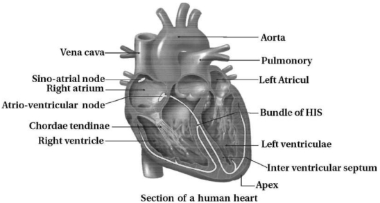
Draw a labelled diagram showing the internal structure of the human heart.
Solution
(N/A) The human heart is a muscular organ that pumps blood throughout the body. Its internal structure consists of four chambers: two upper atria and two lower ventricles.
Key components include:
$1$. $Aorta$: The main artery carrying oxygenated blood to the body.
$2$. $Vena$ $cava$: Large veins returning deoxygenated blood to the right atrium.
$3$. $Sino-atrial$ $node$ $(SAN)$: The natural pacemaker of the heart.
$4$. $Atrio-ventricular$ $node$ $(AVN)$: Relays electrical impulses from the atria to the ventricles.
$5$. $Chordae$ $tendineae$: Fibrous cords that anchor the heart valves.
$6$. $Bundle$ $of$ $His$: Part of the cardiac conduction system.
$7$. $Interventricular$ $septum$: The wall separating the left and right ventricles.
The provided diagram illustrates these structures clearly.
0 likes
View Solution272
Easy
Explain in short: $Lub$ and $Dub$ sounds.
Solution
(N/A) During each cardiac cycle,two prominent sounds are produced which can be easily heard through a stethoscope.
The first heart sound $(lub)$ is associated with the closure of the tricuspid and bicuspid valves.
The second heart sound $(dub)$ is associated with the closure of the semilunar valves.
These sounds are of clinical diagnostic significance.
The first heart sound $(lub)$ is associated with the closure of the tricuspid and bicuspid valves.
The second heart sound $(dub)$ is associated with the closure of the semilunar valves.
These sounds are of clinical diagnostic significance.
1 likes
View Solution273
Easy
Differentiate between Atrium and Ventricle.
Solution
(N/A)
| Atria (Auricles) | Ventricles |
|---|---|
| $(1)$ Located in the upper,broader part of the heart. | $(1)$ Located towards the lower end of the heart. |
| $(2)$ Possess thin muscular walls. | $(2)$ Possess thick,muscular walls to pump blood. |
| $(3)$ Smaller in size compared to ventricles. | $(3)$ Larger in size compared to atria. |
| $(4)$ Receive blood from veins (atrial chambers). | $(4)$ Pump blood out to the lungs and the rest of the body. |
0 likes
View Solution274
MediumMCQ
What is the function of nodal muscular tissue?
A
To pump blood to the lungs
B
To generate and conduct action potentials for heart contraction
C
To provide structural support to the heart valves
D
To filter blood impurities
Solution
(B) The nodal muscular tissue in the human heart consists of the $SA$ node and the $AV$ node.
These specialized tissues have the unique property of auto-excitability,meaning they can generate action potentials without any external stimuli.
$1$. The $SA$ node acts as the natural pacemaker,initiating the rhythmic contraction of the heart.
$2$. The $AV$ node receives the impulse from the $SA$ node and conducts it to the bundle of His and Purkinje fibers,ensuring the coordinated contraction of the atria and ventricles.
Therefore,the primary function is to generate and conduct action potentials for heart contraction.
0 likes
View Solution275
Easy
Define / Explain the following terms:
$(1)$ Pacemaker
$(2)$ Erythroblastosis
$(1)$ Pacemaker
$(2)$ Erythroblastosis
Solution
(N/A) $(1)$ The $SA$ node initiates the heartbeat and sends stimulating impulses every $0.80$ seconds to the atria and ventricles. Therefore,the $SA$ node is known as the pacemaker of the heart.
$(2)$ Erythroblastosis fetalis occurs due to $Rh$ incompatibility. If an $Rh$-negative mother carries an $Rh$-positive fetus for the second time,the maternal antibodies produced against the $Rh$ antigen cross the placenta and destroy the fetal $RBCs$. This condition is known as erythroblastosis fetalis.
$(2)$ Erythroblastosis fetalis occurs due to $Rh$ incompatibility. If an $Rh$-negative mother carries an $Rh$-positive fetus for the second time,the maternal antibodies produced against the $Rh$ antigen cross the placenta and destroy the fetal $RBCs$. This condition is known as erythroblastosis fetalis.
0 likes
View Solution276
Easy
Provide the definition/explanation for the following heart valves:
$(1)$ $AV$ valve (Tricuspid)
$(2)$ Mitral (Bicuspid) valve
$(1)$ $AV$ valve (Tricuspid)
$(2)$ Mitral (Bicuspid) valve
Solution
(N/A) $(1)$ The Tricuspid valve is located between the right atrium and the right ventricle.
It prevents the backflow of blood from the right ventricle into the right atrium during ventricular systole.
$(2)$ The Mitral (Bicuspid) valve is located between the left atrium and the left ventricle.
It prevents the backflow of blood from the left ventricle into the left atrium during ventricular systole.
0 likes
View Solution277
Easy
Define/Explain: $(1)$ Semi-lunar valve.
Solution
(N/A) The semi-lunar valves are located at the openings between the right ventricle and the pulmonary artery,and between the left ventricle and the aorta.
These valves consist of three pocket-like flaps that open when the ventricles contract.
Their primary function is to prevent the backflow of blood into the ventricles from the pulmonary artery and the aorta during ventricular diastole (relaxation).
These valves consist of three pocket-like flaps that open when the ventricles contract.
Their primary function is to prevent the backflow of blood into the ventricles from the pulmonary artery and the aorta during ventricular diastole (relaxation).
0 likes
View Solution278
EasyMCQ
Why do cardiac muscles never get fatigued?
A
They are involuntary in nature.
B
They have a high density of mitochondria and a rich blood supply.
C
They are branched and striated.
D
They are connected by intercalated discs.
Solution
(B) Cardiac muscles are specialized muscles that form the contractile tissue of the heart.
They are characterized by being involuntary,striated,and branched,allowing for coordinated contraction.
Cardiac muscles never get fatigued primarily because they possess a very high density of $mitochondria$ to ensure a continuous supply of $ATP$ through aerobic respiration.
Additionally,they receive a rich and constant blood supply,which provides the necessary oxygen and nutrients to sustain their continuous,rhythmic,and non-fatiguing activity throughout an individual's life.
0 likes
View Solution279
MediumMCQ
Describe the structure of myocardial tissue.
A
Striated,involuntary,branched,and uninucleated cells with intercalated discs.
B
Non-striated,involuntary,spindle-shaped,and uninucleated cells.
C
Striated,voluntary,cylindrical,and multinucleated cells.
D
Striated,involuntary,unbranched,and multinucleated cells.
Solution
(A) Myocardial tissue,also known as cardiac muscle tissue,is a specialized type of muscle found only in the heart.
$1$. Structure: The cells are cylindrical,branched,and typically uninucleated (though some may be binucleated).
$2$. Striations: They exhibit light and dark bands (striations) similar to skeletal muscle,indicating the presence of actin and myosin filaments.
$3$. Intercalated Discs: $A$ unique feature of cardiac muscle is the presence of intercalated discs,which are specialized cell junctions that allow for rapid communication and synchronized contraction of the heart muscle.
$4$. Function: It is involuntary in nature,meaning it contracts rhythmically without conscious control.
0 likes
View Solution280
Easy
The walls of ventricles are much thicker than atria. Explain.
Solution
(N/A) The walls of the ventricles are thicker because they are responsible for pumping blood forcefully into different parts of the body,which requires higher pressure.
The wall of the left ventricle is significantly thicker (about three times) than that of the right ventricle because it must pump blood to the entire systemic circulation,whereas the right ventricle only pumps blood to the lungs.
The wall of the left ventricle is significantly thicker (about three times) than that of the right ventricle because it must pump blood to the entire systemic circulation,whereas the right ventricle only pumps blood to the lungs.
0 likes
View Solution281
Medium
Differentiate between: Tricuspid and bicuspid valve.
Solution
(N/A)
| Tricuspid valve | Bicuspid valve |
|---|---|
| $(1)$ It separates the right atrium from the right ventricle. | $(1)$ It separates the left atrium from the left ventricle. |
| $(2)$ It consists of $3$ cusps (flaps). | $(2)$ It consists of $2$ cusps (flaps). |
| $(3)$ It is also known as the right atrioventricular valve. | $(3)$ It is also known as the mitral valve or left atrioventricular valve. |
0 likes
View Solution282
Easy
Write the feature that distinguishes between the two: Sino-atrial node and Atrio-ventricular node.
Solution
(N/A) $SA$ node: It is a specialized patch of nodal tissue present in the right upper corner of the right atrium. It acts as the natural pacemaker of the heart as it initiates the rhythmic contractile activity of the heart.
$AV$ node: It is a specialized mass of cardiac tissue located in the lower left corner of the right atrium close to the atrio-ventricular septum. It receives the action potential from the $SA$ node and delays it slightly before transmitting it to the bundle of His.
$AV$ node: It is a specialized mass of cardiac tissue located in the lower left corner of the right atrium close to the atrio-ventricular septum. It receives the action potential from the $SA$ node and delays it slightly before transmitting it to the bundle of His.
0 likes
View Solution283
MediumMCQ
Which part of the heart is responsible for initiating and maintaining its rhythmic activity?
A
Atrioventricular node ($AV$ node)
B
Sinoatrial node ($SA$ node)
C
Bundle of His
D
Purkinje fibers
Solution
(B) The $SA$ node (Sinoatrial node) is a specialized patch of cardiac muscle fibers located in the right atrium.
It acts as the natural pacemaker of the heart because it has the unique ability to generate action potentials without any external stimuli.
This electrical impulse initiates the rhythmic contraction of the heart and maintains its rhythmic activity throughout life.
It acts as the natural pacemaker of the heart because it has the unique ability to generate action potentials without any external stimuli.
This electrical impulse initiates the rhythmic contraction of the heart and maintains its rhythmic activity throughout life.
0 likes
View Solution284
Easy
Draw a diagram of the human heart and label the $SAN$,$AVN$,$AV$ bundle,bundle of His,and Purkinje fibres.
Solution
(N/A) The human heart contains a specialized conducting system that initiates and maintains the rhythmic contraction of the heart.
$1$. $SAN$ (Sino-Atrial Node): Located in the right upper corner of the right atrium. It acts as the natural pacemaker.
$2$. $AVN$ (Atrio-Ventricular Node): Located in the lower left corner of the right atrium close to the atrio-ventricular septum.
$3$. $AV$ bundle (Atrio-Ventricular bundle): $A$ bundle of nodal fibres that continues from the $AVN$ and passes through the atrio-ventricular septa.
$4$. Bundle of His: The $AV$ bundle divides into right and left branches that run along the interventricular septum.
$5$. Purkinje fibres: These are minute fibres that arise from the bundle of His and spread throughout the ventricular musculature on their respective sides.
0 likes
View Solution285
MediumMCQ
Analogy type questions:
$(1)$ Right atrium and right corner : $SA$ node :: Corner of right atrium : .......
$(2)$ First heart sound : $Lubb$ :: Second heart sound : ...........
$(1)$ Right atrium and right corner : $SA$ node :: Corner of right atrium : .......
$(2)$ First heart sound : $Lubb$ :: Second heart sound : ...........
A
$(1) AV$ node,$(2) Dubb$
B
$(1) Bundle$ of $His$,$(2) Dubb$
C
$(1) AV$ node,$(2) Murmur$
D
$(1) Purkinje$ fibers,$(2) Dubb$
Solution
(A) $(1)$ The $SA$ node (Sinoatrial node) is located in the upper wall of the right atrium. The $AV$ node (Atrioventricular node) is located in the lower left corner of the right atrium, close to the atrioventricular septum.
$(2)$ The first heart sound $(Lubb)$ is produced by the closure of the atrioventricular valves. The second heart sound $(Dubb)$ is produced by the closure of the semilunar valves.
$(2)$ The first heart sound $(Lubb)$ is produced by the closure of the atrioventricular valves. The second heart sound $(Dubb)$ is produced by the closure of the semilunar valves.
0 likes
View Solution286
EasyMCQ
Select the correct option:
$(1)$ The pericardium is a double-layered membrane surrounding the heart.
$(2)$ The $SA$ node has a higher rate of impulse generation compared to the $AV$ node.
$(1)$ The pericardium is a double-layered membrane surrounding the heart.
$(2)$ The $SA$ node has a higher rate of impulse generation compared to the $AV$ node.
A
Only $(1)$ is correct.
B
Only $(2)$ is correct.
C
Both $(1)$ and $(2)$ are correct.
D
Both $(1)$ and $(2)$ are incorrect.
Solution
(C) $(1)$ The heart is protected by a double-walled membranous bag called the pericardium,enclosing the pericardial fluid.
$(2)$ The $SA$ node (Sino-atrial node) is the natural pacemaker of the heart,capable of generating the maximum number of action potentials ($70-75$ per minute),which is higher than the $AV$ node (Atrio-ventricular node). Therefore,both statements are correct.
$(2)$ The $SA$ node (Sino-atrial node) is the natural pacemaker of the heart,capable of generating the maximum number of action potentials ($70-75$ per minute),which is higher than the $AV$ node (Atrio-ventricular node). Therefore,both statements are correct.
0 likes
View Solution287
EasyMCQ
Choose the correct option: The $AV$ node / $SA$ node is called the pacemaker.
A
$SA$ node
B
$AV$ node
C
Both $SA$ and $AV$ node
D
None of these
Solution
(A) The $SA$ node (Sinoatrial node) is known as the natural pacemaker of the heart.
It is located in the right atrium and is responsible for initiating the electrical impulses that trigger heart contractions.
Therefore,the correct option is $A$.
It is located in the right atrium and is responsible for initiating the electrical impulses that trigger heart contractions.
Therefore,the correct option is $A$.
0 likes
View Solution288
MediumMCQ
Why do cardiac muscles never get fatigued?
A
They are controlled by the somatic nervous system.
B
They contain a large number of mitochondria and myoglobin.
C
They are voluntary in nature.
D
They have a short refractory period.
Solution
(B) Cardiac muscles do not get fatigued because they possess a high density of mitochondria,which provide a continuous supply of $ATP$ through aerobic respiration.
Additionally,they have an abundant supply of myoglobin for oxygen storage and are connected by intercalated discs that allow for synchronized contraction.
While they are involuntary,their resistance to fatigue is primarily due to their specialized metabolic adaptations rather than just their neural control.
0 likes
View Solution289
EasyMCQ
Which scientist discovered the circulation of blood?
A
Karl Landsteiner
B
William Harvey
C
William Bateson
D
None of these
Solution
(B) William Harvey is credited with the discovery of the systemic circulation of blood in the human body.
He published his findings in his book titled 'Exercitatio Anatomica de Motu Cordis et Sanguinis in Animalibus' in $1628$.
Karl Landsteiner is known for the discovery of human blood groups ($ABO$ blood group system).
William Bateson is known for coining the term 'Genetics'.
He published his findings in his book titled 'Exercitatio Anatomica de Motu Cordis et Sanguinis in Animalibus' in $1628$.
Karl Landsteiner is known for the discovery of human blood groups ($ABO$ blood group system).
William Bateson is known for coining the term 'Genetics'.
0 likes
View Solution290
MediumMCQ
When cardiac muscle cells are damaged by a heart attack,they are usually replaced by
A
connective tissue cells
B
new smooth muscle cells
C
new cardiac muscle cells
D
epithelial cells
Solution
(A) Cardiac muscle cells,or cardiac myocytes,are the specialized muscle cells that form the heart muscle. Unlike some other tissues,cardiac muscle cells have very limited regenerative capacity. When these cells are damaged during a myocardial infarction (heart attack),the body cannot replace them with new cardiac muscle cells. Instead,the damaged area is repaired through the process of fibrosis,where the dead cells are replaced by fibrous connective tissue cells,forming a scar.
0 likes
View Solution291
MediumMCQ
Gap junctions are characteristically found in
A
Skeletal muscles
B
Cardiac muscles
C
Multi-unit smooth muscles
D
Striated muscles
Solution
(B) Gap junctions are specialized intercellular connections that allow molecules,ions,and electrical impulses to pass directly between adjacent cells.
In cardiac muscle tissue,these junctions are located within the intercalated discs.
The presence of gap junctions allows the cardiac muscle cells to function as a functional syncytium,meaning that an electrical signal (wave of depolarization) can spread rapidly across the entire tissue.
This synchronization ensures that the heart muscle contracts as a single unit,which is essential for effective blood pumping.
Therefore,gap junctions are a characteristic feature of cardiac muscles.
0 likes
View Solution292
EasyMCQ
Which vein contains the oxygenated blood in humans?
A
Cardiac vein
B
Hepato pancreatic vein
C
Portal vein
D
Pulmonary vein
Solution
(D) The pulmonary vein is the only vein in the human body that carries oxygenated blood instead of deoxygenated blood.
It transports oxygenated blood from the lungs to the left atrium of the heart.
From the left atrium,the blood flows into the left ventricle.
The left ventricle then pumps this oxygenated blood to the rest of the body.
It transports oxygenated blood from the lungs to the left atrium of the heart.
From the left atrium,the blood flows into the left ventricle.
The left ventricle then pumps this oxygenated blood to the rest of the body.
0 likes
View Solution293
EasyMCQ
Which of the following statements is incorrect?
A
Heart is endodermal in origin
B
Human heart is situated between the two lungs,slightly tilted to the left
C
Heart is a double-walled membranous bag
D
Human heart has two atria and two ventricles
Solution
(A) The heart is mesodermal in origin. Therefore,the statement that the heart is endodermal in origin is incorrect.
0 likes
View Solution294
MediumMCQ
The walls of the ventricles are much thicker than those of the atria because
A
They have to pump the blood
B
They have to receive the blood
C
They are present below the atria
D
They have to store the blood
Solution
(A) The walls of the ventricles are much thicker than those of the atria because the ventricles are responsible for pumping blood out of the heart into the pulmonary artery and the aorta.
This process requires generating high pressure to circulate blood throughout the body and to the lungs.
In contrast,the atria only function to receive blood from the veins and pass it into the ventricles,which requires significantly less muscular force,hence they are thinner.
This process requires generating high pressure to circulate blood throughout the body and to the lungs.
In contrast,the atria only function to receive blood from the veins and pass it into the ventricles,which requires significantly less muscular force,hence they are thinner.
0 likes
View Solution295
EasyMCQ
Which one has the thickest wall?
A
Right auricle
B
Right ventricle
C
Left auricle
D
Left ventricle
Solution
(D) The myocardium $(wall)$ of the left ventricle is approximately three times thicker than that of the right ventricle.
This is because the ventricles pump blood out of the heart with significant force.
The right ventricle pumps blood to the lungs via the pulmonary artery,while the left ventricle pumps blood to the entire body via the aorta.
Since the left ventricle must generate enough pressure to overcome systemic resistance and circulate blood throughout the entire body,it requires a much thicker muscular wall.
0 likes
View Solution296
MediumMCQ
The branches of the nodal tissue,which give rise to minute fibres throughout the ventricular musculature of the respective sides are called
A
Sino-atrial node
B
Atrio-ventricular node
C
Purkinje fibres
D
Bundle of His
Solution
(C) The nodal tissue in the heart consists of the $SA$ node,$AV$ node,$AV$ bundle,and Purkinje fibres.
The $AV$ bundle (Bundle of His) passes through the atrioventricular septum and divides into right and left branches.
These branches give rise to minute fibres that spread throughout the ventricular musculature of the respective sides,which are known as Purkinje fibres.
Purkinje fibres conduct the cardiac impulses rapidly throughout the ventricles,ensuring that the heart muscle contracts in a coordinated and efficient manner.
0 likes
View Solution297
MediumMCQ
Coronary heart disease is due to the inadequate blood supply to
A
Heart ventricle
B
Heart auricle
C
Heart volume
D
Heart muscles
Solution
(D) Coronary circulation refers to the circulation of blood within the heart muscle itself. Coronary heart disease occurs when there is an insufficient supply of oxygenated blood to the heart muscles,typically due to the narrowing or blockage of the coronary arteries.
0 likes
View Solution298
EasyMCQ
$SAN$ can generate impulses at a rate of:
A
$70-75\; min^{-1}$
B
$50-55\; min^{-1}$
C
$100-150\; min^{-1}$
D
$35-40\; min^{-1}$
Solution
(A) The heart exhibits auto-rhythmicity,which is the ability to contract spontaneously. The mammalian heart is myogenic,meaning the heartbeat originates from an electrical potential wave called a cardiac impulse,which arises from the sinoatrial node $(SAN)$ and spreads across the cardiac chambers.
The $SAN$ is located in the wall of the right atrium near the opening of the superior vena cava and generates impulses at a rate of approximately $70-75$ times per minute.
From the $SAN$,the cardiac impulse travels to the atrioventricular node $(AVN)$,then passes to the $AV$ bundle (Bundle of His) and its branches,finally reaching the Purkinje fibers in the ventricles.
The Bundle of His provides the only route for the transmission of the excitation wave from the atria to the ventricles. Purkinje fibers conduct these impulses significantly faster than the surrounding cells,ensuring that the heart muscle contracts in the most efficient manner.
0 likes
View Solution299
MediumMCQ
Impulse of heart beat originates from
A
$SA-$node
B
$AV-$node
C
Vagus nerve
D
Cardiac nerve
Solution
(A) $SA-$node (sino-atrial node) is a group of specialized cardiac muscle cells,which have the property of generating rhythmic excitatory waves.
It is also called the pacemaker of the heart as it generates the electrical impulse for all the chambers of the heart to contract.
It is also called the pacemaker of the heart as it generates the electrical impulse for all the chambers of the heart to contract.
0 likes
View SolutionBody Fluids and Circulations — Structure and function of heart · Frequently Asked Questions
1Are these Body Fluids and Circulations questions useful for JEE and NEET?
Yes. All questions in this section are mapped to JEE Main and NEET exam patterns. Previous year questions from JEE Main, NEET, GUJCET and state-level exams are included with full solutions.
2Can I switch to Hindi or Gujarati for these questions?
Yes. Use the language tabs in the hero section or the sidebar to view the same questions and solutions in English, Hindi or Gujarati.
3How do I generate a question paper from this subtopic?
Use the Vedclass Exam Paper Generator — select the chapter and subtopic, set difficulty, and generate Sets A, B, C, D automatically. First 3 chapters of every subject are free.
Vedclass Products
For Students
Vedclass Test Series
Mock tests in real JEE/NEET style with performance analysis. 5-day free trial.
Start Free TrialFor Teachers
Exam Paper Generator
Generate Set A/B/C/D papers from this chapter in 2 minutes. 3 chapters free.
Try FreeFor Institutes
Online Exam Module
Live online exams with unlimited students, 360° analytics & white-label branding.
See DemoFor Teachers & Institutes
Generate a Body Fluids and Circulations Exam Paper in 2 Minutes
Select subtopic & difficulty — Sets A, B, C, D auto-generated with No Repeat logic.
First 3 chapters of every subject are free — no payment required.