
A English
Structure and function of heart Questions in English
Class 11 Biology · Body Fluids and Circulations · Structure and function of heart
410+
Questions
English
Language
100%
With Solutions
Showing 10 of 410 questions in English
401
EasyMCQ
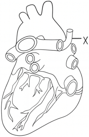
The blood vessel indicated by '$X$' is
A
Pulmonary vein
B
Inferior vena-cava
C
Coronary vein
D
Superior vena-cava
Solution
(D) Based on the anatomical diagram of the human heart,the vessel labeled '$X$' is the Superior Vena Cava.
It is the large vein that carries deoxygenated blood from the upper parts of the body (head,neck,and arms) into the right atrium of the heart.
The other options are incorrect because:
- Pulmonary veins carry oxygenated blood from the lungs to the left atrium.
- The Inferior Vena Cava carries deoxygenated blood from the lower parts of the body to the right atrium.
- Coronary veins drain blood from the heart muscle itself into the coronary sinus.
0 likes
View Solution402
EasyMCQ
Bradycardia indicates slow heart rate, generally below . . . . . . per minute. (in $beats$)
A
$70$
B
$75$
C
$80$
D
$60$
Solution
(D) Bradycardia is a condition characterized by a slower than normal heart rate.
In a healthy adult, the normal resting heart rate typically ranges from $60$ to $100$ beats per minute.
When the heart rate falls below $60$ beats per minute, it is clinically defined as bradycardia.
Therefore, the correct option is $D$.
In a healthy adult, the normal resting heart rate typically ranges from $60$ to $100$ beats per minute.
When the heart rate falls below $60$ beats per minute, it is clinically defined as bradycardia.
Therefore, the correct option is $D$.
0 likes
View Solution403
EasyMCQ
Mitral valve and tricuspid valve are attached to the papillary muscles by . . . . . . .
A
Corpus callosum
B
Columnae carnae
C
Crura cerebri
D
Chordae tendineae
Solution
(D) The $AV$ (atrioventricular) valves, which include the tricuspid valve and the mitral (bicuspid) valve, are anchored to the papillary muscles of the ventricular walls by fibrous cords known as $Chordae \text{ } tendineae$.
These structures prevent the valves from inverting into the atria during ventricular contraction (systole).
These structures prevent the valves from inverting into the atria during ventricular contraction (systole).
0 likes
View Solution404
EasyMCQ
Muscular ridges at the inner surface of the ventricles are called:
A
chordae tendinae
B
interventricular septum
C
papillary muscle
D
trabeculae carneae
Solution
(D) The inner surface of the ventricles is not smooth but contains irregular muscular ridges known as $trabeculae$ $carneae$.
These ridges help in the contraction of the ventricles and prevent the suction effect of the blood against the ventricular walls.
$Chordae$ $tendinae$ are the fibrous cords that attach the valves to the papillary muscles.
$Papillary$ $muscles$ are conical projections from the ventricular walls that anchor the $chordae$ $tendinae$.
$Interventricular$ $septum$ is the wall that separates the left and right ventricles.
0 likes
View Solution405
EasyMCQ
The nodal tissue located in the lower left corner of the right atrium is
A
$SA$ Node
B
$AV$ Node
C
$AV$ bundle
D
Purkinje fibres
Solution
(B) The human heart contains specialized cardiac musculature called nodal tissue.
$1$. The $SA$ Node (Sino-atrial node) is present in the right upper corner of the right atrium.
$2$. The $AV$ Node (Atrio-ventricular node) is located in the lower left corner of the right atrium,close to the atrio-ventricular septum.
$3$. The $AV$ bundle continues from the $AV$ node and passes through the atrio-ventricular septa.
$4$. Purkinje fibres are specialized muscle fibres that arise from the $AV$ bundle and spread throughout the ventricular musculature.
Therefore,the correct answer is $AV$ Node.
0 likes
View Solution406
EasyMCQ
The discovery of blood circulation was made by which scientist?
A
George Gamow
B
Karl Landsteiner
C
Norman Borlaug
D
William Harvey
Solution
(D) The discovery of blood circulation was made by $William \ Harvey$ in the year $1628$. He demonstrated that blood circulates throughout the body in a closed system,pumped by the heart. Therefore,the correct option is $D$.
0 likes
View Solution407
EasyMCQ
The typical 'lub-dub' sounds heard during a heartbeat are produced due to:
A
Closure of semilunar valves
B
Closure of bicuspid and tricuspid valves
C
Closure of bicuspid and tricuspid valves followed by semilunar valves
D
Opening of bicuspid and tricuspid valves followed by semilunar valves
Solution
(C) The correct answer is $C$.
During a cardiac cycle,two prominent heart sounds are produced.
The first heart sound,known as 'lub',is produced due to the closure of the atrioventricular valves (tricuspid and bicuspid valves) at the beginning of ventricular systole.
The second heart sound,known as 'dub',is produced due to the closure of the semilunar valves at the beginning of ventricular diastole.
Therefore,the sequence of sounds is the closure of bicuspid and tricuspid valves followed by the closure of semilunar valves.
0 likes
View Solution408
EasyMCQ
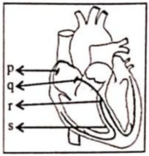
Identify the option showing the correct labeling for $p$,$q$,$r$ and $s$ with reference to the conducting system of the human heart.
A
$p$-Interventricular septum,$q$-$AVN$,$r$-Bundle of His,$s$-$SAN$
B
$p$-$SAN$,$q$-$AVN$,$r$-Bundle of His,$s$-Interventricular septum
C
$p$-$AVN$,$q$-$SAN$,$r$-Interventricular septum,$s$-Bundle of His
D
$p$-Bundle of His,$q$-$SAN$,$r$-Interventricular septum,$s$-$AVN$
Solution
(B) The correct option is $(B)$.
In the human heart's conducting system:
$p$ represents the Sino-Atrial Node $(SAN)$,which is located in the right atrium.
$q$ represents the Atrio-Ventricular Node $(AVN)$,located near the base of the interatrial septum.
$r$ represents the Bundle of His,which originates from the $AVN$ and passes through the interventricular septum.
$s$ represents the Interventricular septum,the wall separating the two ventricles.
0 likes
View Solution409
EasyMCQ
Left auricle receives pure blood from the:
A
pulmonary veins
B
pulmonary artery
C
superior vena cava
D
inferior vena cava
Solution
(A) Pure (oxygenated) blood from the lungs is carried back to the heart by the pulmonary veins,which empty into the left auricle (atrium).
From there,the blood moves to the left ventricle to be pumped to the rest of the body.
From there,the blood moves to the left ventricle to be pumped to the rest of the body.
0 likes
View Solution410
EasyMCQ
The frequency of heartbeat in our body is maintained by:
A
$AV$ Node
B
$SA$ Node
C
Node of Ranvier
D
Chordae tendineae
Solution
(B) The correct answer is $B$.
The sinoatrial $(SA)$ node,often referred to as the natural pacemaker of the heart,is located in the right atrium.
It generates rhythmic electrical impulses that initiate each heartbeat.
These impulses spread through the heart muscle,causing the atria to contract and setting the pace for the heart rate,thereby maintaining the frequency of heartbeats.
0 likes
View SolutionBody Fluids and Circulations — Structure and function of heart · Frequently Asked Questions
1Are these Body Fluids and Circulations questions useful for JEE and NEET?
Yes. All questions in this section are mapped to JEE Main and NEET exam patterns. Previous year questions from JEE Main, NEET, GUJCET and state-level exams are included with full solutions.
2Can I switch to Hindi or Gujarati for these questions?
Yes. Use the language tabs in the hero section or the sidebar to view the same questions and solutions in English, Hindi or Gujarati.
3How do I generate a question paper from this subtopic?
Use the Vedclass Exam Paper Generator — select the chapter and subtopic, set difficulty, and generate Sets A, B, C, D automatically. First 3 chapters of every subject are free.
Vedclass Products
For Students
Vedclass Test Series
Mock tests in real JEE/NEET style with performance analysis. 5-day free trial.
Start Free TrialFor Teachers
Exam Paper Generator
Generate Set A/B/C/D papers from this chapter in 2 minutes. 3 chapters free.
Try FreeFor Institutes
Online Exam Module
Live online exams with unlimited students, 360° analytics & white-label branding.
See DemoFor Teachers & Institutes
Generate a Body Fluids and Circulations Exam Paper in 2 Minutes
Select subtopic & difficulty — Sets A, B, C, D auto-generated with No Repeat logic.
First 3 chapters of every subject are free — no payment required.