
A English
Human excretory system Questions in English
Class 11 Biology · Excretory Products and their Elimination · Human excretory system
164+
Questions
English
Language
100%
With Solutions
Showing 50 of 164 questions in English
51
MediumMCQ
What is the wall of Bowman's capsule in the nephron made of?
A
Cuboidal epithelium
B
Columnar epithelium
C
Squamous epithelium
D
Glandular epithelium
Solution
(C) The Bowman's capsule is a cup-shaped structure that surrounds the glomerulus in the nephron.
Its wall is composed of a single layer of flattened cells known as squamous epithelium,specifically referred to as podocytes in the visceral layer.
This thin,flat structure facilitates the ultrafiltration of blood.
Its wall is composed of a single layer of flattened cells known as squamous epithelium,specifically referred to as podocytes in the visceral layer.
This thin,flat structure facilitates the ultrafiltration of blood.
0 likes
View Solution52
MediumMCQ
Which one of the following correctly explains the function of a specific part of the human nephron?
A
Podocytes : create minute spaces (slit pores) for the filtration of blood into the Bowman's capsule
B
Henle's loop : most reabsorption of the major substances from the glomerular filtrate
C
Distal convoluted tubule : reabsorption of $K^+$ ions into the surrounding blood capillaries
D
Afferent arteriole : carries the blood away from the glomerulus towards renal vein.
Solution
(A) : The visceral layer of Bowman's capsule surrounds the glomerulus and is composed of special type of cells,the podocytes.
The podocytes are so called because they possess foot-like processes (projections),the pedicels.
The space between pedicels are called slit pores (= filtration slits) through which the glomerular filtrate filters.
The podocytes are so called because they possess foot-like processes (projections),the pedicels.
The space between pedicels are called slit pores (= filtration slits) through which the glomerular filtrate filters.
0 likes
View Solution53
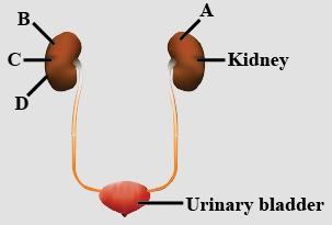
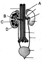
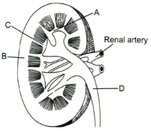
MediumMCQ
The figure shows the human urinary system with structures labelled $A$ to $D$. Select the option which correctly identifies them and gives their characteristics and/or functions.
A
$C$ - Medulla: inner zone of the kidney and contains complete nephrons.
B
$D$ - Cortex: outer part of the kidney and does not contain any part of the nephrons.
C
$A$ - Adrenal gland: located at the anterior part of the kidney. It secretes catecholamines which stimulate glycogen breakdown.
D
$B$ - Pelvis: broad funnel-shaped space inner to the hilum, directly connected to the loops of Henle.
Solution
(C) : In the given figure, $A$ is the adrenal gland which secretes two catecholamines; adrenaline (epinephrine) and noradrenaline (norepinephrine). Adrenaline increases the conversion of glycogen to glucose, providing quick energy for the "fight or flight" response. $B$ is the renal pelvis, which is a sac-like cavity of the kidney leading to the ureters; it is not directly connected to the loop of Henle. $C$ is the medulla, the inner region of the kidney containing the loop of Henle, collecting ducts, and ducts of Bellini. $D$ is the cortex, which has proximal and distal convoluted tubules and contains Malpighian corpuscles.
0 likes
View Solution54
MediumMCQ
Choose the correct sequence of urine transport.
A
$DCTs \rightarrow$ Collecting duct $\rightarrow$ Medullary pyramid $\rightarrow$ Calyces $\rightarrow$ Renal pelvis.
B
Collecting duct $\rightarrow$ Medullary pyramid $\rightarrow$ Calyces $\rightarrow$ $DCTs \rightarrow$ Renal pelvis.
C
Medullary pyramid $\rightarrow$ Collecting duct $\rightarrow$ $DCTs \rightarrow$ Calyces $\rightarrow$ Renal pelvis.
D
$DCTs \rightarrow$ Calyces $\rightarrow$ Renal pelvis $\rightarrow$ Collecting tubule $\rightarrow$ Medullary pyramid.
Solution
(A) The process of urine formation and transport follows a specific anatomical pathway within the kidney.
$1$. Urine is formed in the nephron and enters the Distal Convoluted Tubule $(DCT)$.
$2$. From the $DCT$,urine flows into the Collecting Duct.
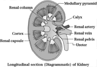
$3$. Multiple collecting ducts converge within the Medullary pyramids.
$4$. Urine then drains into the Calyces (minor and major calyces).
$5$. Finally,it reaches the Renal pelvis before passing into the ureter.
Therefore,the correct sequence is: $DCTs \rightarrow$ Collecting duct $\rightarrow$ Medullary pyramid $\rightarrow$ Calyces $\rightarrow$ Renal pelvis.
$1$. Urine is formed in the nephron and enters the Distal Convoluted Tubule $(DCT)$.
$2$. From the $DCT$,urine flows into the Collecting Duct.
$3$. Multiple collecting ducts converge within the Medullary pyramids.
$4$. Urine then drains into the Calyces (minor and major calyces).
$5$. Finally,it reaches the Renal pelvis before passing into the ureter.
Therefore,the correct sequence is: $DCTs \rightarrow$ Collecting duct $\rightarrow$ Medullary pyramid $\rightarrow$ Calyces $\rightarrow$ Renal pelvis.
0 likes
View Solution55
MediumMCQ
What are the correct dimensions (Length $-$ Width $-$ Thickness) for a kidney of an adult human?
A
$10$ to $12 \,cm$ $-$ $5$ to $7 \,cm$ $-$ $2$ to $3 \,cm$
B
$5$ to $7 \,cm$ $-$ $10$ to $12 \,cm$ $-$ $2$ to $3 \,cm$
C
$2$ to $3 \,cm$ $-$ $5$ to $7 \,cm$ $-$ $10$ to $12 \,cm$
D
$10$ to $12 \,cm$ $-$ $2$ to $3 \,cm$ $-$ $5$ to $7 \,cm$
Solution
(A) The human kidney is a bean-shaped organ.
In an adult human,the dimensions of the kidney are typically:
Length: $10$ to $12 \,cm$
Width: $5$ to $7 \,cm$
Thickness: $2$ to $3 \,cm$
Therefore,the correct sequence is $10$ to $12 \,cm$ (Length) $-$ $5$ to $7 \,cm$ (Width) $-$ $2$ to $3 \,cm$ (Thickness).
In an adult human,the dimensions of the kidney are typically:
Length: $10$ to $12 \,cm$
Width: $5$ to $7 \,cm$
Thickness: $2$ to $3 \,cm$
Therefore,the correct sequence is $10$ to $12 \,cm$ (Length) $-$ $5$ to $7 \,cm$ (Width) $-$ $2$ to $3 \,cm$ (Thickness).
0 likes
View Solution56
MediumMCQ
Which of the following is not a part of a kidney?
A
Peritubular capillaries
B
Convoluted tubules
C
Collecting ducts
D
Urinary bladder
Solution
(D) The human excretory system consists of a pair of kidneys,a pair of ureters,a urinary bladder,and a urethra.
$1$. The kidney is the primary organ of excretion,containing functional units called nephrons.
$2$. Nephrons consist of a glomerulus and renal tubules (which include proximal convoluted tubule,loop of Henle,and distal convoluted tubule).
$3$. Peritubular capillaries are blood vessels that surround the renal tubules within the kidney.
$4$. Collecting ducts are also located within the kidney and receive filtrate from the nephrons.
$5$. The urinary bladder is a muscular sac that stores urine and is located outside the kidney,connected to it via the ureters. Therefore,the urinary bladder is not a part of the kidney.
0 likes
View Solution57
MediumMCQ
The figure shows the human urinary system with structures labeled $A$ to $D$. Select the option which correctly identifies them and gives their characteristics and/or functions.
A
$A$: Adrenal gland - located at the anterior part of the kidney. It secretes catecholamines which stimulate glycogenolysis.
B
$B$: Pelvis - broad funnel-shaped space inner to the hilum,directly connected to the loops of Henle.
C
$C$: Medulla - inner zone of the kidney and contains complex nephrons.
D
$D$: Cortex - outer part of the kidney and does not contain any part of the nephrons.
Solution
(A) Based on the provided diagram of the human urinary system:
$A$ represents the Adrenal gland,which is located at the anterior part of the kidney. It secretes catecholamines (like adrenaline) which stimulate glycogenolysis.
$B$ represents the Renal pelvis,which is a broad funnel-shaped space inner to the hilum. It is not directly connected to the loops of Henle but to the major calyces.
$C$ represents the Medulla,which is the inner zone of the kidney. It contains the loops of Henle and collecting ducts.
$D$ represents the Cortex,which is the outer part of the kidney. It contains the Malpighian corpuscles,proximal convoluted tubules $(PCT)$,and distal convoluted tubules $(DCT)$.
Therefore,the correct identification and description is given in option $A$.
0 likes
View Solution58
MediumMCQ
Inner to the hilum of the kidney,there is a broad funnel-shaped space called
A
Renal pelvis
B
Medulla
C
Cortex
D
Adrenal gland
Solution
(A) The kidney has an indentation on its concave side known as the hilum.
Inner to the hilum,there is a broad,funnel-shaped space called the renal pelvis.
Projections of the renal pelvis are called calyces.
The cortex and medulla are the two main zones of the kidney,but they are not funnel-shaped spaces.
Therefore,the correct answer is $A$.
Inner to the hilum,there is a broad,funnel-shaped space called the renal pelvis.
Projections of the renal pelvis are called calyces.
The cortex and medulla are the two main zones of the kidney,but they are not funnel-shaped spaces.
Therefore,the correct answer is $A$.
0 likes
View Solution59
MediumMCQ
Identify the odd one out.
A
$DCT$
B
$PCT$
C
Loop of Henle
D
Pancreas
Solution
(D) $DCT$ (Distal Convoluted Tubule),$PCT$ (Proximal Convoluted Tubule),and Loop of Henle are all integral parts of the nephron,which is the structural and functional unit of the kidney.
Pancreas is a glandular organ that is part of the digestive and endocrine systems,not the excretory system (urinary system).
Pancreas is a glandular organ that is part of the digestive and endocrine systems,not the excretory system (urinary system).
0 likes
View Solution60
MediumMCQ
Which of the following statements is correct?
A
In cortical nephrons,the loop of Henle is too short and extends only very little into the medulla.
B
In juxtamedullary nephrons,the loop of Henle is too short and runs deep into the medulla.
C
In cortical nephrons,the loop of Henle is very long and extends only very little into the medulla.
D
In juxtamedullary nephrons,the loop of Henle is very long and extends only very little into the medulla.
Solution
(A) Nephrons are of two types based on the position of the loop of Henle:
$1$. Cortical nephrons: In these,the loop of Henle is very short and extends only a little into the medulla. These constitute the majority of nephrons.
$2$. Juxtamedullary nephrons: In these,the loop of Henle is very long and runs deep into the medulla. These are important for concentrating urine.
Therefore,option $A$ is the correct statement.
0 likes
View Solution61
MediumMCQ
Which one of the following correctly explains the function of a specific part of the human nephron?
A
Podocytes: Create minute spaces (slit pores) for the filtration of blood into the Bowman's capsule.
B
Henle's loop: Most reabsorption of the major substances from the glomerular filtrate.
C
Distal convoluted tubule: Reabsorption of $K^+$ ions from the surrounding blood capillaries.
D
Afferent arteriole: Carries the blood away from the glomerulus towards the renal vein.
Solution
(A) The correct option is $A$.
$1$. Podocytes are specialized cells in the inner layer of the Bowman's capsule that form slit pores,which allow for the filtration of blood.
$2$. Henle's loop is primarily responsible for the concentration of urine,not the reabsorption of most substances (which occurs in the Proximal Convoluted Tubule).
$3$. The Distal Convoluted Tubule $(DCT)$ is involved in the conditional reabsorption of $Na^+$ and water,and the secretion of $H^+$,$K^+$,and ammonia,rather than the reabsorption of $K^+$.
$4$. The Afferent arteriole brings blood into the glomerulus,while the Efferent arteriole carries blood away from it.
0 likes
View Solution62
MediumMCQ
Which of the following statements is incorrect?
A
In most of the nephrons,the loop of Henle is short and extends only a short distance into the medulla.
B
In some nephrons,the loop of Henle is very long and extends deep into the medulla.
C
The Bowman's capsule and the tuft of capillaries are known as renal corpuscles.
D
The bunch of small blood capillaries formed by the afferent arteriole is called the glomerulus.
Solution
(C, D) The correct answer is $C$ and $D$. However,in the context of identifying the most incorrect statement regarding terminology:
$1$. Option $A$ is correct: Cortical nephrons have short loops of Henle.
$2$. Option $B$ is correct: Juxtamedullary nephrons have long loops of Henle.
$3$. Option $C$ is incorrect: The Bowman's capsule and the tuft of capillaries (glomerulus) together are known as the 'Renal Corpuscle' or 'Malpighian body',not 'Renal granules'.
$4$. Option $D$ is incorrect: The glomerulus is a tuft of capillaries formed by the 'afferent' arteriole,not the 'efferent' arteriole.
0 likes
View Solution63
EasyMCQ
In every adult human kidney,the dimensions are: length $A$ $cm$,width $B$ $cm$,and thickness $C$ $cm$. Identify the correct values for $A-B-C$.
A
$10$ to $12$,$5$ to $7$,$2$ to $3$
B
$8$ to $10$,$5$ to $6$,$1$ to $2$
C
$5$ to $7$,$10$ to $12$,$1$ to $2$
D
$5$ to $7$,$3$ to $4$,$2$ to $3$
Solution
(A) According to the $NCERT$ textbook,the human kidneys are reddish-brown,bean-shaped structures situated between the levels of the last thoracic and third lumbar vertebra close to the dorsal inner wall of the abdominal cavity.
Each kidney of an adult human measures $10-12$ $cm$ in length,$5-7$ $cm$ in width,and $2-3$ $cm$ in thickness with an average weight of $120-170$ $g$.
Therefore,the correct values are $A = 10-12$,$B = 5-7$,and $C = 2-3$.
Each kidney of an adult human measures $10-12$ $cm$ in length,$5-7$ $cm$ in width,and $2-3$ $cm$ in thickness with an average weight of $120-170$ $g$.
Therefore,the correct values are $A = 10-12$,$B = 5-7$,and $C = 2-3$.
0 likes
View Solution64
MediumMCQ
Which of the following organs is not present in a pair in the human body?
A
Kidney
B
Lungs
C
Urinary bladder
D
Ureter
Solution
(C) In the human excretory system,the kidneys and ureters are present in pairs.
Humans have two kidneys and two ureters.
Lungs are also present in a pair (left and right lung).
The urinary bladder is a single,muscular sac-like structure that stores urine before it is excreted from the body.
Therefore,the urinary bladder is not in a pair.
Humans have two kidneys and two ureters.
Lungs are also present in a pair (left and right lung).
The urinary bladder is a single,muscular sac-like structure that stores urine before it is excreted from the body.
Therefore,the urinary bladder is not in a pair.
0 likes
View Solution65
MediumMCQ
The cortex extends in between the medullary pyramids as renal columns called
A
Calyces
B
Nephrons
C
Renal pelvis
D
Column of Bertini
Solution
(D) The human kidney is divided into two main zones: an outer cortex and an inner medulla. The medulla is divided into a few conical masses called medullary pyramids projecting into the calyces. The cortex extends in between the medullary pyramids as renal columns,which are known as the $Column$ $of$ $Bertini$.
0 likes
View Solution66
MediumMCQ
Choose the incorrect sentence$(s)$.
$(I)$ Malpighian corpuscles,$PCT$ and $DCT$ of the nephron are situated in the medulla of the kidney.
$(II)$ The loop of Henle dips into the medulla.
$(III)$ In juxtamedullary nephrons,the loop of Henle is too short and extends very little into the medulla.
$(IV)$ Vasa recta is absent or highly reduced in cortical nephrons.
A
$I, II$
B
$II, IV$
C
$I, III$
D
$III, IV$
Solution
(C) Statement $(I)$ is incorrect because the Malpighian corpuscle,$PCT$,and $DCT$ are located in the cortex region of the kidney,not the medulla.
Statement $(II)$ is correct; the loop of Henle extends into the medulla.
Statement $(III)$ is incorrect because in juxtamedullary nephrons,the loop of Henle is very long and extends deep into the medulla. Short loops are characteristic of cortical nephrons.
Statement $(IV)$ is correct; vasa recta is absent or highly reduced in cortical nephrons.
Therefore,statements $(I)$ and $(III)$ are incorrect.
0 likes
View Solution67
MediumMCQ
In some of the nephrons,the loop of Henle is very long and runs deep into the medulla. These nephrons are called:
A
Malpighian tubules
B
Cortical nephrons
C
Juxtamedullary nephrons
D
Vasa recta
Solution
(C) Nephrons are classified into two types based on the position of their glomeruli and the length of the loop of Henle:
$1$. Cortical nephrons: These have a short loop of Henle that extends only slightly into the medulla.
$2$. Juxtamedullary nephrons: These have a very long loop of Henle that runs deep into the medulla. This structural adaptation is crucial for the concentration of urine.
Therefore,the correct answer is Juxtamedullary nephrons.
0 likes
View Solution68
MediumMCQ
............ cells of Bowman's capsule called .......... are arranged in an intricate manner so as to leave some minute spaces called slit pores.
A
Endothelium,podocytes
B
Epithelium,podocytes
C
Basement membrane,$JGA$ cells
D
Epithelium,$JGA$ cells
Solution
(B) The Bowman's capsule is a double-walled cup-like structure that encloses the glomerulus.
It consists of an inner visceral layer and an outer parietal layer.
The visceral layer is composed of specialized epithelial cells known as $podocytes$.
These $podocytes$ are arranged in an intricate manner,leaving minute spaces between them known as $filtration$ $slits$ or $slit$ $pores$.
These pores are essential for the process of ultrafiltration,allowing blood to be filtered into the lumen of the Bowman's capsule.
0 likes
View Solution69
EasyMCQ
The kidney of an adult rabbit is .............
A
Pronephros
B
Metanephros
C
Mesonephros
D
Opisthonephros
Solution
(B) The kidneys in vertebrates are classified based on their embryonic development and structural complexity into three types: Pronephros,Mesonephros,and Metanephros.
$1$. Pronephros is the most primitive type,found in the embryos of all vertebrates and in adult cyclostomes.
$2$. Mesonephros is found in the embryos of higher vertebrates (amniotes) and in adult fish and amphibians.
$3$. Metanephros is the most advanced type of kidney,which is found in the adults of higher vertebrates,including reptiles,birds,and mammals (such as the rabbit).
Therefore,the kidney of an adult rabbit is Metanephros.
0 likes
View Solution70
MediumMCQ
Which of the following is not a part of the nephron?
A
Glomerulus
B
Loop of Henle
C
Distal convoluted tubule
D
Collecting duct
Solution
(D) nephron consists of two major parts: the renal corpuscle and the renal tubule.
$1$. The renal corpuscle includes the Glomerulus and Bowman's capsule.
$2$. The renal tubule consists of the Proximal Convoluted Tubule $(PCT)$,Loop of Henle,and Distal Convoluted Tubule $(DCT)$.
$3$. The collecting duct is not considered a part of the nephron itself,as multiple nephrons drain their filtrate into a single collecting duct.
0 likes
View Solution71
EasyMCQ
The basic functional unit of the human kidney is .......
A
Nephron
B
Pyramid
C
Nephridia
D
Loop of Henle
Solution
(A) The basic functional and structural unit of the human kidney is the $Nephron$. Each kidney contains approximately $1$ million nephrons. $A$ nephron consists of a renal corpuscle (glomerulus and Bowman's capsule) and a renal tubule. The $Loop$ $of$ $Henle$ is a part of the nephron,not the entire unit. $Nephridia$ are excretory organs found in annelids like earthworms. Therefore,the correct answer is $Nephron$.
0 likes
View Solution72
MediumMCQ
Which of the following is $NOT$ a part of the renal pyramid?
A
Convoluted tubules
B
Loop of Henle
C
Collecting ducts
D
Peritubular capillaries
Solution
(A) The renal pyramid is a conical structure located in the renal medulla of the kidney.
It primarily contains the loops of Henle and the collecting ducts that transport urine toward the renal pelvis.
Convoluted tubules (proximal and distal) are located in the renal cortex,not within the renal pyramids.
Therefore,convoluted tubules are not a part of the renal pyramid.
It primarily contains the loops of Henle and the collecting ducts that transport urine toward the renal pelvis.
Convoluted tubules (proximal and distal) are located in the renal cortex,not within the renal pyramids.
Therefore,convoluted tubules are not a part of the renal pyramid.
0 likes
View Solution73
MediumMCQ
In which of the following is the minimum content of urea present?
A
Hepatic portal vein
B
Portal vein
C
Renal vein
D
Vena cava
Solution
(C) The renal vein carries blood away from the kidneys after the process of filtration and reabsorption.
Since the kidneys are responsible for removing nitrogenous wastes like urea from the blood,the blood exiting the kidneys via the renal vein contains the lowest concentration of urea compared to other major blood vessels in the body.
Therefore,the renal vein has the minimum content of urea.
Since the kidneys are responsible for removing nitrogenous wastes like urea from the blood,the blood exiting the kidneys via the renal vein contains the lowest concentration of urea compared to other major blood vessels in the body.
Therefore,the renal vein has the minimum content of urea.
0 likes
View Solution74
Easy
Name the following:
Cortical portions projecting between the medullary pyramids in the human kidney.
Cortical portions projecting between the medullary pyramids in the human kidney.
Solution
(N/A) The cortical portions projecting between the medullary pyramids in the human kidney are known as the $Columns$ $of$ $Bertini$ (or renal columns).
These structures represent the extensions of the renal cortex into the medulla,situated between the renal pyramids.
These structures represent the extensions of the renal cortex into the medulla,situated between the renal pyramids.
0 likes
View Solution75
Easy
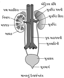
Describe the human excretory system.
Solution
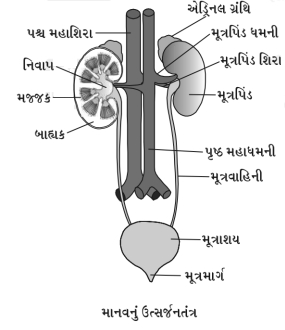
(N/A) In humans,the excretory system consists of a pair of kidneys,a pair of ureters,a urinary bladder,and a urethra.
Kidney: It is a reddish-brown,bean-shaped structure.
$(i)$ Location: It is situated between the levels of the last thoracic and third lumbar vertebra,close to the dorsal inner wall of the abdominal cavity.
(ii) Size: Each kidney of an adult human measures $10-12 \ cm$ in length,$5-7 \ cm$ in width,and $2-3 \ cm$ in thickness.
(iii) Weight: The average weight is $120-170 \ g$.
Both kidneys are not situated on the same plane; the right kidney is slightly lower than the left because the liver is situated above it on the right side of the thoracic cavity.
The external surface of the kidney is convex,and the internal surface is concave.
Internal structure of the kidney: $A$ longitudinal section ($L$.$S$.) of the kidney shows a large opening at the concave surface called the hilum.
Through the hilum,the ureter,blood vessels,and nerves enter.
Inner to the hilum is a broad,funnel-shaped space called the renal pelvis.
Projections of the renal pelvis are called calyces.
The outer layer of the kidney is a tough capsule.
Inside the kidney,there are two zones: an outer cortex and an inner medulla.
The medulla is divided into a few conical masses (medullary pyramids) projecting into the calyces.
The cortex extends in between the medullary pyramids as renal columns called columns of Bertini.
Each kidney has nearly one million complex tubular structures called nephrons.
0 likes
View Solution76
Easy
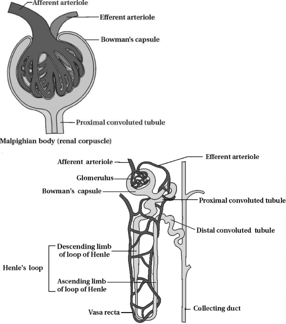
Describe the internal structure of a nephron.
Solution
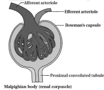
(N/A) Each nephron consists of two major parts:
$(1)$ Malpighian body (Renal corpuscle) and $(2)$ Renal tubule.
$(1)$ Malpighian body: It is a complex structure formed by the glomerulus and Bowman's capsule.
$(i)$ Glomerulus: It is a tuft of capillaries formed by the afferent arteriole,which is a fine branch of the renal artery. Blood from the glomerulus is carried away by an efferent arteriole. The diameter of the efferent arteriole is smaller than that of the afferent arteriole,which helps in creating the pressure required for filtration.
$(ii)$ Bowman's capsule: It is a cup-shaped,double-walled sac that encloses the glomerulus. The outer wall is formed of squamous epithelium,while the inner visceral layer contains specialized cells called podocytes. These cells are arranged in an intricate manner to leave some spaces called filtration slits or slit pores,which allow for the filtration of blood.
$(2)$ Renal tubule: The tubule begins with a proximal convoluted tubule $(PCT)$,followed by the Henle's loop,which is a hairpin-shaped structure with descending and ascending limbs. The ascending limb continues as a highly coiled distal convoluted tubule $(DCT)$.
- The $DCTs$ of many nephrons open into a straight tube called the collecting duct.
- Many collecting ducts converge and open into the renal pelvis through the medullary pyramids in the calyces.
- Urine formation occurs in the nephron,while the collecting duct primarily transports and concentrates the urine before it is eliminated.
0 likes
View Solution77
Easy
Write a short note on the types of nephrons.
Solution
(N/A) Based on their location in the kidney,nephrons are classified into two types:
$(A)$ Juxtamedullary Nephron: These constitute about $15\%$ of the total nephrons. They are large in size,and their Henle's loops are very long,extending deep into the medulla. They are associated with well-developed vasa recta.
$(B)$ Cortical Nephron: These constitute about $85\%$ of the total nephrons. Their Henle's loops are very short and extend only a little into the medulla. In these nephrons,the vasa recta is either absent or highly reduced.
0 likes
View Solution78
Medium
Mention the location,size,and weight of the kidney.
Solution
(N/A) $(i)$ Location: The kidneys are situated between the levels of the last thoracic and third lumbar vertebra,close to the dorsal inner wall of the abdominal cavity.
$(ii)$ Size: Each kidney of an adult human measures $10-12 \text{ cm}$ in length,$5-7 \text{ cm}$ in width,and $2-3 \text{ cm}$ in thickness.
$(iii)$ Weight: The average weight of a kidney is $120-170 \text{ g}$.
$(ii)$ Size: Each kidney of an adult human measures $10-12 \text{ cm}$ in length,$5-7 \text{ cm}$ in width,and $2-3 \text{ cm}$ in thickness.
$(iii)$ Weight: The average weight of a kidney is $120-170 \text{ g}$.
0 likes
View Solution79
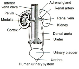
Medium
Draw a labeled diagram showing the various parts of the human excretory system.
Solution
(N/A) The human excretory system consists of the following structures:
$1$. $A$ pair of kidneys: Bean-shaped organs responsible for filtering blood and forming urine.
$2$. $A$ pair of ureters: Ducts that carry urine from the kidneys to the urinary bladder.
$3$. Urinary bladder: $A$ muscular sac that stores urine until it is voided.
$4$. Urethra: $A$ tube through which urine is expelled from the body.
$5$. Associated blood vessels: Renal artery (supplies blood to kidneys) and renal vein (drains blood from kidneys),along with the dorsal aorta and inferior vena cava.
$6$. Adrenal glands: Endocrine glands located on the superior aspect of each kidney.
0 likes
View Solution80
EasyMCQ
Distinguish between: Juxtamedullary and Cortical nephrons.
A
Juxtamedullary nephrons
B
Cortical nephrons
Solution
(N/A) Juxtamedullary nephrons: These constitute about $15\%$ of the total nephrons. These nephrons are larger in size. Their loops are associated with the $Vasa \ recta$. Blood first passes through the glomerulus and then flows through the $Vasa \ recta$ of the Loop of Henle. In these units,the Loop of Henle is very long and extends deep into the medulla.
$(B)$ Cortical nephrons: The Loop of Henle is short and extends only a little into the medulla. They constitute about $85\%$ of the total nephrons. The $Vasa \ recta$ is absent or highly reduced. The efferent arteriole emerging from the glomerulus forms a fine capillary network around the renal tubule,called the $Peritubular \ capillary$ network. $A$ minute vessel from this network runs parallel to the Loop of Henle to form a '$U$' shaped $Vasa \ recta$.
$(B)$ Cortical nephrons: The Loop of Henle is short and extends only a little into the medulla. They constitute about $85\%$ of the total nephrons. The $Vasa \ recta$ is absent or highly reduced. The efferent arteriole emerging from the glomerulus forms a fine capillary network around the renal tubule,called the $Peritubular \ capillary$ network. $A$ minute vessel from this network runs parallel to the Loop of Henle to form a '$U$' shaped $Vasa \ recta$.
0 likes
View Solution81
Medium
What is the difference between an afferent arteriole and an efferent arteriole?
Solution
(N/A)
| Afferent arteriole | Efferent arteriole |
|---|---|
| $(1)$ $A$ branch of the renal artery that enters the Bowman's capsule. | $(1)$ Formed by the union of glomerular capillaries that exit the Bowman's capsule. |
| $(2)$ It has a thicker wall. | $(2)$ It has a thinner wall. |
| $(3)$ Its diameter is wider. | $(3)$ Its diameter is narrower. |
| $(4)$ The blood flowing in it contains a higher concentration of excretory wastes. | $(4)$ The blood flowing in it is filtered and contains concentrated plasma proteins and blood cells. |
0 likes
View Solution82
Easy
Provide definitions/explanations for the following terms:
$(1)$ Excretion
$(2)$ Renal pelvis
$(1)$ Excretion
$(2)$ Renal pelvis
Solution
(N/A) $(1)$ Excretion: The biological process by which nitrogenous metabolic wastes and other unwanted substances are eliminated from the body.
$(2)$ Renal pelvis: $A$ broad,funnel-shaped space located inner to the hilum of the kidney,which collects urine before it passes into the ureter.
$(2)$ Renal pelvis: $A$ broad,funnel-shaped space located inner to the hilum of the kidney,which collects urine before it passes into the ureter.
0 likes
View Solution83
Easy
Provide definitions/explanations for the following terms:
$(1)$ Calyces
$(2)$ Renal pyramids
$(1)$ Calyces
$(2)$ Renal pyramids
Solution
(N/A) $(1)$ Calyces: These are cup-like extensions or projections of the renal pelvis that collect urine from the renal papillae.
$(2)$ Renal pyramids: These are cone-shaped structures found in the renal medulla of the kidney,which contain the loops of Henle and collecting ducts.
$(2)$ Renal pyramids: These are cone-shaped structures found in the renal medulla of the kidney,which contain the loops of Henle and collecting ducts.
0 likes
View Solution84
Easy
Provide definitions/explanations for the following terms:
$(1)$ Column of Bertini
$(2)$ Excretory unit (nephron)
$(1)$ Column of Bertini
$(2)$ Excretory unit (nephron)
Solution
(N/A) $(1)$ The renal cortex extends in between the medullary pyramids as renal columns,which are known as the columns of Bertini.
$(2)$ The nephron is the structural and functional unit of the kidney,responsible for the filtration of blood and the formation of urine.
$(2)$ The nephron is the structural and functional unit of the kidney,responsible for the filtration of blood and the formation of urine.
0 likes
View Solution85
Easy
Provide definitions/explanations for the following terms:
$(1)$ Malpighian body
$(2)$ Sebaceous gland
$(1)$ Malpighian body
$(2)$ Sebaceous gland
Solution
(N/A) $(1)$ Malpighian body: It is a functional component of the nephron in the kidney. It is formed by the combination of the glomerulus and the Bowman's capsule.
$(2)$ Sebaceous gland: It is a typical exocrine gland present in the skin of mammals. It secretes a mixture of wax,sterols,fatty acids,and hydrocarbons,collectively known as sebum,which helps in keeping the skin moist and waterproof.
$(2)$ Sebaceous gland: It is a typical exocrine gland present in the skin of mammals. It secretes a mixture of wax,sterols,fatty acids,and hydrocarbons,collectively known as sebum,which helps in keeping the skin moist and waterproof.
0 likes
View Solution86
Easy
Show the structure of a renal corpuscle with the help of a diagram.
Solution
(N/A) $(1)$ Malpighian Body: It is a complex structure formed of the glomerulus and Bowman's capsule.
$(i)$ Glomerulus: It is a tuft of capillaries formed by the afferent arteriole,which is a fine branch of the renal artery. Blood from the glomerulus is carried away by the efferent arteriole.
The diameter of the efferent arteriole is smaller than that of the afferent arteriole. Blood is filtered in the glomerulus.
$(ii)$ Bowman's Capsule: One side of the closed uriniferous tubule invaginates to form a cup-shaped,double-walled sac-like structure known as Bowman's capsule.
The outer wall of Bowman's capsule is formed of squamous epithelium,while the inner side possesses specific cells called podocytes. These cells are arranged in a special manner to form filtration slits (pores),which allow molecules to pass through them.
Bowman's capsule and the glomerulus together are termed the Malpighian body.
0 likes
View Solution87
Easy
Label the parts in the following diagram.
Solution
(N/A) The diagram represents the Malpighian body (renal corpuscle) of a nephron. The parts are labeled as follows:
$1$. Afferent arteriole: The vessel that brings blood into the glomerulus.
$2$. Efferent arteriole: The vessel that carries blood away from the glomerulus.
$3$. Bowman's capsule: The cup-shaped structure that encloses the glomerulus.
$4$. Glomerulus: $A$ tuft of capillaries formed by the afferent arteriole.
0 likes
View Solution88
Medium
Describe the structure of a human kidney with the help of a labelled diagram.
Solution
(N/A) The internal structure of the human kidney can be described as follows:
$1$. External Features: The kidney is a bean-shaped organ. On its concave surface,there is a notch called the hilum,through which the renal artery,renal vein,and ureter enter or exit the kidney.
$2$. Renal Pelvis: Inner to the hilum is a broad,funnel-shaped space called the renal pelvis,with projections called calyces.
$3$. Capsule: The outer layer of the kidney is covered by a tough,fibrous connective tissue capsule.
$4$. Internal Zones: Inside the kidney,there are two distinct zones: an outer cortex and an inner medulla.
$5$. Medullary Pyramids: The medulla is divided into several conical masses known as medullary pyramids,which project into the calyces.
$6$. Renal Columns: The cortex extends in between the medullary pyramids as renal columns,which are known as the columns of Bertini.
$7$. Nephrons: Each kidney contains nearly one million complex tubular structures called nephrons,which are the functional units of the kidney.
0 likes
View Solution89
Medium
Analogy type question:
$(1)$ Inner concave notch of kidney : Hilum :: Broad funnel-shaped part of renal pelvis : ............. .
$(2)$ Renal tubule begins with a double-walled cup-like structure called Bowman's capsule :: Bowman's capsule along with glomerulus : ............. .
$(1)$ Inner concave notch of kidney : Hilum :: Broad funnel-shaped part of renal pelvis : ............. .
$(2)$ Renal tubule begins with a double-walled cup-like structure called Bowman's capsule :: Bowman's capsule along with glomerulus : ............. .
Solution
(N/A) $(1)$ Renal calyx (or Calyces)
$(2)$ Malpighian body (or Renal corpuscle)
Explanation:
$(1)$ The hilum is the notch on the inner concave side of the kidney. Similarly,the renal pelvis projects into funnel-shaped structures called calyces.
$(2)$ The renal tubule starts with Bowman's capsule. The combination of Bowman's capsule and the enclosed glomerulus is collectively known as the Malpighian body or renal corpuscle.
0 likes
View Solution90
MediumMCQ
Choose the correct option:
$(1)$ $ANF$ is secreted when blood pressure decreases / blood pressure increases.
$(2)$ Renal pyramids are located in the renal cortex / renal medulla.
$(1)$ $ANF$ is secreted when blood pressure decreases / blood pressure increases.
$(2)$ Renal pyramids are located in the renal cortex / renal medulla.
A
$(1)$ Blood pressure decreases,$(2)$ Renal cortex
B
$(1)$ Blood pressure increases,$(2)$ Renal medulla
C
$(1)$ Blood pressure decreases,$(2)$ Renal medulla
D
$(1)$ Blood pressure increases,$(2)$ Renal cortex
Solution
(B) $(1)$ $ANF$ (Atrial Natriuretic Factor) is a peptide hormone secreted by the atrial wall of the heart in response to an increase in blood flow or blood pressure to promote vasodilation and excretion of sodium.
$(2)$ The renal medulla contains conical masses called renal pyramids,which project into the calyces.
$(2)$ The renal medulla contains conical masses called renal pyramids,which project into the calyces.
0 likes
View Solution91
Medium
Draw a neat and labelled diagram of the human excretory system.
Solution
(N/A) In humans,the excretory system consists of a pair of kidneys,a pair of ureters,a urinary bladder,and a urethra.
Kidney: Reddish-brown,bean-shaped structure.
$(i)$ Location: It is situated between the levels of the last thoracic and third lumbar vertebra,close to the dorsal inner wall of the abdominal cavity.
$(ii)$ Size: Each kidney of an adult human measures $10-12 \text{ cm}$ in length,$5-7 \text{ cm}$ in width,and $2-3 \text{ cm}$ in thickness.
$(iii)$ Weight: Average weight is $120-170 \text{ g}$.
Human Urinary System:
Both kidneys are not situated on the same plane; the right kidney is slightly on a lower plane than the left because the liver is situated above it in the right side of the thoracic cavity.
The external surface of the kidney is convex,and the internal surface is concave.
Internal structure of the kidney: The longitudinal section $(L.S.)$ of the kidney shows,at the concave surface,a large opening called the Hilum.
Through the hilum,the ureter,blood vessels,and nerves enter.
Inner to the hilum is a broad,funnel-shaped space called the renal pelvis.
Projections of the renal pelvis are called calyces.
The outer layer of the kidney is a tough capsule.
Inside the kidney,there are two zones: an outer cortex and an inner medulla.
The medulla is divided into a few conical masses (medullary pyramids) projecting into the calyces.
The cortex extends in between the medullary pyramids as renal columns called columns of Bertini.
Each kidney has nearly one million complex tubular structures called nephrons.
0 likes
View Solution92
Medium
Draw a neat and labelled diagram of an excretory unit (nephron).
Solution
(N/A) Each nephron consists of two main parts:
$(1)$ Malpighian body (Renal corpuscle) and $(2)$ Renal tubule.
Each nephron is approximately $3 \ cm$ in length and $20-30 \ \mu m$ in width.
$(1)$ Malpighian body: It is a complex structure formed by the glomerulus and Bowman's capsule.
$(i)$ Glomerulus: It is a tuft of capillaries formed by the afferent arteriole,which is a fine branch of the renal artery. Blood from the glomerulus is carried away by the efferent arteriole.
- The diameter of the efferent arteriole is smaller than that of the afferent arteriole,which helps in the filtration of blood in the glomerulus.
$(ii)$ Bowman's capsule: It is a cup-shaped,double-walled sac that encloses the glomerulus. The outer wall is composed of squamous epithelium,while the inner wall contains specialized cells called podocytes. These cells are arranged in an intricate manner to form filtration slits (or filtration pores),which allow the passage of molecules.
$(2)$ Renal tubule: The part of the nephron immediately following the Malpighian body is the proximal convoluted tubule $(PCT)$,which is a highly coiled structure.
$(i)$ Loop of Henle: It is a hairpin-shaped region following the $PCT$. It consists of a descending limb and an ascending limb.
$(ii)$ Distal convoluted tubule $(DCT)$: The ascending limb continues into another highly coiled tubular region called the $DCT$.
- The $DCT$ of many nephrons opens into a straight tube called the collecting duct.
- Many collecting ducts converge and open into the renal pelvis through medullary pyramids in the calyces.
- Urine formation occurs in the nephron,while the collecting duct transports the urine to the renal pelvis for elimination.
0 likes
View Solution93
MediumMCQ
Part of the kidney through which the ureter, blood vessels and nerves enter is
A
Renal cortex
B
Renal medulla
C
Hilum
D
Urethra
Solution
(C) Towards the center of the inner concave surface of the kidney, there is a notch called the $Hilum$ through which the ureter, blood vessels, and nerves enter the kidney.
Inner to the $Hilum$ is a broad, funnel-shaped space called the $Renal \text{ } pelvis$ with projections called $Calyces$.
Inner to the $Hilum$ is a broad, funnel-shaped space called the $Renal \text{ } pelvis$ with projections called $Calyces$.
0 likes
View Solution94
MediumMCQ
Malpighian body or renal corpuscle is/are:
A
Bowman's capsule
B
Glomerulus
C
Both $(a)$ and $(b)$
D
Proximal convoluted tubule
Solution
(C) The Malpighian body,also known as the renal corpuscle,is the filtration unit of the kidney.
It consists of two main structures: the glomerulus and the Bowman's capsule.
The glomerulus is a tuft of capillaries formed by the afferent arteriole,and the Bowman's capsule is a double-walled cup-like structure that surrounds the glomerulus.
Therefore,both $(a)$ and $(b)$ are correct components of the Malpighian body.
0 likes
View Solution95
EasyMCQ
The primary or main excretory organ in humans is:
A
Skin
B
Lung
C
Kidney
D
Spleen
Solution
(C) The primary excretory organs are specialized structures dedicated to the function of excretion.
In humans,the kidneys are the main excretory organs responsible for filtering the blood,removing metabolic wastes like urea,and maintaining water and electrolyte balance.
In humans,the kidneys are the main excretory organs responsible for filtering the blood,removing metabolic wastes like urea,and maintaining water and electrolyte balance.
0 likes
View Solution96
MediumMCQ
Identify $A$ to $D$ in the given structure and choose the correct option accordingly.
A
$A-$Calyx,$B-$Cortex,$C-$Renal column,$D-$Ureter
B
$A-$Calyx,$B-$Cortex,$C-$Renal column,$D-$Urethra
C
$A-$Urethra,$B-$Cortex,$C-$Renal column,$D-$Calyx
D
$A-$Urethra,$B-$Calyx,$C-$Renal column,$D-$Cortex
Solution
(A) Based on the anatomical structure of the human kidney:
$A$ points to the Calyx (minor calyx),which collects urine from the renal papillae.
$B$ points to the Cortex,the outer granular layer of the kidney.
$C$ points to the Renal column (Column of Bertini),which is the extension of the cortex between the renal pyramids.
$D$ points to the Ureter,the tube that carries urine from the kidney to the urinary bladder.
Therefore,the correct identification is $A-$Calyx,$B-$Cortex,$C-$Renal column,$D-$Ureter.
0 likes
View Solution97
MediumMCQ
In humans,the majority of nephrons have their loop of Henle in the:
A
Cortical region of the kidney
B
Medullary region of the kidney
C
Both $(a)$ and $(b)$
D
Pelvis region of the kidney
Solution
(A) In the human kidney,there are two types of nephrons: cortical nephrons and juxtamedullary nephrons.
About $80\%$ of the nephrons are cortical nephrons,which have a very short loop of Henle that extends only slightly into the medulla,meaning the majority of the loop is located in the cortical region of the kidney.
Only about $20\%$ of the nephrons are juxtamedullary nephrons,which have a long loop of Henle that dips deep into the medulla.
0 likes
View Solution98
MediumMCQ
The structural and functional unit of the human kidney is:
A
Nephron
B
Ureter
C
Loop of Henle
D
Bowman's capsule
Solution
(A) The nephron is the structural and functional unit of the human kidney. Each kidney contains approximately $1$ million nephrons,which are responsible for the filtration of blood and the formation of urine. While the Loop of Henle and Bowman's capsule are parts of the nephron,the nephron itself is the complete unit.
0 likes
View Solution99
EasyMCQ
Bowman's capsule is mainly found in
A
Glomerulus
B
Uriniferous tubule
C
Nephron
D
Malpighian corpuscle
Solution
(D) Each nephron or uriniferous tubule is composed of two main parts: the Malpighian body and the renal tubule.
The Malpighian body consists of the glomerulus and Bowman's capsule.
Bowman's capsule is a double-walled,cup-shaped structure that encloses the glomerulus,forming the initial part of the nephron.
The Malpighian body consists of the glomerulus and Bowman's capsule.
Bowman's capsule is a double-walled,cup-shaped structure that encloses the glomerulus,forming the initial part of the nephron.
0 likes
View Solution100
EasyMCQ
In humans,the primary excretory system consists of:
$I.$ Pair of kidneys
$II.$ One pair of ureters
$III.$ Urinary bladder
$IV.$ Urethra
$V.$ Skin
$VI.$ Lungs
$VII.$ Liver
$I.$ Pair of kidneys
$II.$ One pair of ureters
$III.$ Urinary bladder
$IV.$ Urethra
$V.$ Skin
$VI.$ Lungs
$VII.$ Liver
A
$I, II, III$ and $IV$
B
$III, IV, V$ and $VI$
C
$II, III, IV$ and $V$
D
$I, II, III, IV, V, VI$ and $VII$
Solution
(A) The human excretory system is primarily responsible for the removal of nitrogenous wastes from the body.
It consists of the following structures:
$(i)$ $A$ pair of kidneys: These filter the blood to produce urine.
$(ii)$ $A$ pair of ureters: These transport urine from the kidneys to the urinary bladder.
$(iii)$ Urinary bladder: This stores urine until it is voided.
$(iv)$ Urethra: This is the tube through which urine is expelled from the body.
Therefore,the correct components are $I, II, III$ and $IV$.
0 likes
View SolutionExcretory Products and their Elimination — Human excretory system · Frequently Asked Questions
1Are these Excretory Products and their Elimination questions useful for JEE and NEET?
Yes. All questions in this section are mapped to JEE Main and NEET exam patterns. Previous year questions from JEE Main, NEET, GUJCET and state-level exams are included with full solutions.
2Can I switch to Hindi or Gujarati for these questions?
Yes. Use the language tabs in the hero section or the sidebar to view the same questions and solutions in English, Hindi or Gujarati.
3How do I generate a question paper from this subtopic?
Use the Vedclass Exam Paper Generator — select the chapter and subtopic, set difficulty, and generate Sets A, B, C, D automatically. First 3 chapters of every subject are free.
Vedclass Products
For Students
Vedclass Test Series
Mock tests in real JEE/NEET style with performance analysis. 5-day free trial.
Start Free TrialFor Teachers
Exam Paper Generator
Generate Set A/B/C/D papers from this chapter in 2 minutes. 3 chapters free.
Try FreeFor Institutes
Online Exam Module
Live online exams with unlimited students, 360° analytics & white-label branding.
See DemoFor Teachers & Institutes
Generate a Excretory Products and their Elimination Exam Paper in 2 Minutes
Select subtopic & difficulty — Sets A, B, C, D auto-generated with No Repeat logic.
First 3 chapters of every subject are free — no payment required.