
A English
Muscles Questions in English
Class 11 Biology · Locomotion and Movement · Muscles
365+
Questions
English
Language
100%
With Solutions
Showing 50 of 365 questions in English
251
MediumMCQ
In the thin filament of skeletal muscle fibre,a small globular protein that masks the active sites for myosin on the $F-$actin,is
A
$G-$actin
B
Actin
C
Tropomyosin
D
Troponin
Solution
(D) The thin filament of skeletal muscle fibre is composed of three distinct proteins: actin,tropomyosin,and troponin.
Troponin is a complex of three globular proteins that masks the active binding sites for myosin on the $F-$actin filaments in the resting state.
Tropomyosin is a fibrous protein that runs along the $F-$actin filaments.
Therefore,the small globular protein that masks the active sites is troponin.
Troponin is a complex of three globular proteins that masks the active binding sites for myosin on the $F-$actin filaments in the resting state.
Tropomyosin is a fibrous protein that runs along the $F-$actin filaments.
Therefore,the small globular protein that masks the active sites is troponin.
0 likes
View Solution252
MediumMCQ
Myofilaments or myofibrils are
A
Obliquely arranged filaments of muscle fibre
B
Parallely arranged filaments of muscle fibre
C
Horizontally arranged filaments of muscle fibre
D
Radially arranged filaments of muscle fibre
Solution
(B) Myofilaments or myofibrils are parallely arranged filaments of muscle fibre.
Each muscle fibre is lined by a plasma membrane called the sarcolemma,which encloses the sarcoplasm.
$A$ muscle fibre is a syncytium because the sarcoplasm contains many nuclei.
The endoplasmic reticulum,i.e.,the sarcoplasmic reticulum of the muscle fibres,is the storehouse of calcium ions.
$A$ characteristic feature of the muscle fibre is the presence of a large number of parallely arranged filaments in the sarcoplasm,which are called myofilaments or myofibrils.
0 likes
View Solution253
MediumMCQ
The storehouse of calcium ions in the muscle fibre is
A
Endoplasmic reticulum
B
Golgi body
C
Mitochondria
D
Lysosomes
Solution
(A) The storehouse of calcium ions in the muscle fibre is the sarcoplasmic reticulum.
In muscle fibres,the endoplasmic reticulum $(ER)$ is specifically referred to as the sarcoplasmic reticulum.
Each muscle fibre is enclosed by a plasma membrane called the sarcolemma,which contains the sarcoplasm.
Muscle fibres are syncytial,meaning the sarcoplasm contains many nuclei.
$A$ characteristic feature of muscle fibres is the presence of a large number of parallelly arranged filaments in the sarcoplasm,known as myofilaments or myofibrils.
0 likes
View Solution254
MediumMCQ
Neuromuscular junction is a junction between
A
Two neurons and muscles
B
Sensory neurons and muscles
C
Motor neurons and sarcolemma of muscles
D
Sensory neurons and sarcolemma of muscles
Solution
(C) The junction between a motor neuron and the sarcolemma of the muscle fibre is called the neuromuscular junction or motor end plate.
$A$ neural signal reaching this junction releases a neurotransmitter,$Acetylcholine$,which generates an action potential in the sarcolemma.
$A$ neural signal reaching this junction releases a neurotransmitter,$Acetylcholine$,which generates an action potential in the sarcolemma.
0 likes
View Solution255
MediumMCQ
$A$ sarcomere in the myofibrils of muscle is found in between
A
$2$ $M$-lines
B
$2$ $Z$-lines
C
$2$ $H$-lines
D
$2$ $A$-bands
Solution
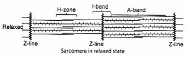
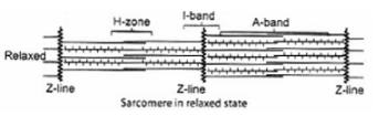
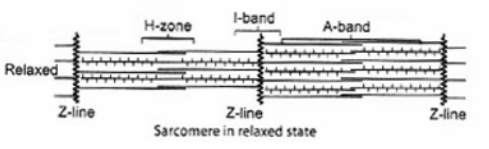
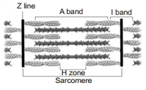
(B) The correct answer is $2$ $Z$-lines.
In the myofibrils of muscle,the thick filaments lie parallel to one another,and thin filaments are present in an orderly array between the thick filaments.
In the centre of the $I$-band,there is an elastic fibre called the $Z$-line which bisects it.
The portion of the myofibril between two successive $Z$-lines is considered as the functional unit of contraction and is called a sarcomere.
In the middle of the $A$-band,a comparatively less dark zone called the $H$-zone is present,and the $M$-line is present in the middle of the $H$-zone.
0 likes
View Solution256
MediumMCQ
In a $A$ state,the edge of thin filaments on either side of thick filaments $B$ overlap the free ends of $C$ filaments leaving the central part of thick filaments. This central part of thick filament,not overlapped by thin filaments is called $D$ zone.
Choose the correct options to fill the gaps $A, B, C$ and $D$,so as to complete the given $NCERT$ statement.
Choose the correct options to fill the gaps $A, B, C$ and $D$,so as to complete the given $NCERT$ statement.
A
$A-$resting,$B-$partially,$C-$thick,$D-H$
B
$A-$resting,$B-$partially,$C-$thin,$D-A$
C
$A-$resting,$B-$partially,$C-$thin,$D-H$
D
$A-$resting,$B-$partially,$C-$thick,$D-M$
Solution
(C) According to the sliding filament theory described in $NCERT$,in a resting state,the edges of thin filaments on either side of the thick filaments partially overlap the free ends of the thin filaments,leaving the central part of the thick filaments.
This central part of the thick filament,which is not overlapped by thin filaments,is called the $H$-zone.
Therefore,the correct sequence is $A-$resting,$B-$partially,$C-$thin,$D-H$.
This central part of the thick filament,which is not overlapped by thin filaments,is called the $H$-zone.
Therefore,the correct sequence is $A-$resting,$B-$partially,$C-$thin,$D-H$.
0 likes
View Solution257
EasyMCQ
Sliding filament theory was given by
A
$AF$ Huxley and $T$ Huxley
B
Leeuwenhoek and Hooke
C
$AF$ Huxley and $HE$ Huxley
D
$AF$ Huxley and Robert Hooke
Solution
(C) The sliding filament theory was proposed by $AF$ Huxley and $HE$ Huxley in $1954$.
This theory explains the mechanism of muscle contraction,describing how actin and myosin filaments slide past each other to shorten the sarcomere without the filaments themselves changing in length.
This theory explains the mechanism of muscle contraction,describing how actin and myosin filaments slide past each other to shorten the sarcomere without the filaments themselves changing in length.
0 likes
View Solution258
MediumMCQ
In which of the following,growth is possible through increase in volume?
A
Cartilage
B
Striated muscle
C
Nerve fiber
D
Lens of eye
Solution
(B) Growth in biological systems occurs through various mechanisms.
$1$. Cartilage grows primarily through the secretion of extracellular matrix.
$2$. Striated muscles grow through hypertrophy,which is an increase in the volume of individual muscle fibers.
$3$. Nerve fibers grow through the extension and elongation of axons and dendrites.
$4$. The lens of the eye grows through the multiplication of cells (hyperplasia).
Therefore,growth through an increase in volume is characteristic of striated muscles.
0 likes
View Solution259
MediumMCQ
Striated appearance of the myofibrils is due to
A
Actin proteins
B
Myosin proteins
C
Both $(a)$ and $(b)$
D
None of these
Solution
(C) Each myofibril has alternate dark and light bands on it.
$A$ detailed study of the myofibril has established that the striated appearance is due to the distribution pattern of two important contractile proteins,$i.e.$,actin and myosin.
Actin forms the thin filaments (light bands),while myosin forms the thick filaments (dark bands),creating the characteristic striated pattern.
0 likes
View Solution260
MediumMCQ
Globular head with a short arm and a tail are the two important parts of
A
$F-actin$
B
$G-actin$
C
Tropomyosin
D
Meromyosin
Solution
(D) Each myosin (thick) filament is a polymerized protein. Many monomeric proteins called meromyosins constitute one thick filament. Each meromyosin has two important parts: a globular head with a short arm and a tail. The head with the short arm is known as Heavy Meromyosin $(HMM)$,and the tail is known as Light Meromyosin $(LMM)$.
0 likes
View Solution261
MediumMCQ
Aerobic muscles are called $...A...$ and anaerobic muscles are called $...B....$ Here $A$ and $B$ refer to:
A
$A-$red fibres; $B-$white fibres
B
$A-$white fibres; $B-$red fibres
C
$A-$white fibres; $B-$black fibres
D
$A-$red fibres; $B-$black fibres
Solution
(A) Muscles contain a red-coloured oxygen-storing pigment called myoglobin.
Myoglobin content is high in some muscles,which gives them a reddish appearance; these are called red fibres.
Red fibres contain a large number of mitochondria,which utilize the stored oxygen for $ATP$ production,hence they are known as aerobic muscles.
Conversely,muscles with a low quantity of myoglobin appear pale or whitish and are called white fibres.
White fibres have fewer mitochondria and rely more on anaerobic processes for energy,hence they are known as anaerobic muscles.
0 likes
View Solution262
EasyMCQ
Latissimus dorsi muscles are
A
Muscles of forearm
B
Muscles of lower jaw
C
Muscles of chest
D
Muscles of shoulder
Solution
(D) The $Latissimus$ $dorsi$ is a pair of large,triangular muscles located in the thoracic and lumbar regions of the back.
These muscles are responsible for the extension,adduction,and medial rotation of the arm.
Additionally,they play a key role in pulling the shoulder back and down.
These muscles are responsible for the extension,adduction,and medial rotation of the arm.
Additionally,they play a key role in pulling the shoulder back and down.
0 likes
View Solution263
MediumMCQ
$I-$bands of myofibrils are bisected by
A
$A-$bands
B
$H-$zone
C
$Z-$lines
D
$M-$lines
Solution
(C) The $I-$band (isotropic band) consists of thin actin filaments.
These bands are bisected by an elastic fiber called the $Z-$line,which runs through the center of each $I-$band.
The $Z-$line serves as the attachment point for actin filaments and defines the boundaries of a sarcomere.
These bands are bisected by an elastic fiber called the $Z-$line,which runs through the center of each $I-$band.
The $Z-$line serves as the attachment point for actin filaments and defines the boundaries of a sarcomere.
0 likes
View Solution264
MediumMCQ
During muscle contraction:
A
Thick filaments slide over thin filaments
B
$I$-band gets reduced
C
Both $(a)$ and $(b)$
D
None of the above
Solution
(C) During muscle contraction,the sliding filament theory explains that thin filaments slide over thick filaments. As a result,the $Z$-lines move closer together,causing the $I$-band to shorten or reduce in length,while the $A$-band maintains its original length.
0 likes
View Solution265
MediumMCQ
The region at the ends of the $A$-band of two adjoining sarcomeres is called
A
$H$-zone
B
$Z$-line
C
$I$-band
D
$M$-line
Solution
(B) The $I$-band is an isotropic band that contains thin filaments. The $Z$-line is a dark fibrous membrane that bisects the $I$-band. The region between two successive $Z$-lines is known as a sarcomere. The $A$-band contains thick filaments. The ends of the $A$-band of two adjoining sarcomeres are separated by the $I$-band,which contains the $Z$-line.
0 likes
View Solution266
MediumMCQ
Identify the muscle which represents the following characteristics and choose the correct option accordingly.
$I.$ Transportation of food through the digestive tract.
$II.$ Transportation of gametes through the genital tract.
$I.$ Transportation of food through the digestive tract.
$II.$ Transportation of gametes through the genital tract.
A
Skeletal muscles
B
Visceral muscles
C
Cardiac muscles
D
Striated muscles
Solution
(B) The correct answer is Visceral muscles.
Visceral muscles are located in the inner walls of hollow visceral organs of the body,such as the alimentary canal and the reproductive tract.
They do not exhibit any striations and are smooth in appearance; hence,they are called smooth muscles or non-striated muscles.
Their activities are not under the voluntary control of the nervous system and are therefore called involuntary muscles.
They assist in physiological processes such as the transportation of food through the digestive tract and the movement of gametes through the genital tract.
0 likes
View Solution267
MediumMCQ
Muscle contains a red-coloured oxygen-containing pigment called
A
Haemoglobin
B
Myoglobin
C
Haemocyanin
D
Both $(a)$ and $(b)$
Solution
(B) Muscle contains a red-coloured oxygen-containing pigment called myoglobin.
Myoglobin is an iron- and oxygen-binding protein found in the muscle tissue of vertebrates.
It is present in higher concentrations in red muscle fibres compared to white muscle fibres,which helps in aerobic respiration.
Myoglobin is an iron- and oxygen-binding protein found in the muscle tissue of vertebrates.
It is present in higher concentrations in red muscle fibres compared to white muscle fibres,which helps in aerobic respiration.
0 likes
View Solution268
MediumMCQ
Contraction of the muscles takes place by the sliding of
A
Thick filament over thin filament
B
Thin filament over thick filament
C
Thin filament over thin filament
D
Thick filament over thick filament
Solution
(B) The correct answer is $B$. The mechanism of muscle contraction is best explained by the sliding filament theory. This theory states that the contraction of a muscle fibre occurs when the thin filaments (actin) slide over the thick filaments (myosin),thereby shortening the sarcomere.
0 likes
View Solution269
MediumMCQ
Cross arms of the myosin monomer consist of:
A
Outward projection of $G-$actin filament
B
Outward projection of the head region of meromyosin
C
Outward projection of the tail region of meromyosin
D
Both $(b)$ and $(c)$
Solution
(B) The cross arm consists of the outward projection of the head region of meromyosin.
Each myosin (thick filament) is a polymerized protein consisting of many monomeric proteins called meromyosin.
Each meromyosin has two important parts: a globular head with a short arm and a tail.
The globular head with the short arm is called Heavy Meromyosin $(HMM)$,while the tail is called Light Meromyosin $(LMM)$.
The $HMM$ component,which includes the head and the short arm,projects outwards at regular distances and angles from the surface of the polymerized myosin filament; this structure is known as the cross arm.
The globular head acts as an active $ATPase$ enzyme and contains binding sites for $ATP$ and active sites for actin.
0 likes
View Solution270
MediumMCQ
Which of the following statements about the molecular arrangement of actin and myosin are correct?
$I.$ Each actin (thin filament) is made up of $2$ $F$ (filamentous) actins.
$II.$ $F$-actin is the polymer of $G$ (globular) actin.
$III.$ $2$ $F$-actins are twisted into a helix.
$IV.$ Two strands of tropomyosin (protein) lie in the grooves of $F$-actin.
The correct option is:
$I.$ Each actin (thin filament) is made up of $2$ $F$ (filamentous) actins.
$II.$ $F$-actin is the polymer of $G$ (globular) actin.
$III.$ $2$ $F$-actins are twisted into a helix.
$IV.$ Two strands of tropomyosin (protein) lie in the grooves of $F$-actin.
The correct option is:
A
$I$ and $II$
B
$III$ and $IV$
C
$I$ and $IV$
D
All except $IV$
Solution
(D) Statement $I$ is correct: Each thin filament (actin) is composed of two $F$-actin filaments.
Statement $II$ is correct: $F$-actin is a polymer of monomeric $G$-actin (globular actin) proteins.
Statement $III$ is correct: These two $F$-actin filaments are twisted into a double helix structure.
Statement $IV$ is correct: Two filaments of another protein,tropomyosin,run close to the $F$-actin throughout its length.
Since all statements $I, II, III,$ and $IV$ are correct,none of the provided options perfectly match the standard biological fact. However,based on the structure of the question,if we evaluate the options provided,there is a discrepancy. Re-evaluating: All statements are factually correct according to $NCERT$. Given the options,if the question implies selecting the set of correct statements,all are correct.
0 likes
View Solution271
MediumMCQ
During muscle contraction,$ATP$ provides energy for
A
Cross-bridge detachment
B
Building up action potential
C
Releasing $Ca^{2+}$ from sarcoplasmic reticulum
D
Cross-bridge attachment of myosin to actin
Solution
(A) During muscle contraction,$ATP$ is essential for the detachment of the myosin head from the actin filament.
$1$. When an $ATP$ molecule binds to the myosin head,it causes the cross-bridge to detach from the actin filament.
$2$. Subsequently,the hydrolysis of $ATP$ into $ADP$ and inorganic phosphate $(Pi)$ provides the energy required to 'cock' the myosin head into its high-energy position.
$3$. This energized myosin head then forms a new cross-bridge with the actin filament,leading to the power stroke that causes muscle contraction.
Therefore,the energy provided by $ATP$ is primarily utilized for the detachment of the cross-bridge and the subsequent preparation for the next cycle.
0 likes
View Solution272
MediumMCQ
What is the location of troponin in the process of muscle contraction?
A
Attached to myosin filament
B
Attached to tropomyosin
C
Attached to myosin cross bridge
D
Attached to $T$-tubule
Solution
(B) The proteins troponin and tropomyosin are closely associated with actin filaments. Troponin is a complex of three polypeptide subunits: $T_{n}C$,$T_{n}I$,and $T_{n}T$. Specifically,$T_{n}T$ binds to tropomyosin,$T_{n}I$ binds to actin,and $T_{n}C$ binds to $Ca^{2+}$ ions. Therefore,troponin is attached to tropomyosin on the actin filament.
0 likes
View Solution273
EasyMCQ
Visceral muscles are also called
A
Smooth muscles
B
Non-striated muscles
C
Involuntary muscles
D
All of these
Solution
(D) Visceral muscles are located in the inner walls of hollow visceral organs of the body,such as the alimentary canal and the reproductive tract.
They do not exhibit any striations and appear smooth under a microscope,hence they are called smooth muscles or non-striated muscles.
Their activities are not under the voluntary control of the nervous system; therefore,they are called involuntary muscles.
They assist in various physiological processes,such as the transportation of food through the digestive tract and the movement of gametes through the genital tract.
0 likes
View Solution274
MediumMCQ
Arrange the following steps of muscle contraction in the sequence of events occurring first:
$I.$ Receptor sites on sarcolemma
$II.$ Nerve impulse
$III.$ Release of $Ca^{2+}$
$IV.$ Acetylcholine release
$V.$ Shortening of sarcomere
$VI.$ Synaptic cleft
$VII.$ Spread of impulse over sarcolemma on $T$-tubule
The correct option is:
$I.$ Receptor sites on sarcolemma
$II.$ Nerve impulse
$III.$ Release of $Ca^{2+}$
$IV.$ Acetylcholine release
$V.$ Shortening of sarcomere
$VI.$ Synaptic cleft
$VII.$ Spread of impulse over sarcolemma on $T$-tubule
The correct option is:
A
$II \rightarrow IV \rightarrow VI \rightarrow I \rightarrow VII \rightarrow III \rightarrow V$
B
$II \rightarrow IV \rightarrow I \rightarrow VI \rightarrow VII \rightarrow III \rightarrow V$
C
$II \rightarrow IV \rightarrow I \rightarrow VI \rightarrow VII \rightarrow V \rightarrow III$
D
$IV \rightarrow II \rightarrow I \rightarrow VI \rightarrow VII \rightarrow V \rightarrow III$
Solution
(A) The sequence of events in muscle contraction is as follows:
$1$. $A$ nerve impulse arrives at the neuromuscular junction $(II)$.
$2$. This triggers the release of the neurotransmitter acetylcholine $(IV)$.
$3$. Acetylcholine diffuses across the synaptic cleft $(VI)$.
$4$. It binds to receptor sites on the sarcolemma $(I)$.
$5$. This generates an action potential that spreads over the sarcolemma and into the $T$-tubules $(VII)$.
$6$. The impulse triggers the release of $Ca^{2+}$ ions from the sarcoplasmic reticulum $(III)$.
$7$. $Ca^{2+}$ binds to troponin,leading to the sliding of filaments and the shortening of the sarcomere $(V)$.
Thus,the correct sequence is $II \rightarrow IV \rightarrow VI \rightarrow I \rightarrow VII \rightarrow III \rightarrow V$.
0 likes
View Solution275
MediumMCQ
Arrange the given steps of muscle contraction in the series of events from first to last.
$I.$ Myosin head binds to the exposed active site on actin to form a cross bridge.
$II.$ The $Z$-line attached to these actin are also pulled inwards,thereby causing shortening of sarcomere,also called contraction.
$III.$ This pulls the attached actin filaments towards the centre of $A$-band.
The correct option is:
A
$I \rightarrow II \rightarrow III$
B
$III \rightarrow II \rightarrow I$
C
$I \rightarrow III \rightarrow II$
D
$III \rightarrow I \rightarrow II$
Solution
(C) The process of muscle contraction occurs in the following sequence:
$1.$ By utilizing the energy from $ATP$ hydrolysis,the myosin head binds to the exposed active sites on actin to form a cross bridge $(I)$.
$2.$ This cross bridge formation pulls the attached actin filaments towards the centre of the $A$-band $(III)$.
$3.$ The $Z$-line attached to these actin filaments is also pulled inwards,thereby causing the shortening of the sarcomere,which is known as contraction $(II)$.
Therefore,the correct sequence is $I \rightarrow III \rightarrow II$.
0 likes
View Solution276
MediumMCQ
Relaxation of the muscle takes place due to
$I.$ Pumping of $Ca^{2+}$ ions into the sarcoplasmic reticulum
$II.$ Presence of $ATP$
$III.$ Conformational changes in troponin and masking of the actin filament
Which of the following options contains the correct statements?
$I.$ Pumping of $Ca^{2+}$ ions into the sarcoplasmic reticulum
$II.$ Presence of $ATP$
$III.$ Conformational changes in troponin and masking of the actin filament
Which of the following options contains the correct statements?
A
$I$ and $III$
B
$I$ and $II$
C
$II$ and $III$
D
$I, II,$ and $III$
Solution
(D) Muscle relaxation involves the following steps:
$1$. The $Ca^{2+}$ ions are actively pumped back into the sarcoplasmic reticulum,which lowers the cytosolic concentration of $Ca^{2+}$.
$2$. Due to the decrease in $Ca^{2+}$ concentration,the $Ca^{2+}$ ions dissociate from troponin.
$3$. This causes a conformational change in the troponin-tropomyosin complex,which leads to the masking of the active binding sites on the actin filament.
$4$. Consequently,the cross-bridge cycle stops,and the muscle relaxes.
$5$. $ATP$ is required for the detachment of the myosin head from the actin filament and for the active transport of $Ca^{2+}$ back into the sarcoplasmic reticulum. Therefore,all three statements are correct.
0 likes
View Solution277
MediumMCQ
The head of a myosin monomer consists of:
$I.$ Actin binding sites
$II.$ $ATP$ binding sites
$III.$ $ADP$ binding sites
$IV.$ $AMP$ binding sites
Select the correct options:
$I.$ Actin binding sites
$II.$ $ATP$ binding sites
$III.$ $ADP$ binding sites
$IV.$ $AMP$ binding sites
Select the correct options:
A
$I$ and $II$
B
$III$ and $IV$
C
$I$ and $IV$
D
$II$ and $IV$
Solution
(A) Each myosin (thick filament) is a polymerized protein. Many monomeric proteins called meromyosin constitute one thick filament.
Each meromyosin has two important parts: a globular head with a short arm and a tail. The former is called heavy meromyosin $(HMM)$ and the latter is called light meromyosin $(LMM)$.
The $HMM$ component,i.e.,the head and short arm,projects outwards at a regular distance and angle from the surface of the polymerized myosin filament and is called the cross-arm.
The globular head acts as an active $ATPase$ enzyme and contains binding sites for $ATP$ and active sites for actin.
0 likes
View Solution278
MediumMCQ
Skeletal muscles are closely associated with the $...A...$ components of the body. They have $... B...$ appearance under the microscope and hence are called $...C...$ muscles.
Choose the correct options to fill $A, B$ and $C$,so as to complete the given $NCERT$ statement.
Choose the correct options to fill $A, B$ and $C$,so as to complete the given $NCERT$ statement.
A
$A-$muscular,$B-$stripped,$C-$striated
B
$A-$visceral,$B-$stripped,$C-$striated
C
$A-$skeletal,$B-$stripped,$C-$striated
D
$A-$microfibrillar,$B-$stripped,$C-$striated
Solution
(C) According to the $NCERT$ textbook,skeletal muscles are closely associated with the skeletal components of the body.
They have a striped appearance under the microscope due to the presence of alternating light and dark bands,and hence are called striated muscles.
Therefore,the correct sequence is $A-$skeletal,$B-$stripped,$C-$striated.
They have a striped appearance under the microscope due to the presence of alternating light and dark bands,and hence are called striated muscles.
Therefore,the correct sequence is $A-$skeletal,$B-$stripped,$C-$striated.
0 likes
View Solution279
MediumMCQ
The set of ions necessary for muscle contraction is
A
$Ca^{2+}$ and $Mg^{2+}$
B
$Na^{+}$ and $Mg^{2+}$
C
$Na^{+}$ and $K^{+}$
D
$Na^{+}$ and $Ca^{2+}$
Solution
(A) $Ca^{2+}$ and $Mg^{2+}$ ions are essential for muscle contraction.
$Ca^{2+}$ ions bind to troponin, which exposes the active sites on actin filaments for myosin head attachment.
$Mg^{2+}$ ions act as a cofactor for the enzyme myosin ATPase, which is required for the hydrolysis of $ATP$ to provide energy for the contraction process.
Reaction: $\text{ATP} + H_{2}O \xrightarrow{Mg^{2+}/Ca^{2+}} \text{ADP} + P_{i} + \text{Energy}$.
0 likes
View Solution280
MediumMCQ
Which of the following statements are false regarding the muscle structure?
$I.$ In the centre of each $I$-band is an elastic fibre ($Z$-line) which bisects it.
$II.$ Thin filaments are firmly attached to the $Z$-line.
$III.$ $M$-line is a fibrous membrane in the middle of $A$-band.
$IV.$ $A$ sarcomere comprises one full $A$-band and two half $I$-bands.
$I.$ In the centre of each $I$-band is an elastic fibre ($Z$-line) which bisects it.
$II.$ Thin filaments are firmly attached to the $Z$-line.
$III.$ $M$-line is a fibrous membrane in the middle of $A$-band.
$IV.$ $A$ sarcomere comprises one full $A$-band and two half $I$-bands.
A
$I$ and $II$
B
$III$ and $IV$
C
$II$ and $III$
D
None of these
Solution
(D) Statement $I$ is correct: The $I$-band (isotropic band) contains actin filaments and is bisected by an elastic fibre called the $Z$-line.
Statement $II$ is correct: Thin filaments (actin) are anchored to the $Z$-line.
Statement $III$ is correct: The $M$-line is a thin fibrous membrane that holds the thick filaments (myosin) together in the centre of the $A$-band.
Statement $IV$ is correct: $A$ sarcomere is defined as the portion of a myofibril between two successive $Z$-lines,which consists of one full $A$-band and two half $I$-bands.
Since all statements are true,none of the statements are false.
0 likes
View Solution281
MediumMCQ
Select the correct statement with reference to muscle structure.
$I.$ Each myosin is a polymerized protein.
$II.$ Many meromyosin constitute one thick filament (myosin).
$III.$ Each meromyosin's tail is called heavy meromyosin $(HMM)$ and head is called light meromyosin $(LMM)$.
$IV.$ The globular head is an active $ATPase$ enzyme and has binding sites for $ATP$ and active sites for actin.
Choose the option with correct statements.
A
All except $I$
B
All except $IV$
C
All except $III$
D
All except $II$
Solution
(C) Analysis of statements regarding muscle structure:
$I.$ Correct: Myosin is a polymerized protein made up of many meromyosin units.
$II.$ Correct: Many meromyosin molecules aggregate to form one thick filament (myosin).
$III.$ Incorrect: The head of meromyosin is called heavy meromyosin $(HMM)$ and the tail is called light meromyosin $(LMM)$. The statement provided is reversed.
$IV.$ Correct: The globular head of myosin acts as an active $ATPase$ enzyme and contains binding sites for $ATP$ and active sites for actin.
Therefore,statements $I, II,$ and $IV$ are correct,while $III$ is incorrect. Thus,the correct option is $C$.
0 likes
View Solution282
MediumMCQ
Which of the following statements is true with reference to the structure of a muscle fibre?
A
$H-$zone is present in the middle of $A-$band.
B
$A-$band is present in the middle of sarcomere.
C
$M-$line is present in the middle of $H-$zone.
D
All of the above.
Solution
(D) All of the above statements are correct.
$1$. The thick filaments lie parallel to one another,and thin filaments are present in an orderly array between the thick filaments.
$2$. In the centre of the $I-$band,there is a band of amorphous material called the $Z-$line.
$3$. In the middle of the $A-$band,a comparatively less dark zone called the $H-$zone is present.
$4$. The area between two successive $Z-$lines is called a sarcomere,and the $A-$band is located in the middle of this sarcomere.
$5$. The $M-$line is a thin fibrous membrane that holds the thick filaments together and is present in the middle of the $H-$zone.
0 likes
View Solution283
MediumMCQ
$I.$ Sarcoplasmic reticulum are abundant
$II.$ Myoglobin content is high
$III.$ Sarcoplasmic reticulum are moderate
$IV.$ Aerobic muscles
$V.$ Depends on anaerobic respiration for energy
$VI.$ Less myoglobin content
Select the option with correct statements for red muscle.
$II.$ Myoglobin content is high
$III.$ Sarcoplasmic reticulum are moderate
$IV.$ Aerobic muscles
$V.$ Depends on anaerobic respiration for energy
$VI.$ Less myoglobin content
Select the option with correct statements for red muscle.
A
$I, II \; \text{and} \; IV$
B
$I, II \; \text{and} \; III$
C
$II, III \; \text{and} \; IV$
D
$II \; \text{and} \; IV$
Solution
(D) Red muscle fibers are characterized by a high content of myoglobin, which gives them a reddish appearance. They are also known as aerobic muscles because they rely primarily on aerobic respiration for energy production. Additionally, they possess a moderately developed sarcoplasmic reticulum compared to white muscle fibers. Therefore, statements $II$ (high myoglobin content) and $IV$ (aerobic muscles) are correct. Statement $III$ is also technically correct as they have moderate sarcoplasmic reticulum, but in the context of standard multiple-choice questions focusing on primary characteristics, $II$ and $IV$ are the most defining features. However, looking at the options provided, $II$ and $IV$ is the most accurate selection.
0 likes
View Solution284
MediumMCQ
Arrange the following steps of muscle relaxation in the correct chronological sequence:
$I.$ $Ca^{2+}$ ions are pumped back to the sarcoplasmic reticulum.
$II.$ $Z$-lines return to their original position.
$III.$ Masking of actin filaments occurs.
$IV.$ Relaxation of muscles.
$I.$ $Ca^{2+}$ ions are pumped back to the sarcoplasmic reticulum.
$II.$ $Z$-lines return to their original position.
$III.$ Masking of actin filaments occurs.
$IV.$ Relaxation of muscles.
A
$I \rightarrow II \rightarrow III \rightarrow IV$
B
$I \rightarrow III \rightarrow II \rightarrow IV$
C
$IV \rightarrow III \rightarrow II \rightarrow I$
D
$IV \rightarrow III \rightarrow I \rightarrow II$
Solution
(B) The process of muscle relaxation occurs in the following sequence:
$1$. $Ca^{2+}$ ions are pumped back into the sarcoplasmic reticulum,which leads to a decrease in their concentration in the sarcoplasm $(I)$.
$2$. This decrease in $Ca^{2+}$ concentration causes the troponin to move back,resulting in the masking of the active sites on the actin filaments $(III)$.
$3$. Due to the masking of active sites,the cross-bridges break,and the $Z$-lines return to their original position $(II)$.
$4$. Finally,the muscle fiber returns to its relaxed state $(IV)$.
Therefore,the correct sequence is $I \rightarrow III \rightarrow II \rightarrow IV$.
0 likes
View Solution285
MediumMCQ
Choose the correct properties of muscle fibres.
$I.$ Muscle fibre is lined by the plasma membrane called sarcolemma.
$II.$ Cytoplasm of the muscle fibre is called tonoplasm.
$III.$ Sarcolemma of the muscle fibre encloses the sarcoplasm.
$IV.$ Muscle fibre is a syncytium.
Select the correct option.
$I.$ Muscle fibre is lined by the plasma membrane called sarcolemma.
$II.$ Cytoplasm of the muscle fibre is called tonoplasm.
$III.$ Sarcolemma of the muscle fibre encloses the sarcoplasm.
$IV.$ Muscle fibre is a syncytium.
Select the correct option.
A
All except $II$
B
All except $I$
C
All except $III$
D
All except $IV$
Solution
(A) Each muscle fibre is lined by the plasma membrane called sarcolemma,which encloses the sarcoplasm.
Statement $I$ is correct because the plasma membrane of a muscle fibre is indeed called the sarcolemma.
Statement $II$ is incorrect because the cytoplasm of a muscle fibre is called sarcoplasm,not tonoplasm (tonoplasm is the membrane of the vacuole in plant cells).
Statement $III$ is correct because the sarcolemma encloses the sarcoplasm.
Statement $IV$ is correct because a muscle fibre is a syncytium,meaning it contains many nuclei within a single cell membrane.
Therefore,all statements are correct except $II$.
0 likes
View Solution286
MediumMCQ
Based on their location in the human body or animals,the types of muscles are:
$I.$ Skeletal
$II.$ Visceral
$III.$ Cardiac
$IV.$ Non-visceral
The correct option is:
$I.$ Skeletal
$II.$ Visceral
$III.$ Cardiac
$IV.$ Non-visceral
The correct option is:
A
$I$ and $II$
B
$II$ and $IV$
C
$I, II,$ and $III$
D
$I, III,$ and $IV$
Solution
(C) Muscles are classified based on different criteria,such as their location,appearance,and the nature of the regulation of their activities.
Based on their location in the body,muscles are classified into three types:
$1.$ Skeletal muscles: These are primarily attached to the bones of the skeletal system.
$2.$ Visceral muscles: These are located in the inner walls of hollow visceral organs of the body like the alimentary canal and reproductive tract.
$3.$ Cardiac muscles: These are the muscles of the heart.
Therefore,$I, II,$ and $III$ are the correct types based on location.
0 likes
View Solution287
MediumMCQ
Muscle fatigue occurs due to the accumulation of:
A
$CO_2$
B
Myosin ATPase
C
Lactic acid
D
Creatine phosphate
Solution
(C) Muscle fatigue occurs due to the accumulation of lactic acid in the muscle fibers. When muscles undergo strenuous exercise,the oxygen supply becomes insufficient for aerobic respiration. Consequently,the muscles switch to anaerobic respiration (glycolysis),which results in the production and accumulation of lactic acid,leading to fatigue.
0 likes
View Solution288
MediumMCQ
Which of the following structures contract and relax rhythmically to produce movement?
A
Flagella
B
Cilia
C
Muscles
D
Pseudopodia
Solution
(C) Muscles are specialized tissues that possess the unique property of contractility. They contract and relax rhythmically in response to nerve impulses,which generates the force required for body movement and locomotion.
0 likes
View Solution289
EasyMCQ
$A$ human body contains how many muscles?
A
$640$
B
$639$
C
$600$
D
$700$
Solution
(B) The human body contains approximately $639$ skeletal muscles. While estimates can vary slightly depending on how individual muscles are defined or grouped,$639$ is the widely accepted figure in human anatomy and physiology textbooks.
0 likes
View Solution290
MediumMCQ
$A$ collagenous connective tissue layer holds the muscle bundles together.
A
Perimysium
B
Endomysium
C
Epimysium
D
Fascia
Solution
(D) Each skeletal muscle is made up of a number of muscle bundles or fascicles held together by a common collagenous connective tissue layer called fascia.
0 likes
View Solution291
MediumMCQ
$A$ bundle of muscle fibre is called
A
Fascia
B
Glenoid cavity
C
Myocyte
D
Fasciculus
Solution
(D) single bundle of muscle fibres is known as a fasciculus (plural: fasciculi).
Each muscle is made up of many bundles of muscle fibres called fasciculi,which are held together by a common connective tissue layer called fascia.
Each muscle is made up of many bundles of muscle fibres called fasciculi,which are held together by a common connective tissue layer called fascia.
0 likes
View Solution292
EasyMCQ
The sheath covering the bundle of muscle fibres is
A
Epimysium
B
Endomysium
C
Perimysium
D
Mesoderm
Solution
(C) In a muscle,the muscle fibres are organized into bundles called fascicles.
$1$. $Epimysium$: The connective tissue sheath that surrounds the entire muscle.
$2$. $Perimysium$: The connective tissue sheath that surrounds a bundle of muscle fibres (fascicle).
$3$. $Endomysium$: The connective tissue sheath that surrounds individual muscle fibres.
$4$. $Mesoderm$: The middle germ layer of an embryo from which muscles are derived.
Therefore,the sheath covering the bundle of muscle fibres is the $Perimysium$.
0 likes
View Solution293
MediumMCQ
The plasma membrane of the muscle fibre is called
A
Sarcoplasma
B
Sarcolemma
C
Sarcoplasmic Reticulum
D
Syncytial
Solution
(B) Sarcolemma: The plasma membrane of the muscle fibre.
Sarcoplasma: The cytoplasm of the muscle fibre.
Sarcoplasmic Reticulum: The endoplasmic reticulum of the muscle fibre,which stores calcium ions.
Syncytial: $A$ condition where a cell contains multiple nuclei,often formed by the fusion of cells.
Sarcoplasma: The cytoplasm of the muscle fibre.
Sarcoplasmic Reticulum: The endoplasmic reticulum of the muscle fibre,which stores calcium ions.
Syncytial: $A$ condition where a cell contains multiple nuclei,often formed by the fusion of cells.
0 likes
View Solution294
MediumMCQ
The dark band present on a myofibril is known as:
A
Isotropic band
B
Anisotropic band
C
Hensen's zone
D
$M$-line
Solution
(B) The dark band present on a myofibril is called the Anisotropic band,also known as the $A$-band. It contains both thick myosin filaments and thin actin filaments,which overlap in this region,giving it a dark appearance under a microscope.
0 likes
View Solution295
MediumMCQ
$M-$line passes through the centre of
A
$Z-$disc
B
$I-$band
C
$HMM$
D
$H-$zone
Solution
(D) The $M-$line is a thin,dark fibrous protein structure located in the center of the $H-$zone of a sarcomere.
It serves to anchor and hold the thick myosin filaments in place during muscle contraction.
The $H-$zone is the central region of the $A-$band where only thick filaments are present.
It serves to anchor and hold the thick myosin filaments in place during muscle contraction.
The $H-$zone is the central region of the $A-$band where only thick filaments are present.
0 likes
View Solution296
EasyMCQ
The structural and functional unit of myofibril which contracts to cause movement is called
A
Sarcolemma
B
Sarcomere
C
Fascia
D
Myosin
Solution
(B) The structural and functional unit of a myofibril is known as a sarcomere.
It is defined as the segment of a myofibril located between two successive $Z-$lines.
When the muscle contracts,these sarcomeres shorten,leading to the movement of the muscle fiber.
It is defined as the segment of a myofibril located between two successive $Z-$lines.
When the muscle contracts,these sarcomeres shorten,leading to the movement of the muscle fiber.
0 likes
View Solution297
MediumMCQ
$A$ sarcomere consists of
A
One $A$-band and one $I$-band
B
Half $A$-band and two half $I$-bands
C
Half $A$-band and one $I$-band
D
One $A$-band and two half $I$-bands
Solution
(D) sarcomere is defined as the structural and functional unit of a muscle fiber,which is the segment of a myofibril located between two successive $Z$-lines.
It consists of one complete $A$-band in the center and two halves of $I$-bands on either side of the $A$-band,each adjacent to a $Z$-line.
It consists of one complete $A$-band in the center and two halves of $I$-bands on either side of the $A$-band,each adjacent to a $Z$-line.
0 likes
View Solution298
MediumMCQ
Which of the protein is not a part of thin myofilament?
A
Myosin
B
Actin
C
Troponin
D
Tropomyosin
Solution
(A) The thick myofilaments are primarily composed of the protein $Myosin$.
Thin myofilaments are composed of three main proteins: $Actin$,$Tropomyosin$,and $Troponin$.
Therefore,$Myosin$ is not a part of the thin myofilament.
Thin myofilaments are composed of three main proteins: $Actin$,$Tropomyosin$,and $Troponin$.
Therefore,$Myosin$ is not a part of the thin myofilament.
0 likes
View Solution299
MediumMCQ
The monomeric protein which polymerises to form myosin is
A
Actin
B
Meromyosin
C
Tropomyosin
D
Troponin
Solution
(B) Each myosin filament is a polymerised protein. Many monomeric proteins called meromyosins polymerise to form one thick filament of myosin. Each meromyosin has two important parts,a globular head with a short arm and a tail.
0 likes
View Solution300
MediumMCQ
Select the correct option regarding the components of myosin.
A
$HMM = Tail + Short \; arm$
B
$LMM = Tail + Head$
C
$HMM = Head + Short \; arm$
D
$HMM = Head$
Solution
(C) Each myosin molecule consists of two parts: a globular head with a short arm and a tail.
$1$. The globular head with the short arm is known as Heavy Meromyosin $(HMM)$.
$2$. The tail portion is known as Light Meromyosin $(LMM)$.
Therefore,the correct option is $HMM = Head + Short \; arm$.
$1$. The globular head with the short arm is known as Heavy Meromyosin $(HMM)$.
$2$. The tail portion is known as Light Meromyosin $(LMM)$.
Therefore,the correct option is $HMM = Head + Short \; arm$.
0 likes
View SolutionLocomotion and Movement — Muscles · Frequently Asked Questions
1Are these Locomotion and Movement questions useful for JEE and NEET?
Yes. All questions in this section are mapped to JEE Main and NEET exam patterns. Previous year questions from JEE Main, NEET, GUJCET and state-level exams are included with full solutions.
2Can I switch to Hindi or Gujarati for these questions?
Yes. Use the language tabs in the hero section or the sidebar to view the same questions and solutions in English, Hindi or Gujarati.
3How do I generate a question paper from this subtopic?
Use the Vedclass Exam Paper Generator — select the chapter and subtopic, set difficulty, and generate Sets A, B, C, D automatically. First 3 chapters of every subject are free.
Vedclass Products
For Students
Vedclass Test Series
Mock tests in real JEE/NEET style with performance analysis. 5-day free trial.
Start Free TrialFor Teachers
Exam Paper Generator
Generate Set A/B/C/D papers from this chapter in 2 minutes. 3 chapters free.
Try FreeFor Institutes
Online Exam Module
Live online exams with unlimited students, 360° analytics & white-label branding.
See DemoFor Teachers & Institutes
Generate a Locomotion and Movement Exam Paper in 2 Minutes
Select subtopic & difficulty — Sets A, B, C, D auto-generated with No Repeat logic.
First 3 chapters of every subject are free — no payment required.