
A English
Muscles Questions in English
Class 11 Biology · Locomotion and Movement · Muscles
365+
Questions
English
Language
100%
With Solutions
Showing 50 of 365 questions in English
301
MediumMCQ
$ATP$ binding site is located on the
A
Tropomyosin
B
Actin
C
Myosin
D
Troponin
Solution
(C) The $ATP$ binding site is located on the head of the myosin molecule. The myosin head also possesses an $ATPase$ enzyme activity,which hydrolyzes $ATP$ to provide the energy required for muscle contraction.
0 likes
View Solution302
MediumMCQ
The anaerobic contraction of skeletal muscle causes deposition or accumulation of
A
Gluconic acid
B
Lactic acid
C
Pyruvic acid
D
Hydrochloric acid
Solution
(B) During strenuous exercise,the oxygen supply to the skeletal muscles becomes insufficient for aerobic respiration.
As a result,the muscles undergo anaerobic respiration.
This process leads to the conversion of pyruvic acid into lactic acid via the enzyme lactate dehydrogenase.
The accumulation of lactic acid in the muscle fibers causes muscle fatigue and pain.
As a result,the muscles undergo anaerobic respiration.
This process leads to the conversion of pyruvic acid into lactic acid via the enzyme lactate dehydrogenase.
The accumulation of lactic acid in the muscle fibers causes muscle fatigue and pain.
0 likes
View Solution303
MediumMCQ
Select the true statement.
A
$A-$band is present in the middle of the sarcomere.
B
$H-$zone is present in the middle of the $A-$band.
C
$M-$line is present in the middle of the $H-$zone.
D
All of these.
Solution
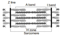
(D) The sarcomere is the functional unit of muscle contraction.
In the structure of a sarcomere,the $A-$band (anisotropic band) is located in the central region.
The $H-$zone is a lighter central region within the $A-$band where only thick filaments (myosin) are present.
The $M-$line is a thin,dark line that runs through the center of the $H-$zone,anchoring the thick filaments.
Therefore,all the given statements are correct.
0 likes
View Solution304
MediumMCQ
During muscle contraction in a skeletal muscle fibre,$Ca^{2+}$ combines with
A
$TpT$
B
$TpC$
C
$TpI$
D
Tropomyosin
Solution
(B) During muscle contraction,the $Ca^{2+}$ ions released into the sarcoplasm bind to the $TpC$ (Troponin $C$) subunit of the troponin complex.
This binding induces a conformational change in the troponin complex,which in turn moves the tropomyosin molecule away from the active sites on the actin filament.
This exposes the myosin-binding sites on actin,allowing the formation of cross-bridges between actin and myosin,which leads to muscle contraction.
0 likes
View Solution305
MediumMCQ
In a contracted skeletal muscle fibre,
A
A-band અદ્રશ્ય થાય છે
B
I-band ઘટે છે
C
M-line લંબાય છે
D
H-zone લંબાય છે
Solution
(C) During the contraction of a skeletal muscle fibre,the sliding filament theory explains the process:
$1$. The $A-$band length remains constant because the length of myosin filaments does not change.
$2$. The $I-$band shortens as the actin filaments slide over the myosin filaments.
$3$. The $H-$zone shortens or may disappear completely during maximal contraction.
$4$. The $M-$line remains visible as the central attachment point for myosin filaments.
Therefore,none of the options $A, B, C, D$ are strictly correct as written,but based on standard textbook questions of this type,the $H-$zone shortens and the $I-$band shortens. If the question implies what happens to the $H-$zone,it shortens. Given the options provided,there is a common error in such questions; however,the $A-$band is the only structure that remains constant in length.
0 likes
View Solution306
MediumMCQ
Which of the following ions is essential for muscular contraction?
A
$Na^{+}, Ca^{2+}$
B
$Mg^{2+}, Ca^{2+}$
C
$Mg^{2+}, K^{+}$
D
$K^{+}, Na^{+}$
Solution
(B) $Mg^{2+}$ and $Ca^{2+}$ ions are essential for muscular contraction.
$1$. $Mg^{2+}$ ions are required for the polymerization of $G-\text{actin}$ into $F-\text{actin}$ filaments.
$2$. $Ca^{2+}$ ions bind to the $TpC$ subunit of troponin,which causes a conformational change that displaces the troponin-tropomyosin complex,thereby exposing the active myosin-binding sites on the actin filament.
0 likes
View Solution307
MediumMCQ
In which of the following functions are white muscles not used?
A
Moving of eye balls
B
Fast and strenuous work for short duration
C
For sustained work at a slow rate for a prolonged duration
D
Fast flights as in sparrows
Solution
(C) White muscle fibres,also known as fast-twitch fibres,contain less myoglobin and fewer mitochondria. They are adapted for rapid,powerful contractions but fatigue quickly.
They are primarily used for:
$1$. Moving of eye balls.
$2$. Fast and strenuous work for short duration.
$3$. Fast flights as in sparrows.
They are not used for sustained work at a slow rate for a prolonged duration,as this function is performed by red muscle fibres (slow-twitch fibres),which are rich in myoglobin and mitochondria and are resistant to fatigue.
0 likes
View Solution308
MediumMCQ
The backward bending of the shank is worked out by
A
Gluteus maximus
B
Quadriceps femoris muscles
C
Adductor group of muscles
D
Gastrocnemius and hamstrings
Solution
(D) The backward bending of the shank (lower leg) at the knee joint is known as flexion.
This movement is primarily performed by the hamstring muscles located at the back of the thigh.
The gastrocnemius muscle also assists in this action by acting across the knee joint.
Therefore,the correct answer is $D$.
This movement is primarily performed by the hamstring muscles located at the back of the thigh.
The gastrocnemius muscle also assists in this action by acting across the knee joint.
Therefore,the correct answer is $D$.
0 likes
View Solution309
MediumMCQ
Which one is not the character of red skeletal muscle?
A
Smaller diameter
B
More mitochondria
C
More sarcoplasmic reticulum
D
More blood capillaries
Solution
(C) Red muscle fibres are characterized by a smaller diameter,a higher number of mitochondria,and a greater density of blood capillaries compared to white muscle fibres.
They contain less sarcoplasmic reticulum because they rely more on aerobic respiration rather than rapid,short-term bursts of contraction that require quick calcium release.
Therefore,having 'more sarcoplasmic reticulum' is not a characteristic of red skeletal muscle.
They contain less sarcoplasmic reticulum because they rely more on aerobic respiration rather than rapid,short-term bursts of contraction that require quick calcium release.
Therefore,having 'more sarcoplasmic reticulum' is not a characteristic of red skeletal muscle.
0 likes
View Solution310
MediumMCQ
Least blood supply is present in which of the following?
A
Skeletal muscle
B
Smooth muscle
C
Cardiac muscle
D
None of these
Solution
(B) The blood supply to muscle tissues varies based on their metabolic activity and function.
Cardiac muscle requires a constant and high supply of oxygen and nutrients,thus it has a very rich blood supply.
Skeletal muscles also have a significant blood supply to support contraction and recovery.
Smooth muscles,which are found in the walls of hollow organs,generally have the least blood supply compared to cardiac and skeletal muscles,as their metabolic demands are lower and they often exhibit sustained,slow contractions.
0 likes
View Solution311
MediumMCQ
Which of the following muscles are not under the voluntary control of the nervous system?
A
Pharynx
B
Urinary bladder
C
Anterior end of oesophagus
D
Tongue
Solution
(B) The muscles of the urinary bladder are composed of involuntary smooth muscle fibers,which are not under the voluntary control of the nervous system.
In contrast,the pharynx,the anterior end of the oesophagus,and the tongue are composed of skeletal muscles,which are under the voluntary control of the somatic nervous system.
In contrast,the pharynx,the anterior end of the oesophagus,and the tongue are composed of skeletal muscles,which are under the voluntary control of the somatic nervous system.
0 likes
View Solution312
MediumMCQ
$A : Ca^{2+}$ plays an important role in muscle contraction.
$R : Ca^{2+}$ combines with the troponin chain,displacing tropomyosin and allowing the myosin head to combine with actin to form an actomyosin complex.
$R : Ca^{2+}$ combines with the troponin chain,displacing tropomyosin and allowing the myosin head to combine with actin to form an actomyosin complex.
A
Assertion and Reason both are correct and Reason is the correct explanation of Assertion.
B
Assertion and Reason both are correct but Reason is not the correct explanation of Assertion.
C
Assertion is correct,but Reason is incorrect.
D
Both Assertion and Reason are incorrect.
Solution
(A) $Ca^{2+}$ ions play a crucial role in muscle contraction. When $Ca^{2+}$ is released into the sarcoplasm,it binds to the $TpC$ subunit of troponin. This binding induces a conformational change in the troponin complex,which in turn displaces tropomyosin from the active sites on the actin filament. This exposure allows the myosin heads to bind to the actin,forming an actomyosin complex,which initiates the contraction process.
0 likes
View Solution313
DifficultMCQ
$A$: All muscles follow the 'All or None' principle.
$R$: All muscles contract either fully or not at all depending upon the availability of a threshold stimulus.
$R$: All muscles contract either fully or not at all depending upon the availability of a threshold stimulus.
A
Assertion and Reason both are correct and Reason is the correct explanation of Assertion.
B
Assertion and Reason both are correct but Reason is not the correct explanation of Assertion.
C
Assertion is correct,but Reason is incorrect.
D
Both Assertion and Reason are incorrect.
Solution
(D) The 'All or None' principle states that a single muscle fibre contracts completely or not at all when stimulated by a threshold stimulus.
However,an entire muscle organ consists of many muscle fibres.
When a stimulus is applied to a whole muscle,the strength of contraction depends on the number of muscle fibres recruited.
Therefore,the whole muscle does not follow the 'All or None' principle; it shows graded responses.
Thus,the Assertion is incorrect,and the Reason is also incorrect because it incorrectly attributes the 'All or None' principle to the entire muscle rather than individual muscle fibres.
0 likes
View Solution314
MediumMCQ
$A$: Muscle tetanus is the phenomenon of sustained contraction of a muscle due to a succession of nerve impulses/stimuli being received by it.
$R$: Many of our daily activities are due to tetanic contraction of muscles,such as holding a book.
$R$: Many of our daily activities are due to tetanic contraction of muscles,such as holding a book.
A
Assertion and Reason both are correct and Reason is the correct explanation of Assertion.
B
Assertion and Reason both are correct but Reason is not the correct explanation of Assertion.
C
Assertion is correct,but Reason is incorrect.
D
Both Assertion and Reason are incorrect.
Solution
(C) : Tetanus in muscle physiology refers to a state of sustained contraction where the muscle fiber does not relax between stimuli because the frequency of nerve impulses is very high. This is a correct statement.
$R$: Many of our daily activities,such as holding a book,require the muscle to maintain a constant state of tension (tonus) rather than a full tetanic contraction. Tetanic contraction is a pathological or experimental state of maximal sustained contraction,whereas holding a book involves sustained muscle tone (tonus) maintained by asynchronous motor unit recruitment. Therefore,the Reason is incorrect.
$R$: Many of our daily activities,such as holding a book,require the muscle to maintain a constant state of tension (tonus) rather than a full tetanic contraction. Tetanic contraction is a pathological or experimental state of maximal sustained contraction,whereas holding a book involves sustained muscle tone (tonus) maintained by asynchronous motor unit recruitment. Therefore,the Reason is incorrect.
0 likes
View Solution315
MediumMCQ
$A$: Application of stimuli repeatedly just at the start of relaxation of a muscle fibre shows an increase in the extent of contraction initially and results in treppe or staircase phenomenon.
$R$: It can be attributed to the summation effect of the sub-threshold stimuli being given again and again.
$R$: It can be attributed to the summation effect of the sub-threshold stimuli being given again and again.
A
Assertion and Reason both are correct and also correct explanation.
B
Assertion and Reason both are correct but not explanation of assertion.
C
Assertion is correct,but Reason is incorrect.
D
Both Assertion and Reason are incorrect.
Solution
(C) The $A$ is correct because the staircase effect (treppe) is a phenomenon where repeated stimulation of a muscle fibre at the onset of relaxation leads to progressively stronger contractions.
This occurs because of the increased availability of $Ca^{2+}$ ions in the sarcoplasm and the warming effect of the muscle,which improves enzyme efficiency.
However,the $R$ is incorrect because treppe is not caused by the summation of sub-threshold stimuli.
Sub-threshold stimuli are too weak to trigger an action potential,whereas treppe occurs with stimuli that are already at or above the threshold level.
0 likes
View Solution316
MediumMCQ
$A$: Latent period is the interval between the application of an appropriate stimulus and the initiation of contraction.
$R$: Latent period is minimum in cardiac muscle fibres.
$R$: Latent period is minimum in cardiac muscle fibres.
A
Assertion and Reason both are correct and Reason is the correct explanation of Assertion.
B
Assertion and Reason both are correct but Reason is not the correct explanation of Assertion.
C
Assertion is correct,but Reason is incorrect.
D
Both Assertion and Reason are incorrect.
Solution
(C) The latent period is the time interval between the application of a stimulus and the start of muscle contraction.
In skeletal muscle,the latent period is very short (approximately $0.01$ seconds).
In cardiac muscle,the latent period is significantly longer due to the prolonged refractory period,which prevents tetanic contraction.
Therefore,the statement that the latent period is minimum in cardiac muscle is incorrect.
Thus,Assertion is correct,but Reason is incorrect.
0 likes
View Solution317
MediumMCQ
During muscular contraction,which of the following events occur?
$(a)$ $'H'$ zone disappears
$(b)$ $'A'$ band widens
$(c)$ $'I'$ band reduces in width
$(d)$ Myosin hydrolyzes $ATP$,releasing the $ADP$ and $Pi$
$(e)$ $Z$-lines attached to actins are pulled inwards
Choose the correct answer from the options given below.
$(a)$ $'H'$ zone disappears
$(b)$ $'A'$ band widens
$(c)$ $'I'$ band reduces in width
$(d)$ Myosin hydrolyzes $ATP$,releasing the $ADP$ and $Pi$
$(e)$ $Z$-lines attached to actins are pulled inwards
Choose the correct answer from the options given below.
A
$(a), (c), (d), (e)$ only
B
$(a), (b), (c), (d)$ only
C
$(b), (c), (d), (e)$ only
D
$(b), (d), (e), (a)$ only
Solution
(A) According to the sliding filament theory of muscle contraction:
$1$. The $'H'$ zone disappears as actin filaments slide over myosin filaments.
$2$. The $'A'$ band (anisotropic band) retains its length because it represents the length of the myosin filaments,which do not shorten.
$3$. The $'I'$ band (isotropic band) reduces in width as actin filaments are pulled towards the center of the sarcomere.
$4$. Myosin heads hydrolyze $ATP$ to $ADP$ and $Pi$,providing energy for the power stroke.
$5$. The $Z$-lines attached to actin filaments are pulled inwards towards the center of the sarcomere,shortening the sarcomere.
Therefore,events $(a), (c), (d),$ and $(e)$ are correct.
0 likes
View Solution318
MediumMCQ
Choose the correct option for the following:
Biceps $\quad$ Iris $\quad$ Heart
Biceps $\quad$ Iris $\quad$ Heart
A
Skeletal muscle $\quad$ Smooth muscle $\quad$ Cardiac muscle
B
Smooth muscle $\quad$ Skeletal muscle $\quad$ Cardiac muscle
C
Striated muscle $\quad$ Skeletal muscle $\quad$ Cardiac muscle
D
Skeletal muscle $\quad$ Striated muscle $\quad$ Cardiac muscle
Solution
(A) $1$. Biceps are voluntary muscles attached to bones,classified as skeletal muscles.
$2$. The iris contains smooth muscles (involuntary) that control the size of the pupil.
$3$. The heart is composed of cardiac muscles,which are specialized involuntary muscles.
Therefore,the correct sequence is: Biceps (Skeletal muscle),Iris (Smooth muscle),Heart (Cardiac muscle).
$2$. The iris contains smooth muscles (involuntary) that control the size of the pupil.
$3$. The heart is composed of cardiac muscles,which are specialized involuntary muscles.
Therefore,the correct sequence is: Biceps (Skeletal muscle),Iris (Smooth muscle),Heart (Cardiac muscle).
0 likes
View Solution319
MediumMCQ
Which muscle shows striations?
A
Skeletal muscle
B
Smooth muscle
C
Cardiac muscle
D
Both $A$ and $C$
Solution
(D) Striations are characteristic features of muscles that contain organized sarcomeres.
$1$. Skeletal muscles are voluntary and striated.
$2$. Cardiac muscles are involuntary and also striated.
$3$. Smooth muscles are non-striated (unstriated) and involuntary.
Therefore,both skeletal and cardiac muscles show striations.
$1$. Skeletal muscles are voluntary and striated.
$2$. Cardiac muscles are involuntary and also striated.
$3$. Smooth muscles are non-striated (unstriated) and involuntary.
Therefore,both skeletal and cardiac muscles show striations.
0 likes
View Solution320
MediumMCQ
Which of the following are the special properties of muscles?
A
Excitability
B
Contractility
C
Extensibility
D
All of the above
Solution
(D) Muscles are specialized tissues of mesodermal origin. They possess several unique properties that allow them to perform their functions effectively:
$1$. Excitability: Muscles respond to stimuli such as chemical,electrical,or mechanical signals.
$2$. Contractility: Muscles can shorten in length in response to stimulation,which is the primary mechanism for movement.
$3$. Extensibility: Muscles have the ability to be stretched beyond their resting length.
$4$. Elasticity: Muscles can return to their original shape and length after being stretched.
Since all the listed options (Excitability,Contractility,and Extensibility) are fundamental properties of muscle tissue,the correct answer is $D$.
0 likes
View Solution321
MediumMCQ
Which muscle is responsible for changes in body posture?
A
Skeletal muscle
B
Smooth muscle
C
Cardiac muscle
D
All of the above
Solution
(A) Skeletal muscles are primarily associated with the skeletal components of the body.
They are involved in locomotory actions and changes in body posture.
These muscles are under voluntary control and are attached to bones via tendons.
Smooth muscles and cardiac muscles are involuntary and are involved in internal organ functions,not in maintaining body posture.
They are involved in locomotory actions and changes in body posture.
These muscles are under voluntary control and are attached to bones via tendons.
Smooth muscles and cardiac muscles are involuntary and are involved in internal organ functions,not in maintaining body posture.
0 likes
View Solution322
MediumMCQ
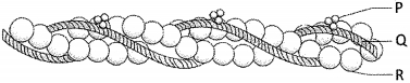
What are $P, Q,$ and $R$ in the given diagram?
$P \quad\quad Q \quad \quad R$
$P \quad\quad Q \quad \quad R$
A
Fascicle $\quad$ Muscle fiber $\quad$ Blood capillary
B
Muscle fiber $\quad$ Fascicle $\quad$ Blood capillary
C
Fascicle $\quad$ Blood capillary $\quad$ Muscle fiber
D
Muscle fiber $\quad$ Blood capillary $\quad$ Fascicle
Solution
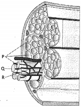
(A) In the structure of a skeletal muscle,the muscle is organized into bundles of muscle fibers called fascicles $(P)$.
Each fascicle contains several muscle fibers $(Q)$.
These fibers are surrounded by a network of blood capillaries $(R)$ to supply oxygen and nutrients.
Each fascicle contains several muscle fibers $(Q)$.
These fibers are surrounded by a network of blood capillaries $(R)$ to supply oxygen and nutrients.
0 likes
View Solution323
MediumMCQ
In the resting state of muscles, where is calcium stored?
A
Cytoplasm
B
Mitochondria
C
Sarcoplasmic reticulum
D
Nucleus
Solution
(C) In muscle cells, the $Sarcoplasmic$ $Reticulum$ $(SR)$ acts as a specialized storage site for calcium ions $(Ca^{2+})$.
During the resting state, calcium ions are sequestered within the $SR$ to prevent muscle contraction.
When an action potential reaches the muscle fiber, these calcium ions are released into the sarcoplasm, which triggers the binding of calcium to troponin, leading to muscle contraction.
During the resting state, calcium ions are sequestered within the $SR$ to prevent muscle contraction.
When an action potential reaches the muscle fiber, these calcium ions are released into the sarcoplasm, which triggers the binding of calcium to troponin, leading to muscle contraction.
0 likes
View Solution324
MediumMCQ
Which bands contain only actin and only myosin filaments,respectively?
A
$A$-band,$I$-band
B
$I$-band,$A$-band
C
$H$-zone,$I$-band
D
$I$-band,$H$-zone
Solution
(D) In the structure of a sarcomere,the $I$-band (Isotropic band) contains only thin actin filaments.
The $A$-band (Anisotropic band) contains both actin and myosin filaments.
The $H$-zone (Hensen's zone) is the central part of the $A$-band that contains only thick myosin filaments.
Therefore,the band containing only actin is the $I$-band,and the region containing only myosin is the $H$-zone.
The $A$-band (Anisotropic band) contains both actin and myosin filaments.
The $H$-zone (Hensen's zone) is the central part of the $A$-band that contains only thick myosin filaments.
Therefore,the band containing only actin is the $I$-band,and the region containing only myosin is the $H$-zone.
0 likes
View Solution325
MediumMCQ
Between two successive $M$-lines,there is/are:
A
One complete $I$-band,two half $A$-bands
B
One complete $A$-band,two half $I$-bands
C
One half $A$-band,one half $I$-band
D
One complete $A$-band,one complete $I$-band
Solution
(B) The functional unit of muscle contraction is the sarcomere.
In a sarcomere,the $A$-band (anisotropic band) is located in the center,containing both actin and myosin filaments.
The $M$-line is a thin dark line that runs through the center of the $A$-band.
The $I$-band (isotropic band) contains only actin filaments and is bisected by the $Z$-line.
When considering the structure between two successive $M$-lines,it spans across two adjacent sarcomeres.
Specifically,the region between two $M$-lines consists of one complete $A$-band (from the first sarcomere),two half $I$-bands (one from each side of the $Z$-line),and the $Z$-line itself.
However,in the context of standard structural definitions of the sarcomere,the region between two $Z$-lines is one sarcomere. The region between two $M$-lines effectively encompasses one full $A$-band and two half $I$-bands.
0 likes
View Solution326
MediumMCQ
Match the following columns:
| Column-$I$ | Column-$II$ |
| $P$. Skeletal muscle | $I$. Biceps |
| $Q$. Smooth muscle | $II$. Iris |
| $R$. Cardiac muscle | $III$. Heart |
A
$(P-III), (Q-II), (R-I)$
B
$(P-I), (Q-II), (R-III)$
C
$(P-II), (Q-I), (R-III)$
D
$(P-III), (Q-I), (R-II)$
Solution
(B) $1$. Skeletal muscles are voluntary muscles attached to bones,such as the biceps $(P-I)$.
$2$. Smooth muscles are involuntary muscles found in internal organs like the iris of the eye $(Q-II)$.
$3$. Cardiac muscles are involuntary muscles found exclusively in the heart $(R-III)$.
Therefore,the correct matching is $(P-I), (Q-II), (R-III)$.
$2$. Smooth muscles are involuntary muscles found in internal organs like the iris of the eye $(Q-II)$.
$3$. Cardiac muscles are involuntary muscles found exclusively in the heart $(R-III)$.
Therefore,the correct matching is $(P-I), (Q-II), (R-III)$.
0 likes
View Solution327
MediumMCQ
How many main types of muscles are there?
A
$2$
B
$3$
C
$4$
D
$5$
Solution
(B) Based on their location,appearance,and nature of regulation,muscles are classified into $3$ main types:
$1$. Skeletal muscles: These are closely associated with the skeletal components of the body and are striated.
$2$. Visceral (Smooth) muscles: These are located in the inner walls of hollow visceral organs and are non-striated.
$3$. Cardiac muscles: These are the muscles of the heart and are striated and involuntary.
0 likes
View Solution328
MediumMCQ
$A$ sarcomere is defined as the segment between:
A
Two $M$-lines
B
Two $Z$-lines
C
$H$-zone
D
$A$-band
Solution
(B) sarcomere is the functional unit of a striated muscle fiber.
It is defined as the portion of a myofibril located between two successive $Z$-lines (or $Z$-discs).
These $Z$-lines are dense protein structures that anchor the actin filaments.
Therefore,the correct option is $B$.
It is defined as the portion of a myofibril located between two successive $Z$-lines (or $Z$-discs).
These $Z$-lines are dense protein structures that anchor the actin filaments.
Therefore,the correct option is $B$.
0 likes
View Solution329
MediumMCQ
In the given diagram,which label represents the $I$-band?
A
$P$
B
$Q$
C
$R$
D
$S$
Solution
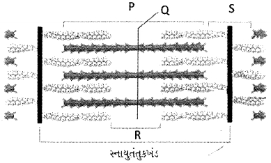
(D) In the structure of a sarcomere:
$P$ represents the $Z$-line (or $Z$-disc),which bisects the $I$-band.
$Q$ represents the $M$-line,which is the central line in the $A$-band.
$R$ represents the $H$-zone,which is the central part of the $A$-band containing only thick filaments.
$S$ represents the $I$-band (isotropic band),which contains only thin filaments and is bisected by the $Z$-line.
Therefore,the correct label for the $I$-band is $S$.
0 likes
View Solution330
MediumMCQ
Protein attached to $M$-line $= ....P....$,Protein attached to $Z$-line $= ....Q....$
A
$P -$ Myosin filament,$Q -$ Actin filament
B
$P -$ Actin filament,$Q -$ Myosin filament
C
$P -$ Myosin filament,$Q -$ Myosin filament
D
$P -$ Actin filament,$Q -$ Actin filament
Solution
(A) In the structure of a sarcomere,the $M$-line is a thin fibrous membrane in the center of the $A$-band where thick filaments (myosin) are anchored.
Conversely,the $Z$-line (or $Z$-disc) is a dark fibrous membrane that bisects the $I$-band,where thin filaments (actin) are firmly attached.
Therefore,the protein/filament attached to the $M$-line is myosin $(P)$ and the protein/filament attached to the $Z$-line is actin $(Q)$.
Conversely,the $Z$-line (or $Z$-disc) is a dark fibrous membrane that bisects the $I$-band,where thin filaments (actin) are firmly attached.
Therefore,the protein/filament attached to the $M$-line is myosin $(P)$ and the protein/filament attached to the $Z$-line is actin $(Q)$.
0 likes
View Solution331
MediumMCQ
Which protein is distributed at regular intervals on the tropomyosin protein?
A
Actin
B
Myosin
C
Troponin
D
$G$-actin
Solution
(C) In the structure of a thin filament (actin filament),two filaments of $F$-actin are helically wound to each other. Two filaments of a protein called tropomyosin also run close to the $F$-actin throughout its length. $A$ complex protein called troponin is distributed at regular intervals on the tropomyosin. Troponin masks the active binding sites for myosin on the actin filaments in the resting state.
0 likes
View Solution332
MediumMCQ
Where are $ATPase$ enzymes located in a muscle fiber?
A
$LMM$ (Light Meromyosin)
B
$HMM$ (Heavy Meromyosin)
C
$F-actin$
D
Troponin
Solution
(B) The myosin filament is a polymerized protein consisting of many monomeric proteins called meromyosins. Each meromyosin has two parts: a globular head with a short arm and a tail. The globular head is an active ATPase enzyme and has binding sites for $ATP$ and active sites for actin. This head and short arm are collectively known as Heavy Meromyosin $(HMM)$,while the tail portion is known as Light Meromyosin $(LMM)$. Therefore,the $ATPase$ enzyme activity is located in the $HMM$ part of the myosin molecule.
0 likes
View Solution333
MediumMCQ
The myosin binding site is located on ..........
A
$F-actin$
B
$Troponin$
C
$Tropomyosin$
D
$Myosin$
Solution
(A) The thin filament (actin filament) is composed of two $F-actin$ polymers,which are helically wound to each other.
Each $F-actin$ is a polymer of monomeric $G-actin$ (globular actin) proteins.
Two filaments of another protein,$tropomyosin$,also run close to the $F-actin$ throughout its length.
$A$ complex protein,$troponin$,is distributed at regular intervals on the $tropomyosin$.
In the resting state,a subunit of $troponin$ masks the active binding sites for $myosin$ on the $actin$ filaments.
Therefore,the active binding sites for $myosin$ are present on the $actin$ (specifically $F-actin$) molecules.
0 likes
View Solution334
MediumMCQ
What does $HMM$ stand for in the context of muscle structure?
A
Tail $+$ Head
B
Short arm $+$ Head
C
Head
D
Tail $+$ Short arm $+$ Head
Solution
(B) In the context of muscle structure,specifically regarding the myosin filament,$HMM$ stands for $Heavy$ $Meromyosin$.
Each myosin molecule consists of two parts: a globular head with a short arm and a tail.
The head and the short arm together are referred to as $Heavy$ $Meromyosin$ $(HMM)$,
while the tail portion is referred to as $Light$ $Meromyosin$ $(LMM)$.
Each myosin molecule consists of two parts: a globular head with a short arm and a tail.
The head and the short arm together are referred to as $Heavy$ $Meromyosin$ $(HMM)$,
while the tail portion is referred to as $Light$ $Meromyosin$ $(LMM)$.
0 likes
View Solution335
MediumMCQ
The actin filament is shown below. Where does $Ca^{++}$ bind during muscle contraction?
A
$P$
B
$Q$
C
$R$
D
$P$ and $Q$
Solution
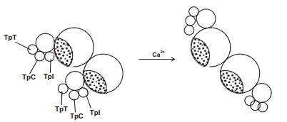
(A) In the given diagram of the actin filament:
$P$ represents the troponin complex.
$Q$ represents the tropomyosin filament.
$R$ represents the $F$-actin (filamentous actin) strand.
During muscle contraction,$Ca^{++}$ ions bind to the troponin complex $(P)$,which causes a conformational change in the troponin-tropomyosin complex,thereby exposing the active binding sites on the actin filaments for the myosin heads to attach. Therefore,the correct answer is $P$.
0 likes
View Solution336
MediumMCQ
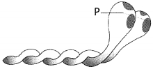
Which of the following activities occurs at the site labeled $P$ in the given diagram of a myosin monomer?
A
Actin binding
B
$ATP$ synthesis
C
$ATP$ hydrolysis
D
Both $A$ and $C$
Solution
(D) The diagram shows a myosin monomer. The head region labeled $P$ contains two important binding sites: one for actin and one for $ATP$.
During muscle contraction,the myosin head binds to actin to form a cross-bridge.
Additionally,the myosin head possesses $ATPase$ activity,which catalyzes the hydrolysis of $ATP$ to provide energy for the contraction process.
Therefore,both actin binding and $ATP$ hydrolysis occur at the myosin head $(P)$.
0 likes
View Solution337
MediumMCQ
Identify the incorrect statement regarding muscle contraction.
A
$A$-band remains constant.
B
$I$-band decreases.
C
Length of filaments decreases.
D
$Z$-lines come closer.
Solution
(C) According to the Sliding Filament Theory,during muscle contraction,the thin filaments (actin) slide over the thick filaments (myosin).
$1$. The $A$-band (anisotropic band) remains constant in length because the myosin filaments do not change their length.
$2$. The $I$-band (isotropic band) decreases in length as the actin filaments move deeper into the $A$-band.
$3$. The $Z$-lines move closer to each other,shortening the sarcomere.
$4$. The length of the individual filaments (actin and myosin) does not change; only their degree of overlap changes. Therefore,the statement that the length of filaments decreases is incorrect.
0 likes
View Solution338
MediumMCQ
The neuromuscular junction or motor end plate is a junction between:
A
$A$ motor neuron and the sarcolemma of a muscle fiber
B
$A$ sensory neuron and the sarcolemma of a muscle fiber
C
$A$ motor neuron and a sensory neuron
D
All of the above
Solution
(A) neuromuscular junction $(NMJ)$ is a specialized chemical synapse between a motor neuron and a muscle fiber.
It allows the motor neuron to transmit a signal to the muscle fiber,causing muscle contraction.
The junction consists of the axon terminal of the motor neuron and the specialized region of the muscle fiber membrane known as the sarcolemma.
Therefore,the correct junction is between a motor neuron and the sarcolemma of a muscle fiber.
0 likes
View Solution339
MediumMCQ
During the mechanism of muscle contraction,$Ca^{2+}$ binds with which of the following?
A
Troponin
B
Tropomyosin
C
$F$-actin
D
Myosin
Solution
(A) During muscle contraction,the signal for contraction is sent by the central nervous system via a motor neuron.
When the action potential reaches the neuromuscular junction,it triggers the release of neurotransmitters,leading to an action potential in the sarcolemma.
This action potential spreads through the $T$-tubules and causes the release of $Ca^{2+}$ ions into the sarcoplasm from the sarcoplasmic reticulum.
These $Ca^{2+}$ ions bind to the $Troponin$ subunit on the actin filaments.
This binding causes a conformational change in the troponin-tropomyosin complex,exposing the active sites on the $F$-actin filaments for the myosin heads to bind,thereby initiating contraction.
0 likes
View Solution340
MediumMCQ
Select the correct option for the following processes in muscle contraction:
Cross-bridge formation $\quad$ Cross-bridge breaking
Cross-bridge formation $\quad$ Cross-bridge breaking
A
$ATP$ absence $\quad$ $ATP$ presence
B
$ATP$ presence $\quad$ $ATP$ absence
C
$ATP$ presence $\quad$ $ATP$ presence
D
$ATP$ absence $\quad$ $ATP$ absence
Solution
(B) During muscle contraction,the cross-bridge formation between actin and myosin filaments occurs when $Ca^{2+}$ ions bind to troponin,exposing the active sites on actin. This step does not require $ATP$ hydrolysis directly.
However,the breaking of the cross-bridge (detachment of the myosin head from actin) requires the binding of a new $ATP$ molecule to the myosin head. The hydrolysis of this $ATP$ then provides the energy for the myosin head to return to its 'cocked' position.
Therefore,cross-bridge formation occurs in the presence of $Ca^{2+}$ (but does not require $ATP$ for the binding itself),while cross-bridge breaking strictly requires the presence of $ATP$.
0 likes
View Solution341
MediumMCQ
Identify the characteristics of red muscle fibers from the following:
$I -$ Many mitochondria
$II -$ Use large amounts of $O_2$ for $ATP$ production
$III -$ Low amount of myoglobin
$IV -$ Less sarcoplasmic reticulum
Which of the above are characteristic of red muscle fibers?
$I -$ Many mitochondria
$II -$ Use large amounts of $O_2$ for $ATP$ production
$III -$ Low amount of myoglobin
$IV -$ Less sarcoplasmic reticulum
Which of the above are characteristic of red muscle fibers?
A
$I, II, III, IV$
B
$I, II$
C
$I, II, IV$
D
$I, II, III$
Solution
(C) Red muscle fibers are characterized by the following features:
$1$. They contain a large number of mitochondria,which can utilize large amounts of $O_2$ stored in them for $ATP$ production.
$2$. They contain a high amount of myoglobin,which gives them a reddish appearance.
$3$. They have a less developed sarcoplasmic reticulum compared to white muscle fibers.
Based on the given statements:
- Statement $I$ (Many mitochondria) is correct.
- Statement $II$ (Use large amounts of $O_2$) is correct.
- Statement $III$ (Low amount of myoglobin) is incorrect,as red muscle fibers have high myoglobin content.
- Statement $IV$ (Less sarcoplasmic reticulum) is correct.
Therefore,the correct characteristics are $I, II,$ and $IV$.
0 likes
View Solution342
MediumMCQ
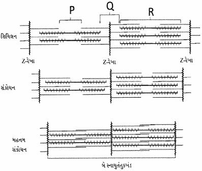
Identify the bands $P, Q,$ and $R$ in the given diagram.
$\quad\quad\quad P \quad\quad\quad Q\quad\quad\quad R$
$\quad\quad\quad P \quad\quad\quad Q\quad\quad\quad R$
A
$H$-band $\quad$ $A$-band $\quad$ $I$-band
B
$H$-band $\quad$ $I$-band $\quad$ $A$-band
C
$I$-band $\quad$ $A$-band $\quad$ $H$-band
D
$A$-band $\quad$ $I$-band $\quad$ $H$-band
Solution
(B) In the structure of a sarcomere:
$1$. The $H$-zone $(P)$ is the central part of the $A$-band where only thick myosin filaments are present.
$2$. The $I$-band $(Q)$ is the light band containing only thin actin filaments,bisected by the $Z$-line.
$3$. The $A$-band $(R)$ is the dark band that contains both actin and myosin filaments.
Therefore,the correct identification is $P = H$-band,$Q = I$-band,and $R = A$-band.
0 likes
View Solution343
MediumMCQ
According to the sliding filament theory:
A
Actin and myosin filaments slide over each other to increase the length of the sarcomere.
B
Length of $A$-band does not change.
C
$I$-band increases in length.
D
The actin filaments slide away from $A$-band resulting in shortening of sarcomere.
Solution
(B) The sliding filament theory states that muscle contraction occurs when thin actin filaments slide over thick myosin filaments.
During this process,the $A$-band remains constant in length because the length of the myosin filaments does not change.
The $I$-band shortens as the actin filaments are pulled toward the center of the sarcomere.
The $H$-zone also shortens or disappears.
Consequently,the overall length of the sarcomere decreases,leading to muscle contraction.
0 likes
View Solution344
MediumMCQ
Which of the following statements are correct regarding skeletal muscle?
$A$. Muscle bundles are held together by a collagenous connective tissue layer called fascicle.
$B$. Sarcoplasmic reticulum of muscle fibre is a storehouse of calcium ions.
$C$. Striated appearance of skeletal muscle fibre is due to the distribution pattern of actin and myosin proteins.
$D$. $M$ line is considered as the functional unit of contraction called sarcomere.
Choose the most appropriate answer from the options given below:
A
$C$ and $D$ only
B
$A, B$ and $C$ only
C
$B$ and $C$ only
D
$A, C$ and $D$ only
Solution
(C) Statement $A$ is incorrect because muscle bundles are held together by a collagenous connective tissue layer called fascia,not fascicle. The bundles themselves are called fascicles.
Statement $B$ is correct because the sarcoplasmic reticulum of muscle fibres is indeed a storehouse of calcium ions,which are essential for muscle contraction.
Statement $C$ is correct because the striated appearance of skeletal muscle fibres is due to the regular distribution pattern of actin and myosin proteins.
Statement $D$ is incorrect because the portion of the myofibril between two successive $Z$-lines is considered the functional unit of contraction,called a sarcomere,not the $M$-line.
Therefore,only statements $B$ and $C$ are correct.
0 likes
View Solution345
DifficultMCQ
Select the correct statements regarding the mechanism of muscle contraction.
$A.$ It is initiated by a signal sent by $\text{CNS}$ via a motor neuron,not a sensory neuron.
$B.$ Neurotransmitter generates an action potential in the sarcolemma.
$C.$ Increased $Ca^{++}$ level leads to the binding of calcium with troponin on actin filaments.
$D.$ Masking of the active site of actin prevents contraction; unmasking leads to contraction.
$E.$ Utilising the energy from $\text{ATP}$ hydrolysis to form a cross-bridge.
A
$B, C$ and $E$ only
B
$C, D$ and $E$ only
C
$A$ and $D$ only
D
$B, D$ and $E$ only
Solution
(A) The mechanism of muscle contraction is explained by the sliding filament theory:
$1$. Muscle contraction is initiated by a signal sent by the $\text{CNS}$ via a motor neuron (not a sensory neuron),making statement $A$ incorrect.
$2$. The motor neuron releases a neurotransmitter (acetylcholine) which generates an action potential in the sarcolemma,making statement $B$ correct.
$3$. This action potential triggers the release of $Ca^{++}$ ions into the sarcoplasm. Increased $Ca^{++}$ levels lead to the binding of calcium with troponin on actin filaments,which exposes the active sites,making statement $C$ correct.
$4$. Masking of the active site prevents contraction,while unmasking leads to contraction,making statement $D$ incorrect.
$5$. The myosin head then utilizes the energy from $\text{ATP}$ hydrolysis to form a cross-bridge with the actin filament,making statement $E$ correct.
Therefore,statements $B, C,$ and $E$ are correct.
0 likes
View Solution346
MediumMCQ
$ATP$ase of the muscle is located on:
A
Actinin
B
Troponin
C
Myosin
D
Actin
Solution
(C) The $ATP$ase enzyme activity in muscle fibers is associated with the head of the $Myosin$ filament.
During muscle contraction,the $Myosin$ head acts as an $ATP$ase,hydrolyzing $ATP$ into $ADP$ and inorganic phosphate $(Pi)$.
This energy release allows the $Myosin$ head to bind to the active sites on the $Actin$ filament,forming cross-bridges and facilitating muscle contraction.
During muscle contraction,the $Myosin$ head acts as an $ATP$ase,hydrolyzing $ATP$ into $ADP$ and inorganic phosphate $(Pi)$.
This energy release allows the $Myosin$ head to bind to the active sites on the $Actin$ filament,forming cross-bridges and facilitating muscle contraction.
0 likes
View Solution347
MediumMCQ
The central part of the thick filament which is not overlapped by thin filaments is called as:
A
$Z-$zone
B
$H-$zone
C
$I-$zone
D
$F-$zone
Solution
(B) In the structure of a sarcomere,the thick filaments (myosin) are located in the center of the $A$-band.
During muscle contraction and relaxation,the thin filaments (actin) extend from the $Z$-lines towards the center of the sarcomere.
The central portion of the thick filament that is not overlapped by the thin filaments is known as the $H-$zone (Hensen's zone).
Therefore,the correct option is $B$.
During muscle contraction and relaxation,the thin filaments (actin) extend from the $Z$-lines towards the center of the sarcomere.
The central portion of the thick filament that is not overlapped by the thin filaments is known as the $H-$zone (Hensen's zone).
Therefore,the correct option is $B$.
0 likes
View Solution348
EasyMCQ
Which of the following is not a part of the thin filament?
A
Troponin
B
Tropomyosin
C
Actin
D
Meromyosin
Solution
(D) The thin filament (also known as the $I$-band) is primarily composed of three proteins: $Actin$,$Tropomyosin$,and $Troponin$.
$Actin$ forms the backbone of the thin filament.
$Tropomyosin$ runs along the $Actin$ filaments to regulate binding sites.
$Troponin$ is a complex of three proteins attached to $Tropomyosin$.
$Meromyosin$ is the structural unit of the thick filament $(Myosin)$,not the thin filament.
$Actin$ forms the backbone of the thin filament.
$Tropomyosin$ runs along the $Actin$ filaments to regulate binding sites.
$Troponin$ is a complex of three proteins attached to $Tropomyosin$.
$Meromyosin$ is the structural unit of the thick filament $(Myosin)$,not the thin filament.
0 likes
View Solution349
EasyMCQ
The "myosin binding site" is present on:
A
Actin protein
B
Myosin protein
C
Tropomyosin protein
D
Troponin protein
Solution
(A) In the structure of a thin filament (actin filament), the $F$-actin is composed of two helical strands of polymerized $G$-actin molecules.
Each $G$-actin monomer has a specific binding site for the myosin head.
In a resting state, these binding sites on the actin filaments are covered by the regulatory protein tropomyosin, which prevents the interaction between actin and myosin.
Therefore, the myosin binding site is located on the actin protein.
0 likes
View Solution350
MediumMCQ
Globular protein which masks active sites on $G$-actin is :-
A
Troponin
B
Tropomyosin
C
Myosin
D
Meromyosin
Solution
(A) The thin filament (actin) consists of two $F$-actin helically wound to each other. Each $F$-actin is a polymer of monomeric $G$-actin (globular actin) proteins. Two filaments of another protein,$Tropomyosin$,also run close to the $F$-actin throughout its length. $A$ complex protein,$Troponin$,is distributed at regular intervals on the $Tropomyosin$. In the resting state,a subunit of $Troponin$ masks the active binding sites for $Myosin$ on the actin filaments.
0 likes
View SolutionLocomotion and Movement — Muscles · Frequently Asked Questions
1Are these Locomotion and Movement questions useful for JEE and NEET?
Yes. All questions in this section are mapped to JEE Main and NEET exam patterns. Previous year questions from JEE Main, NEET, GUJCET and state-level exams are included with full solutions.
2Can I switch to Hindi or Gujarati for these questions?
Yes. Use the language tabs in the hero section or the sidebar to view the same questions and solutions in English, Hindi or Gujarati.
3How do I generate a question paper from this subtopic?
Use the Vedclass Exam Paper Generator — select the chapter and subtopic, set difficulty, and generate Sets A, B, C, D automatically. First 3 chapters of every subject are free.
Vedclass Products
For Students
Vedclass Test Series
Mock tests in real JEE/NEET style with performance analysis. 5-day free trial.
Start Free TrialFor Teachers
Exam Paper Generator
Generate Set A/B/C/D papers from this chapter in 2 minutes. 3 chapters free.
Try FreeFor Institutes
Online Exam Module
Live online exams with unlimited students, 360° analytics & white-label branding.
See DemoFor Teachers & Institutes
Generate a Locomotion and Movement Exam Paper in 2 Minutes
Select subtopic & difficulty — Sets A, B, C, D auto-generated with No Repeat logic.
First 3 chapters of every subject are free — no payment required.