
A English
Muscles Questions in English
Class 11 Biology · Locomotion and Movement · Muscles
365+
Questions
English
Language
100%
With Solutions
Showing 50 of 365 questions in English
151
MediumMCQ
Which one of the following types of muscle fibers will be the first one to undergo fatigue?
A
Slow oxidative fibers
B
Fast oxidative-glycolytic fibers
C
Fast glycolytic fibers
D
Aerobic fibers
Solution
(C) Muscle fibers are classified based on their contraction speed and metabolic pathways.
$1$. $Slow$ $oxidative$ $fibers$ ($Type$ $I$) are rich in myoglobin and mitochondria, making them highly resistant to fatigue.
$2$. $Fast$ $oxidative-glycolytic$ $fibers$ ($Type$ $IIa$) have intermediate properties.
$3$. $Fast$ $glycolytic$ $fibers$ ($Type$ $IIb$) rely primarily on anaerobic glycolysis for energy. They have fewer mitochondria and less myoglobin, which leads to a rapid accumulation of lactic acid and depletion of glycogen stores, causing them to fatigue very quickly compared to other types.
0 likes
View Solution152
MediumMCQ
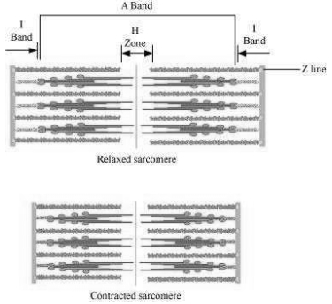
During muscle contraction in humans,the:
A
Actin filaments shorten
B
Sarcomere does not shorten
C
Length of $A$ band remains the same
D
$A, H$ and $I$ bands shorten
Solution
(C) According to the Sliding Filament Theory,muscle contraction occurs when thin filaments (actin) slide over thick filaments (myosin).
$1$. The length of the individual actin and myosin filaments does not change.
$2$. The $A$ band,which represents the length of the myosin filaments,remains constant in length.
$3$. The $I$ band and the $H$ zone shorten as the actin filaments slide toward the center of the sarcomere.
$4$. The sarcomere itself shortens as the $Z$ lines are pulled closer together.
Therefore,the length of the $A$ band remains the same during contraction.
0 likes
View Solution153
MediumMCQ
Identify the odd one out from the following list.
A
Actin
B
Renin
C
Tropomyosin
D
Myosin
Solution
(B) The correct answer is $B$ (Renin).
$1$. Actin,Tropomyosin,and Myosin are the primary contractile proteins found in muscle fibers.
$2$. Actin and Tropomyosin are thin filament proteins,while Myosin is a thick filament protein.
$3$. Renin is an enzyme secreted by the juxtaglomerular cells of the kidney,which plays a role in the renin-angiotensin-aldosterone system $(RAAS)$ to regulate blood pressure.
$4$. Therefore,Renin is the odd one out as it is not a muscle protein.
0 likes
View Solution154
MediumMCQ
The sliding filament theory can be best explained as:
A
Actin and myosin filaments shorten and slide past each other.
B
Actin and myosin filaments do not shorten but rather slide past each other.
C
When myofilaments slide past each other,myosin filaments shorten while actin filaments do not shorten.
D
When myofilaments slide past each other,actin filaments shorten while myosin filaments do not shorten.
Solution
(B) The sliding filament theory states that muscle contraction occurs due to the sliding of thin filaments (actin) over thick filaments (myosin).
During this process,the length of the individual filaments (actin and myosin) remains constant; they do not shorten.
Instead,the filaments slide past each other,which increases the overlap between them,thereby shortening the sarcomere and causing muscle contraction.
During this process,the length of the individual filaments (actin and myosin) remains constant; they do not shorten.
Instead,the filaments slide past each other,which increases the overlap between them,thereby shortening the sarcomere and causing muscle contraction.
0 likes
View Solution155
MediumMCQ
Which part of a muscle cell is the storehouse of calcium ions?
A
Sarcolemma
B
Sarcoplasm
C
Sarcoplasmic reticulum
D
None of the above
Solution
(C) The sarcoplasmic reticulum is a specialized type of smooth endoplasmic reticulum found in smooth and striated muscle fibers.
Its primary function is to store and release calcium ions $(Ca^{2+})$.
When a muscle fiber is stimulated,calcium ions are released from the sarcoplasmic reticulum into the sarcoplasm,which triggers the process of muscle contraction.
Therefore,the correct answer is $C$.
Its primary function is to store and release calcium ions $(Ca^{2+})$.
When a muscle fiber is stimulated,calcium ions are released from the sarcoplasmic reticulum into the sarcoplasm,which triggers the process of muscle contraction.
Therefore,the correct answer is $C$.
0 likes
View Solution156
MediumMCQ
Endomysium is present in which of the following muscles?
A
Skeletal
B
Cardiac
C
Smooth
D
All of these
Solution
(D) Endomysium is the innermost layer of connective tissue that surrounds individual muscle fibers.
It is a characteristic feature of all three types of muscle tissues: skeletal,cardiac,and smooth muscles.
In skeletal muscle,it surrounds each muscle fiber.
In cardiac muscle,it surrounds individual cardiomyocytes.
In smooth muscle,it surrounds individual smooth muscle cells.
Therefore,the correct answer is that it is present in all of these muscle types.
0 likes
View Solution157
MediumMCQ
Which ion binds with troponin during muscle contraction?
A
$HCO_3^-$
B
$Ca^{+2}$
C
$Cl^-$
D
$Na^+$
Solution
(B) During muscle contraction,an action potential travels along the sarcolemma and reaches the $T$-tubules,triggering the release of $Ca^{+2}$ ions from the sarcoplasmic reticulum into the sarcoplasm.
These $Ca^{+2}$ ions bind to the troponin complex on the thin filaments (actin).
This binding causes a conformational change in the troponin-tropomyosin complex,which shifts the tropomyosin away from the active sites on the actin filament.
This exposure of active sites allows the myosin heads to bind to actin,forming cross-bridges and initiating muscle contraction.
0 likes
View Solution158
MediumMCQ
In which of the following muscle components are actin-binding sites present?
A
Troponin
B
Tropomyosin
C
Meromyosin
D
Intercalated disc
Solution
(C) Each myosin filament is a polymerized protein consisting of many monomeric proteins called $Meromyosin$. Each $Meromyosin$ has two important parts: a globular head with a short arm and a tail. The globular head is an active ATPase enzyme and has binding sites for $ATP$ and active sites for $Actin$.
0 likes
View Solution159
MediumMCQ
Lack of relaxation between successive stimuli in sustained muscle contraction is known as
A
Tetanus
B
Paralysis
C
Osteoporosis
D
Fatigue
Solution
(A) In muscle physiology,when a muscle is stimulated repeatedly at a high frequency,it does not get enough time to relax between successive stimuli.
This leads to a state of sustained contraction known as $Tetanus$.
$Paralysis$ refers to the loss of muscle function.
$Osteoporosis$ is a bone disorder characterized by decreased bone mass.
$Fatigue$ is the decline in the ability of a muscle to generate force after prolonged activity.
This leads to a state of sustained contraction known as $Tetanus$.
$Paralysis$ refers to the loss of muscle function.
$Osteoporosis$ is a bone disorder characterized by decreased bone mass.
$Fatigue$ is the decline in the ability of a muscle to generate force after prolonged activity.
1 likes
View Solution160
MediumMCQ
Select the correct option for skeletal muscle.
$(I)$ In human body,it is made of a number of muscle bundles.
$(II)$ Muscle bundles contain a number of muscle fibers and each fiber is lined by the sarcolemma.
$(III)$ In a muscle fiber,the sarcoplasm contains only one nucleus.
$(IV)$ In a muscle fiber,the sarcoplasmic reticulum is the storehouse of calcium ions.
A
Only $I$
B
$I$ and $II$
C
$I, II$ and $III$
D
$I, II$ and $IV$
Solution
(D) Statement $(I)$ is correct: Skeletal muscles are composed of many muscle bundles (fascicles).
Statement $(II)$ is correct: Each muscle bundle contains many muscle fibers,and each muscle fiber is enclosed by a plasma membrane called the sarcolemma.
Statement $(III)$ is incorrect: Muscle fibers are syncytial,meaning the sarcoplasm contains many nuclei (multinucleated).
Statement $(IV)$ is correct: The sarcoplasmic reticulum of the muscle fiber is the storehouse of calcium ions,which are essential for muscle contraction.
Therefore,statements $(I), (II),$ and $(IV)$ are correct.
0 likes
View Solution161
MediumMCQ
$A-$ Visceral muscles do not show any striations and are smooth in appearance. Hence,they are known as non-striated muscles.
$R-$ Their activities are under the voluntary control of the nervous system.
$R-$ Their activities are under the voluntary control of the nervous system.
A
Both $A$ and $R$ are true
B
Both $A$ and $R$ are false
C
$A$ is false,$R$ is true
D
$A$ is true,$R$ is false
Solution
(D) Visceral muscles (smooth muscles) are located in the inner walls of hollow visceral organs like the alimentary canal and reproductive tract.
They do not exhibit striations under a microscope,hence they are called non-striated or smooth muscles.
Their activities are not under the voluntary control of the nervous system; they are involuntary muscles.
Therefore,statement $A$ is true,but statement $R$ is false.
They do not exhibit striations under a microscope,hence they are called non-striated or smooth muscles.
Their activities are not under the voluntary control of the nervous system; they are involuntary muscles.
Therefore,statement $A$ is true,but statement $R$ is false.
0 likes
View Solution162
MediumMCQ
What is the dark line lying in the middle of the $H$-zone called?
A
$M$-line
B
$H$-zone
C
$A$-band
D
$Z$-line
Solution
(A) In the structure of a sarcomere,the $A$-band contains both actin and myosin filaments.
Within the $A$-band,there is a central region where only thick myosin filaments are present,known as the $H$-zone.
The $H$-zone is bisected by a thin,dark fibrous membrane called the $M$-line.
Therefore,the dark line lying in the middle of the $H$-zone is the $M$-line.
Within the $A$-band,there is a central region where only thick myosin filaments are present,known as the $H$-zone.
The $H$-zone is bisected by a thin,dark fibrous membrane called the $M$-line.
Therefore,the dark line lying in the middle of the $H$-zone is the $M$-line.
0 likes
View Solution163
MediumMCQ
Which sentence is not true for muscle contraction?
A
During stimulation when muscle contracts,the length of filaments (thin and thick) does not change but merely slides over one another.
B
During stimulation,the $Z$-lines of the sarcomere come closer to each other.
C
On stimulation,there is no change in the light band.
D
All of the above
Solution
(C) According to the sliding filament theory of muscle contraction:
$1$. The length of the thin and thick filaments remains constant; they slide past each other,which is a correct statement.
$2$. During contraction,the $Z$-lines defining the sarcomere are pulled closer together,which is a correct statement.
$3$. During contraction,the light band ($I$-band) shortens because the actin filaments slide into the $A$-band. Therefore,the statement that there is no change in the light band is incorrect.
Since the question asks for the statement that is not true,option $C$ is the correct answer.
0 likes
View Solution164
MediumMCQ
$A -$ Calcium ions $(Ca^{2+})$ activate the interaction between myosin and actin filaments.
$R -$ The cross-bridge is formed in the presence of $ATP,$ and the breaking of the cross-bridge also requires the binding of a new $ATP$ molecule.
$R -$ The cross-bridge is formed in the presence of $ATP,$ and the breaking of the cross-bridge also requires the binding of a new $ATP$ molecule.
A
$A$ and $R$ both are correct.
B
$A$ and $R$ both are incorrect.
C
$A$ is correct and $R$ is incorrect.
D
$A$ is incorrect and $R$ is correct.
Solution
(C) Assertion $(A)$ is correct: Calcium ions $(Ca^{2+})$ bind to the troponin subunit on actin filaments,which causes a conformational change that exposes the active sites for myosin on the actin filament,thereby activating the interaction.
Reason $(R)$ is incorrect: While the formation of the cross-bridge requires $ATP$ hydrolysis,the breaking or detachment of the cross-bridge specifically requires the binding of a new $ATP$ molecule to the myosin head. The statement claims breaking occurs in the absence of $ATP,$ which is false; in the absence of $ATP,$ the cross-bridge remains locked (a state known as rigor mortis).
Therefore,$A$ is correct and $R$ is incorrect.
0 likes
View Solution165
MediumMCQ
$A-$ Actin filaments occur in two forms: the monomeric $G$-actin and the polymeric $F$-actin.
$R-$ Tropomyosin is a rod-shaped fibrous protein. Tropomyosin forms two helical strands,which are wrapped around the $F$-actin filaments.
$R-$ Tropomyosin is a rod-shaped fibrous protein. Tropomyosin forms two helical strands,which are wrapped around the $F$-actin filaments.
A
$A$ and $R$ both are correct.
B
$A$ and $R$ both are incorrect.
C
$A$ is correct and $R$ is incorrect.
D
$A$ is incorrect and $R$ is correct.
Solution
(D) Assertion $(A)$ is incorrect because $G$-actin (Globular actin) is the monomeric form,while $F$-actin (Filamentous actin) is the polymeric form. The statement incorrectly swapped these terms.
Reason $(R)$ is correct. Tropomyosin is indeed a rod-shaped fibrous protein that forms two helical strands,which run along the length of the $F$-actin filaments to regulate muscle contraction.
Reason $(R)$ is correct. Tropomyosin is indeed a rod-shaped fibrous protein that forms two helical strands,which run along the length of the $F$-actin filaments to regulate muscle contraction.
0 likes
View Solution166
MediumMCQ
In the resting state,a subunit of troponin masks the active binding sites for .......... on the .......... filaments.
A
Myosin,tropomyosin
B
Actin,myosin
C
Actin,troponin
D
Myosin,actin
Solution
(D) In the resting state,the protein complex troponin is distributed at regular intervals on the tropomyosin filament.
Specifically,a subunit of troponin masks the active binding sites for myosin heads on the actin filaments.
This prevents the interaction between actin and myosin,thereby keeping the muscle in a relaxed state.
Specifically,a subunit of troponin masks the active binding sites for myosin heads on the actin filaments.
This prevents the interaction between actin and myosin,thereby keeping the muscle in a relaxed state.
0 likes
View Solution167
MediumMCQ
It is not a characteristic of cardiac muscles.
A
Intercalated disc
B
Light-dark band
C
Contract as a unit
D
Multinucleated cell
Solution
(D) Cardiac muscles are involuntary,striated muscles found in the heart.
$1$. They possess intercalated discs,which are specialized junctions that allow rapid communication between cells.
$2$. They exhibit light and dark bands (striations) due to the arrangement of actin and myosin filaments.
$3$. They contract as a unit because the intercalated discs allow the impulse to spread quickly,ensuring the heart beats in a synchronized manner.
$4$. Cardiac muscle cells are typically uninucleated (or sometimes binucleated),whereas skeletal muscle cells are multinucleated. Therefore,being a multinucleated cell is not a characteristic of cardiac muscle.
0 likes
View Solution168
MediumMCQ
The functional unit of the contractile system for a striated muscle is:
A
Myofibril
B
Sarcomere
C
$Z$-lines
D
Cross bridges
Solution
(B) The functional unit of contraction in striated muscle is the $Sarcomere$.
It is defined as the segment of a myofibril between two successive $Z$-lines.
Each $Sarcomere$ contains the contractile proteins,actin and myosin,arranged in a specific pattern that allows for muscle shortening during contraction.
It is defined as the segment of a myofibril between two successive $Z$-lines.
Each $Sarcomere$ contains the contractile proteins,actin and myosin,arranged in a specific pattern that allows for muscle shortening during contraction.
0 likes
View Solution169
MediumMCQ
Which of the following does not consist solely of involuntary muscles?
A
Muscular layer of blood vessels
B
Muscles of glandular ducts
C
Muscles of the iris
D
Muscles of the urethra
Solution
(D) Involuntary muscles (smooth muscles) are found in the walls of internal organs such as blood vessels,glandular ducts,and the iris. The urethra,however,contains both involuntary smooth muscles (internal urethral sphincter) and voluntary skeletal muscles (external urethral sphincter) to control micturition. Therefore,the urethra does not consist solely of involuntary muscles.
0 likes
View Solution170
MediumMCQ
Which ion is essential for muscle contraction?
A
$Ca^{2+}$
B
$Na^+$
C
$K^+$
D
$Cl^-$
Solution
(A) Muscle contraction is initiated by the release of calcium ions $(Ca^{2+})$ from the sarcoplasmic reticulum into the sarcoplasm.
These $Ca^{2+}$ ions bind to the troponin complex on the actin filaments,which causes a conformational change that exposes the myosin-binding sites on the actin.
This allows the myosin heads to bind to actin,forming cross-bridges and leading to muscle contraction via the sliding filament mechanism.
These $Ca^{2+}$ ions bind to the troponin complex on the actin filaments,which causes a conformational change that exposes the myosin-binding sites on the actin.
This allows the myosin heads to bind to actin,forming cross-bridges and leading to muscle contraction via the sliding filament mechanism.
0 likes
View Solution171
MediumMCQ
Which of the following is a contractile protein of muscle?
A
All of the above
B
Myosin
C
Actin
D
Tubulin
Solution
(A) Muscle contraction is primarily driven by the interaction between two key contractile proteins: $Actin$ and $Myosin$.
$Actin$ forms the thin filaments,while $Myosin$ forms the thick filaments.
These proteins slide past each other during the contraction process,known as the sliding filament theory.
$Tubulin$ is a structural protein involved in the formation of microtubules,not muscle contraction.
Therefore,both $Actin$ and $Myosin$ are contractile proteins,making the correct choice 'All of the above'.
0 likes
View Solution172
MediumMCQ
What is a sarcomere?
A
The region between two $H$-lines
B
The region between two $A$-lines
C
The region between two $I$-bands
D
The region between two $Z$-lines
Solution
(D) sarcomere is the functional unit of a striated muscle fiber.
It is defined as the segment of a myofibril located between two successive $Z$-lines (or $Z$-discs).
These $Z$-lines are dense protein structures that bisect the $I$-bands and serve as the anchor points for actin filaments.
Therefore,the correct option is $D$.
It is defined as the segment of a myofibril located between two successive $Z$-lines (or $Z$-discs).
These $Z$-lines are dense protein structures that bisect the $I$-bands and serve as the anchor points for actin filaments.
Therefore,the correct option is $D$.
0 likes
View Solution173
MediumMCQ
Which of the following statements is correct regarding muscle contraction?
A
The length of the $H$-zone increases.
B
The length of the $A$-band remains constant.
C
The length of the $I$-band increases.
D
The distance between two $Z$-lines increases.
Solution
(B) According to the Sliding Filament Theory of muscle contraction:
$1$. During contraction,the thin filaments slide over the thick filaments,which pulls the $Z$-lines closer together,thereby shortening the sarcomere.
$2$. The $A$-band (Anisotropic band) contains myosin filaments and its length remains constant throughout the contraction process.
$3$. The $I$-band (Isotropic band) and the $H$-zone shorten as the actin filaments move deeper into the $A$-band.
$4$. Therefore,the statement that the length of the $A$-band remains constant is correct.
0 likes
View Solution174
MediumMCQ
The $ATPase$ enzyme required for muscle contraction is located on ..........
A
Actinin
B
Troponin
C
Myosin
D
Actin
Solution
(C) The $ATPase$ enzyme is located on the head of the myosin filament.
During muscle contraction,the myosin head binds to the active site on actin to form a cross-bridge.
The $ATPase$ activity of the myosin head hydrolyzes $ATP$ into $ADP$ and inorganic phosphate $(Pi)$,which provides the energy required for the power stroke and muscle contraction.
During muscle contraction,the myosin head binds to the active site on actin to form a cross-bridge.
The $ATPase$ activity of the myosin head hydrolyzes $ATP$ into $ADP$ and inorganic phosphate $(Pi)$,which provides the energy required for the power stroke and muscle contraction.
0 likes
View Solution175
MediumMCQ
Which contractile protein of skeletal muscle is associated with $ATPase$ activity?
A
Myosin
B
$\alpha$-Actinin
C
Troponin
D
Tropomyosin
Solution
(A) The contractile proteins in skeletal muscle include actin and myosin.
Myosin is a motor protein that forms the thick filaments.
The head of the myosin molecule contains an $ATPase$ enzyme site,which hydrolyzes $ATP$ to provide the energy required for muscle contraction.
Therefore,myosin is the protein associated with $ATPase$ activity.
Myosin is a motor protein that forms the thick filaments.
The head of the myosin molecule contains an $ATPase$ enzyme site,which hydrolyzes $ATP$ to provide the energy required for muscle contraction.
Therefore,myosin is the protein associated with $ATPase$ activity.
0 likes
View Solution176
MediumMCQ
In a skeletal muscle fiber,what is the $H$-zone?
A
The central part of the thick filament (myosin) not overlapped by thin filaments (actin).
B
The absence of myofibrils in the center of the $A$-band.
C
The space between the $Z$-lines.
D
The region where actin filaments overlap with myosin filaments.
Solution
(A) In a skeletal muscle fiber,the $A$-band contains both actin and myosin filaments.
However,the central part of the thick filament (myosin) is not overlapped by thin filaments (actin).
This central,lighter region within the $A$-band is known as the $H$-zone.
Therefore,the $H$-zone represents the portion of the myosin filament that does not overlap with actin filaments.
However,the central part of the thick filament (myosin) is not overlapped by thin filaments (actin).
This central,lighter region within the $A$-band is known as the $H$-zone.
Therefore,the $H$-zone represents the portion of the myosin filament that does not overlap with actin filaments.
0 likes
View Solution177
MediumMCQ
Name the ions responsible for unmasking the active sites on myosin for cross-bridge formation during muscle contraction.
A
Sodium
B
Potassium
C
Calcium
D
Magnesium
Solution
(C) During muscle contraction,the signal for contraction is sent by the central nervous system via a motor neuron.
When the action potential reaches the neuromuscular junction,it triggers the release of neurotransmitters,leading to an action potential in the sarcolemma.
This action potential spreads through the $T$-tubules and triggers the release of $Ca^{2+}$ ions from the sarcoplasmic reticulum into the sarcoplasm.
These $Ca^{2+}$ ions bind to the troponin subunit on the actin filaments.
This binding causes a conformational change that moves tropomyosin away from the active sites on the actin filament,thereby 'unmasking' them.
Once the active sites are exposed,the myosin heads can bind to them to form cross-bridges,initiating the contraction process.
Therefore,$Ca^{2+}$ ions are responsible for this process.
0 likes
View Solution178
MediumMCQ
What is the state of sustained muscle contraction without relaxation between successive stimuli called?
A
Fatigue
B
Tetanus
C
Tonus
D
Spasm
Solution
(B) When a muscle is stimulated by repeated,rapid stimuli,it does not get enough time to relax between the stimuli. This results in a state of sustained,continuous contraction known as $Tetanus$. This is different from the disease tetanus,which is caused by the bacterium $Clostridium$ $tetani$.
0 likes
View Solution179
MediumMCQ
At which site do muscle fibers receive stimulation from a motor neuron?
A
Neuromuscular junction
B
$T$-tubule system
C
Myofibrils
D
Sarcoplasmic reticulum
Solution
(A) The stimulation of muscle fibers by a motor neuron occurs at the neuromuscular junction (also known as the motor end plate).
This is a chemical synapse formed between a motor neuron and a muscle fiber.
When an action potential reaches the axon terminal of the motor neuron,it triggers the release of the neurotransmitter acetylcholine into the synaptic cleft.
This neurotransmitter binds to receptors on the sarcolemma of the muscle fiber,initiating an action potential that leads to muscle contraction.
0 likes
View Solution180
MediumMCQ
Calcium is important in skeletal muscle contraction because it
A
Prevents the formation of bonds between the myosin cross bridge and the actin filament.
B
Binds to troponin to remove the masking of active sites on actin.
C
Detaches the myosin head from the actin filament.
D
Activates the myosin $ATPase$ by binding to it.
Solution
(B) During skeletal muscle contraction,the binding of $Ca^{2+}$ ions to the $Troponin$ complex is a critical step.
In a relaxed state,the active sites on the actin filament are masked by the $Tropomyosin$ protein.
When $Ca^{2+}$ ions are released from the sarcoplasmic reticulum,they bind to $Troponin$.
This binding causes a conformational change in the $Troponin-Tropomyosin$ complex,which shifts the $Tropomyosin$ away from the active sites on the actin filament.
This exposure allows the myosin heads to bind to the actin,forming cross-bridges and initiating muscle contraction.
0 likes
View Solution181
MediumMCQ
Contractile tissues have the following features:
$(i)$ Mesodermal in origin
$(ii)$ They contain stretch receptors
$(iii)$ Rhythmic contractions are seen in them
$(iv)$ They do not fatigue during the life of the animal
Which of the above are characteristics of sphincters?
$(i)$ Mesodermal in origin
$(ii)$ They contain stretch receptors
$(iii)$ Rhythmic contractions are seen in them
$(iv)$ They do not fatigue during the life of the animal
Which of the above are characteristics of sphincters?
A
All the four
B
Only $(i), (ii)$ and $(iii)$
C
Only $(i), (ii)$ and $(iv)$
D
Only $(i), (iii)$ and $(iv)$
Solution
(C) Sphincters are specialized circular muscles that control the passage of substances through body openings.
$(i)$ Sphincters are mesodermal in origin,as they are composed of muscle tissue.
$(ii)$ They contain stretch receptors that help in regulating their tone and contraction based on the presence of contents.
$(iii)$ Rhythmic contractions are characteristic of cardiac muscle or peristaltic movements,not typical sphincters,which generally maintain a tonic state of contraction.
$(iv)$ Sphincters do not fatigue during the life of the animal because they are designed to maintain a constant state of contraction to prevent leakage.
Therefore,the characteristics $(i), (ii),$ and $(iv)$ are associated with sphincters.
0 likes
View Solution182
EasyMCQ
The sensation of fatigue in the muscles after prolonged strenuous physical work is caused by
A
a decrease in the supply of oxygen
B
minor wear and tear of muscle fibres
C
the depletion of glucose
D
the accumulation of lactic acid
Solution
(D) During prolonged strenuous physical work,the demand for $ATP$ in muscle cells increases significantly. When the oxygen supply is insufficient to meet this demand through aerobic respiration,muscle cells switch to anaerobic respiration. In this process,glucose is broken down into lactic acid. The accumulation of lactic acid in the muscle tissue leads to a decrease in $pH$ and interferes with muscle contraction,resulting in the sensation of fatigue.
0 likes
View Solution183
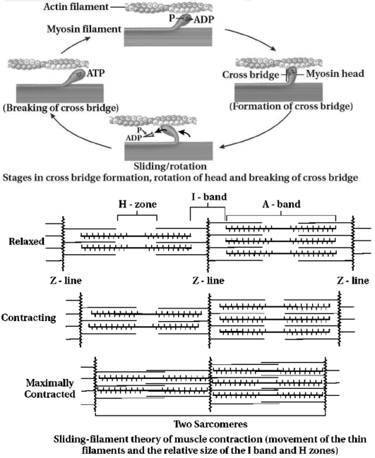
EasyMCQ
The given figure represents the cross-bridge cycle in skeletal muscle. What does step $B$ in the figure represent?
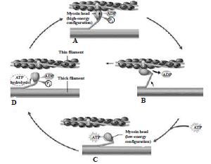
A
Attachment of myosin head to actin forming a cross-bridge.
B
Release of phosphate. Myosin changes shape to pull actin.
C
Attachment of new $ATP$ to myosin head. The cross-bridge detaches.
D
Splitting of $ATP$ into $ADP$ and $Pi$. Myosin cocks into its high-energy conformation.
Solution
(B) Step $A$: Attachment of myosin head to actin forming a cross-bridge.
Step $B$: Release of phosphate. Myosin changes shape to pull actin.
Step $C$: Attachment of new $ATP$ to myosin head. The cross-bridge detaches.
Step $D$: Splitting of $ATP$ into $ADP$ and $Pi$. Myosin cocks into its high-energy conformation.
Step $B$: Release of phosphate. Myosin changes shape to pull actin.
Step $C$: Attachment of new $ATP$ to myosin head. The cross-bridge detaches.
Step $D$: Splitting of $ATP$ into $ADP$ and $Pi$. Myosin cocks into its high-energy conformation.
0 likes
View Solution184
MediumMCQ
Assertion: Fatigue is the inability of a muscle to relax.
Reason: It is due to lactic acid accumulation by repeated contractions.
Reason: It is due to lactic acid accumulation by repeated contractions.
A
If both Assertion and Reason are correct and the Reason is a correct explanation of the Assertion.
B
If both Assertion and Reason are correct but Reason is not a correct explanation of the Assertion.
C
If the Assertion is correct but Reason is incorrect.
D
If both the Assertion and Reason are incorrect.
Solution
(A) Muscle fatigue is defined as the inability of a muscle to contract or relax effectively after prolonged or repeated stimulation.
This condition is primarily caused by the accumulation of lactic acid in the muscle tissue,which occurs due to anaerobic respiration during intense or repeated muscle contractions.
Therefore,both the Assertion and the Reason are correct,and the Reason provides a valid explanation for the phenomenon of muscle fatigue.
0 likes
View Solution185
MediumMCQ
Assertion : The phase of muscle contraction occurs when myosin binds and releases actin.
Reason : Muscle contraction is initiated by a signal sent by the peripheral nervous system via motor neuron.
Reason : Muscle contraction is initiated by a signal sent by the peripheral nervous system via motor neuron.
A
If both Assertion and Reason are correct and the Reason is a correct explanation of the Assertion.
B
If both Assertion and Reason are correct but Reason is not a correct explanation of the Assertion.
C
If the Assertion is correct but Reason is incorrect.
D
If both the Assertion and Reason are incorrect.
Solution
(C) The assertion is correct because muscle contraction involves the formation and breaking of cross-bridges between myosin heads and actin filaments,known as the sliding filament theory.
The reason is incorrect because muscle contraction is initiated by a signal sent by the $Central \ Nervous \ System$ $(CNS)$,not the peripheral nervous system,via a motor neuron.
Therefore,the assertion is correct,but the reason is incorrect.
The reason is incorrect because muscle contraction is initiated by a signal sent by the $Central \ Nervous \ System$ $(CNS)$,not the peripheral nervous system,via a motor neuron.
Therefore,the assertion is correct,but the reason is incorrect.
0 likes
View Solution186
Medium
Draw the diagram of a sarcomere of skeletal muscle showing different regions.
Solution
(N/A) sarcomere is the functional unit of a skeletal muscle fiber. It is defined as the segment of a myofibril between two successive $Z$-lines.
Key components include:
$1$. $Z$-line: $A$ dark,fibrous protein band that bisects the $I$-band.
$2$. $I$-band (Isotropic band): Contains only thin actin filaments.
$3$. $A$-band (Anisotropic band): Contains both thick myosin filaments and overlapping thin actin filaments.
$4$. $M$-line: $A$ thin fibrous membrane in the middle of the $A$-band.
$5$. $H$-zone: The central part of the $A$-band where only thick filaments are present.
0 likes
View Solution187
Medium
Define the sliding filament theory of muscle contraction.
Solution
(N/A) The sliding filament theory explains the process of muscle contraction during which the thin filaments slide over the thick filaments,which shortens the myofibril.
Each muscle fibre has alternate light and dark bands,which contain special contractile proteins called actin and myosin,respectively.
Actin is a thin contractile protein present in the light band,known as the $I$-band,whereas myosin is a thick contractile protein present in the dark band,known as the $A$-band.
There is an elastic fibre called the $Z$-line that bisects each $I$-band. The thin filament is firmly anchored to the $Z$-line.
The central part of the thick filament that is not overlapped by the thin filament is known as the $H$-zone.
During muscle contraction,the myosin heads or cross-bridges come in close contact with the thin filaments.
As a result,the thin filaments are pulled towards the middle of the sarcomere.
The $Z$-line attached to the actin filaments is also pulled,leading to the shortening of the sarcomere.
Hence,the length of the $A$-band remains constant,while the $I$-band shortens and the $H$-zone disappears.
0 likes
View Solution188
Difficult
Describe the important steps in muscle contraction.
Solution
(N/A) During skeletal muscle contraction,the thick filament slides over the thin filament by a repeated binding and release of myosin along the filament. This whole process occurs in a sequential manner.
Step $1$: Muscle contraction is initiated by signals that travel along the axon and reach the neuromuscular junction or motor end plate. The neuromuscular junction is a junction between a neuron and the sarcolemma of the muscle fibre. As a result,Acetylcholine (a neurotransmitter) is released into the synaptic cleft,generating an action potential in the sarcolemma.
Step $2$: The generation of this action potential releases calcium ions $(Ca^{2+})$ from the sarcoplasmic reticulum into the sarcoplasm.
Step $3$: The increased level of calcium ions in the sarcoplasm leads to the activation of actin sites. Calcium ions bind to the troponin on actin filaments and remove the tropomyosin wrapped around actin filaments. Hence,active actin sites are exposed,allowing myosin heads to attach to these sites.
Step $4$: In this stage,the myosin head attaches to the exposed site of actin and forms cross-bridges by utilizing energy from $ATP$ hydrolysis. The actin filaments are pulled toward the center of the $A$-band. As a result,the $H$-zone reduces. This is the stage where the actual contraction of the muscle occurs.
Step $5$: After muscle contraction,the myosin head pulls the actin filament and releases $ADP$ along with inorganic phosphate. New $ATP$ molecules bind to the myosin head,causing it to detach and the cross-bridges to break.
Step $6$: This process of formation and breaking down of cross-bridges continues until the stimulus stops. When the stimulus ceases,calcium ions are pumped back into the sarcoplasmic reticulum. As a result,the concentration of calcium ions in the sarcoplasm decreases,thereby masking the actin filaments again and leading to muscle relaxation.
0 likes
View Solution189
Easy
Give information regarding characteristics of muscles and its types.
Solution
(N/A) $-\quad$ Muscle is a specialised tissue of mesodermal origin.
$\Rightarrow$ It contributes to $40-50 \%$ of the body weight of a human adult.
- Muscles possess special properties like excitability,contractility,extensibility,and elasticity.
- Muscles are classified based on different criteria,such as location,appearance,and nature of regulation.
- Based on their location,there are three types of muscles: $(i)$ Skeletal muscles,$(ii)$ Visceral muscles,and $(iii)$ Cardiac muscles.
$(i)$ Skeletal muscles: These are closely associated with the skeletal components of the body (e.g.,limbs,trunk).
- Under the microscope,they show dark and light stripes,hence they are called striated muscles.
- They are under the control of the voluntary nervous system,hence they are known as voluntary muscles.
- These muscles are involved in locomotory actions and changes in body posture.
$(ii)$ Visceral muscles (Non-striated): These are located in the inner walls of hollow visceral organs of the body,such as the alimentary canal,reproductive tract,and trachea.
- They do not show striations and appear smooth,hence they are called smooth muscles (non-striated muscles).
- Their activities are not under voluntary control,hence they are known as involuntary muscles.
- They assist in functions like the transportation of food through the digestive tract and gametes through the genital tract.
$(iii)$ Cardiac muscles: These are the muscles of the heart. Many cardiac muscle cells assemble in a branching pattern. Cardiac muscles are short,cylindrical,and connected to each other by branching processes.
- These muscles are innervated by the autonomic nervous system,so they are involuntary in nature.
- Contraction of cardiac muscles is rhythmic,rapid,and continuous (they do not get fatigued). They are connected by intercalated discs and receive a rich blood supply.
0 likes
View Solution190
Easy
Discuss the microscopic structure of skeletal muscle tissue.
Solution
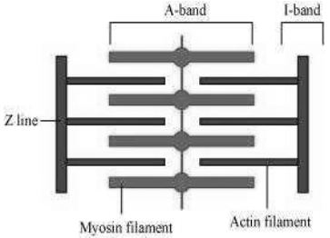
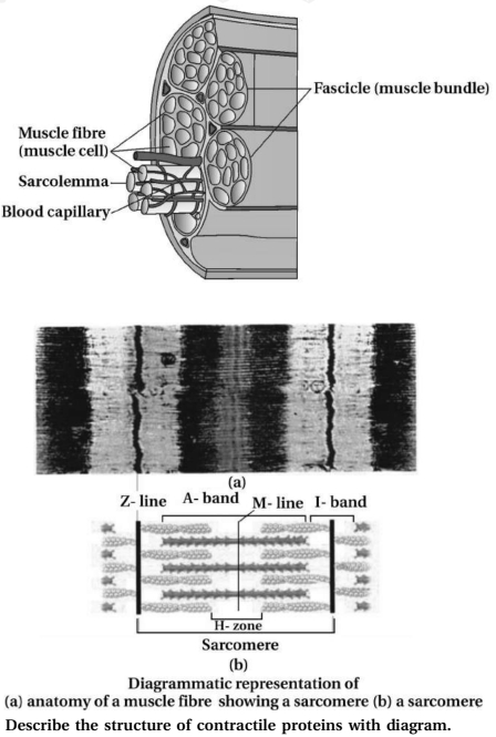
(N/A) Each organised skeletal muscle in our body is made of a number of muscle bundles or fascicles held together by a common collagenous connective tissue layer called fascia. Each muscle bundle contains a number of muscle fibres.
- Each muscle fibre is lined by the plasma membrane called sarcolemma enclosing sarcoplasm.
- Muscle fibre is a syncytium as the sarcoplasm contains many nuclei.
- The endoplasmic reticulum of the muscle fibres (sarcoplasmic reticulum) is the storehouse of calcium ions.
- $A$ characteristic feature of the muscle fibre is the presence of a large number of parallelly arranged filaments in the sarcoplasm called myofilaments or myofibrils.
- Each myofibril has alternate dark and light bands on it. It is due to the distribution pattern of two important proteins - Actin and Myosin.
- The light bands contain actin and is called $I$-band (Isotropic band).
- The dark band is called $A$-band (Anisotropic band) and contains myosin protein.
- Both the proteins are arranged as rod-like structures,parallel to each other and to the longitudinal axis of the myofibrils.
- Actin filaments are thinner as compared to the myosin filaments.
- In the centre of each $I$-band is an elastic fibre called $Z$-line which bisects it. The thin filaments are firmly attached to the $Z$-line.
- The thick filaments in the $A$-band are also held together in the middle of this band by a thin fibrous membrane called $M$-line.
- The $A$ and $I$ bands are arranged alternately throughout the length of the myofibrils.
- The portion of the myofibril between two successive $Z$-lines is considered as the functional unit of contraction and is called a sarcomere.
- In a resting state,the edges of thin filaments on either side of the thick filaments partially overlap the free ends of the thick filaments leaving the central part of the thick filaments. This central part of the thick filament,not overlapped by thin filaments,is called $H$-zone.
0 likes
View Solution191
Easy
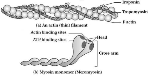
Describe the structure of contractile proteins with a diagram.
Solution
(N/A) Contractile proteins consist of actin (thin) filaments and myosin (thick) filaments.
$1$. Actin Filaments:
- Each actin filament is composed of two '$F$' (filamentous) actins helically wound to each other.
- Each '$F$' actin is a polymer of monomeric '$G$' (globular) actins.
- Two filaments of another protein,tropomyosin,also run close to the '$F$' actins throughout their length.
- $A$ complex protein,troponin,is distributed at regular intervals on the tropomyosin.
- In the resting state,a subunit of troponin masks the active binding sites for myosin on the actin filaments.
$2$. Myosin Filaments:
- Each myosin filament is a polymerised protein consisting of many monomeric proteins called meromyosins.
- Each meromyosin has two important parts: a globular head with a short arm and a tail.
- The head and short arm are called heavy meromyosin $(HMM)$,and the tail is called light meromyosin $(LMM)$.
- The $HMM$ component projects outwards at regular distances and angles from the surface of a polymerised myosin filament and is known as the cross-arm.
- The globular head is an active $ATPase$ enzyme and has binding sites for $ATP$ and active sites for actin.
0 likes
View Solution192
Easy
Describe the mechanism of muscle contraction with a diagram.
Solution
(N/A) The mechanism of muscle contraction is best explained by the sliding filament theory,which states that contraction of a muscle fibre takes place by the sliding of the thin filaments over the thick filaments.
This theory was postulated by $A$.$F$. Huxley and $J$. Jensen.
Muscle contraction is initiated by a signal sent by the central nervous system $(CNS)$ via a motor neuron.
$A$ motor neuron along with the muscle fibres connected to it constitutes a motor unit. The junction between a motor neuron and the sarcolemma of the muscle fibre is called the neuromuscular junction.
$A$ neural signal reaching this junction releases a neurotransmitter (Acetylcholine) which generates an action potential in the sarcolemma. This spreads through the muscle fibre and causes the release of $Ca^{++}$ (calcium ions) into the sarcoplasm.
Increase in $Ca^{++}$ level leads to the binding of $Ca^{++}$ with a subunit of troponin on actin filaments,and thereby removes the masking of active sites for myosin.
Utilising the energy from $ATP$ hydrolysis,the myosin head now binds to exposed active sites on actin to form a cross-bridge.
This pulls the attached actin filaments towards the centre of the '$A$' band. The '$Z$' line attached to these actins are also pulled inwards,thereby causing a shortening of the sarcomere,i.e.,contraction.
It is clear from the above steps that during shortening of the muscle (contraction),the '$I$' bands get reduced,whereas the '$A$' band retains its length.
The myosin,releasing the $ADP$ and $P_i$,goes back to its relaxed state. $A$ new $ATP$ binds and the cross-bridge is broken.
The $ATP$ is again hydrolysed by the myosin head and the cycle of cross-bridge formation and breakage is repeated,causing further sliding. The process continues till the $Ca^{++}$ ions are pumped back to the sarcoplasmic cisternae,resulting in the masking of actin filaments. This causes the return of '$Z$' lines back to their original position (i.e.,relaxation).
The reaction time of the fibres can vary in different muscles. Repeated activation of the muscles can lead to the accumulation of lactic acid due to anaerobic breakdown of glycogen in them,causing fatigue.
0 likes
View Solution193
Easy
Discuss the difference between red muscle and white muscles.
Solution
(N/A) Muscles contain a red-colored $O_{2}$-storing pigment called myoglobin.
Myoglobin content is high in some muscles,which gives them a reddish appearance; these are called red fibers.
Red fibers contain a large number of mitochondria,which utilize the stored oxygen for $ATP$ production,making them aerobic muscles (e.g.,flight muscles in $Aves$).
Conversely,some muscles possess very little myoglobin and appear pale or whitish; these are called white fibers.
White fibers have fewer mitochondria but a high amount of sarcoplasmic reticulum,and they rely on anaerobic processes for energy (e.g.,human eyeball muscles).
0 likes
View Solution194
EasyMCQ
What are the characteristic features of skeletal muscles?
A
They are involuntary and striated.
B
They are voluntary and striated.
C
They are voluntary and non-striated.
D
They are involuntary and non-striated.
Solution
(B) Skeletal muscles are primarily attached to the bones of the skeleton.
They are under the control of the voluntary nervous system,which is why they are also known as voluntary muscles.
Under a microscope,they exhibit a striped or striated appearance due to the arrangement of actin and myosin filaments,hence they are called striated muscles.
These muscles are primarily involved in locomotory actions and changes in body postures.
0 likes
View Solution195
EasyMCQ
In which condition does relaxation occur in muscles?
A
When $Ca^{++}$ ions are pumped back into the sarcoplasmic reticulum.
B
When $ATP$ binds to the myosin head.
C
When the cross-bridge is formed.
D
When the actin filaments slide over myosin.
Solution
(A) Relaxation occurs when $Ca^{++}$ ions are pumped back into the sarcoplasmic reticulum,resulting in the masking of actin filaments.
As the $Ca^{++}$ concentration decreases,the troponin-tropomyosin complex returns to its original position,covering the active sites on the actin filament.
This prevents the formation of cross-bridges,allowing the muscle fiber to return to its original state (relaxation) and the '$Z$' lines to return to their original position.
0 likes
View Solution196
Easy
Differentiate between striated muscle fibers and visceral muscle fibers.
Solution
(N/A)
| Striated muscle fiber | Visceral muscle fiber |
|---|---|
| $(1)$ These are closely associated with the skeleton. | $(1)$ These are found in the walls of internal organs. |
| $(2)$ Contraction and relaxation are very fast. | $(2)$ Contraction and relaxation are very slow. |
| $(3)$ They get fatigued easily. | $(3)$ They do not fatigue easily. |
| $(4)$ Light and dark striations are observed. | $(4)$ No dark and light striations are observed. |
| $(5)$ They are innervated by voluntary nerves. | $(5)$ They are innervated by involuntary nerves. |
0 likes
View Solution197
Easy
Differentiate between Red muscles and White muscles.
Solution
(N/A)
| Red muscles | White muscles |
|---|---|
| $(1)$ Presence of abundant myoglobin gives red colour to muscles. | $(1)$ Myoglobin is scarce,so they appear pale or white in colour. |
| $(2)$ They contain a higher number of mitochondria. | $(2)$ Mitochondria are fewer in number. |
| $(3)$ They store more $O_2$ and synthesize more $ATP$ through aerobic respiration. | $(3)$ They store less $O_2$ and rely more on anaerobic processes,producing less $ATP$. |
| $(4)$ Sarcoplasmic reticulum is comparatively less developed. | $(4)$ Sarcoplasmic reticulum is abundantly observed. |
| $(5)$ Example: Flight muscles of pigeon. | $(5)$ Example: Eyeball muscles. |
0 likes
View Solution198
Easy
Give scientific reasons: Striated muscles get fatigued rapidly.
Solution
(N/A) Striated muscles undergo rapid contraction and relaxation.
$\Rightarrow$ During intense activity,glycogen is broken down anaerobically,which leads to the accumulation of lactic acid.
- Due to the accumulation of lactic acid and the depletion of energy reserves,the muscles become unresponsive to further stimulation.
$=$ This state of temporary inability to contract is known as muscle fatigue.
0 likes
View Solution199
Easy
Give scientific reasons: During flight,birds do not fatigue easily.
Solution
(N/A) Birds do not fatigue easily during flight because their flight muscles are primarily composed of red muscle fibers.
- Red muscle fibers contain a high concentration of myoglobin,which acts as an oxygen-storing pigment.
- These muscles are rich in mitochondria,which facilitate aerobic respiration to produce a large amount of $ATP$.
- Due to the efficient and continuous production of $ATP$ through aerobic pathways,these muscles do not accumulate lactic acid and thus do not fatigue easily,allowing birds to fly for long distances.
0 likes
View Solution200
Easy
Define and explain the following:
$(1)$ Skeletal muscles
$(2)$ Visceral muscles
$(1)$ Skeletal muscles
$(2)$ Visceral muscles
Solution
(N/A) $(1)$ Skeletal muscles: These muscles are closely associated with the skeletal components of the body,such as the head,trunk,and limbs. Due to this association,they are termed skeletal muscles. They are typically striated and under voluntary control.
$(2)$ Visceral muscles: These muscles are located in the inner walls of hollow visceral organs of the body,such as the alimentary canal (digestive system),respiratory tract,and reproductive tract. They are smooth in appearance and are under involuntary control.
$(2)$ Visceral muscles: These muscles are located in the inner walls of hollow visceral organs of the body,such as the alimentary canal (digestive system),respiratory tract,and reproductive tract. They are smooth in appearance and are under involuntary control.
0 likes
View SolutionLocomotion and Movement — Muscles · Frequently Asked Questions
1Are these Locomotion and Movement questions useful for JEE and NEET?
Yes. All questions in this section are mapped to JEE Main and NEET exam patterns. Previous year questions from JEE Main, NEET, GUJCET and state-level exams are included with full solutions.
2Can I switch to Hindi or Gujarati for these questions?
Yes. Use the language tabs in the hero section or the sidebar to view the same questions and solutions in English, Hindi or Gujarati.
3How do I generate a question paper from this subtopic?
Use the Vedclass Exam Paper Generator — select the chapter and subtopic, set difficulty, and generate Sets A, B, C, D automatically. First 3 chapters of every subject are free.
Vedclass Products
For Students
Vedclass Test Series
Mock tests in real JEE/NEET style with performance analysis. 5-day free trial.
Start Free TrialFor Teachers
Exam Paper Generator
Generate Set A/B/C/D papers from this chapter in 2 minutes. 3 chapters free.
Try FreeFor Institutes
Online Exam Module
Live online exams with unlimited students, 360° analytics & white-label branding.
See DemoFor Teachers & Institutes
Generate a Locomotion and Movement Exam Paper in 2 Minutes
Select subtopic & difficulty — Sets A, B, C, D auto-generated with No Repeat logic.
First 3 chapters of every subject are free — no payment required.