
A English
Muscles Questions in English
Class 11 Biology · Locomotion and Movement · Muscles
365+
Questions
English
Language
100%
With Solutions
Showing 50 of 365 questions in English
201
Easy
Define the following terms:
$(1)$ Intercalated disc
$(2)$ Neuromuscular junction
$(1)$ Intercalated disc
$(2)$ Neuromuscular junction
Solution
(N/A) $(1)$ Intercalated disc: These are specialized cell-to-cell junctions that connect adjacent cardiac muscle cells. They facilitate the rapid transmission of electrical impulses,allowing the heart muscle to contract as a single functional unit (syncytium).
$(2)$ Neuromuscular junction: This is the chemical synapse formed between a motor neuron and a muscle fiber. It is the site where the neurotransmitter (acetylcholine) is released to initiate muscle contraction.
$(2)$ Neuromuscular junction: This is the chemical synapse formed between a motor neuron and a muscle fiber. It is the site where the neurotransmitter (acetylcholine) is released to initiate muscle contraction.
0 likes
View Solution202
Easy
Define the following terms:
$(1)$ Sarcomere
$(2)$ Acromion process
$(1)$ Sarcomere
$(2)$ Acromion process
Solution
(N/A) $(1)$ In a muscle fibre,the region between two consecutive $Z$-lines is termed as a sarcomere. Each sarcomere contains one $A$-band and two half $I$-bands. It is the structural and functional unit of a muscle fibre.
$(2)$ The scapula of the pectoral girdle is a large,flat,triangular bone. The flattened,expanded process present on the spine of the scapula is called the acromion process.
$(2)$ The scapula of the pectoral girdle is a large,flat,triangular bone. The flattened,expanded process present on the spine of the scapula is called the acromion process.
0 likes
View Solution203
Easy
Define the following terms:
$(1)$ Acetabulum
$(2)$ Red muscles
$(1)$ Acetabulum
$(2)$ Red muscles
Solution
(N/A) $(1)$ Acetabulum: It is a cup-like cavity formed at the point of fusion of the ilium,ischium,and pubis bones of the pelvic girdle. The head of the femur bone of the thigh articulates with this cavity to form the hip joint.
$(2)$ Red muscles: These are muscle fibers that contain a high concentration of myoglobin,which gives them a reddish appearance. They are rich in mitochondria and are capable of aerobic respiration,allowing them to store more $O_2$ and synthesize $ATP$ efficiently for sustained activity,such as the flight muscles of birds.
$(2)$ Red muscles: These are muscle fibers that contain a high concentration of myoglobin,which gives them a reddish appearance. They are rich in mitochondria and are capable of aerobic respiration,allowing them to store more $O_2$ and synthesize $ATP$ efficiently for sustained activity,such as the flight muscles of birds.
0 likes
View Solution204
Easy
Match the following components with their respective locations and functions:
$(1)$ Synovial fluid
$(2)$ Troponin
$(1)$ Synovial fluid
$(2)$ Troponin
Solution
(N/A) $(1)$ Location: In freely movable joints,a gap is found between the articulating surfaces of two bones,known as the synovial space.
Function: The synovial space is filled with viscous synovial fluid. It lubricates the joints of bones to facilitate easy movement.
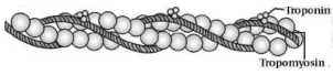
$(2)$ Location: It is a small,complex globular protein distributed along with tropomyosin at definite intervals on the actin filaments.
Function: In the relaxed state,troponin masks the active binding sites for myosin on the actin filaments,preventing contraction until calcium ions are released.
Function: The synovial space is filled with viscous synovial fluid. It lubricates the joints of bones to facilitate easy movement.
$(2)$ Location: It is a small,complex globular protein distributed along with tropomyosin at definite intervals on the actin filaments.
Function: In the relaxed state,troponin masks the active binding sites for myosin on the actin filaments,preventing contraction until calcium ions are released.
0 likes
View Solution205
EasyMCQ
What is the full form of the following terms used in muscle structure?
$(1)$ $A$-disc
$(2)$ $I$-disc
$(1)$ $A$-disc
$(2)$ $I$-disc
A
$(1)$ Anisotropic disc,$(2)$ Isotropic disc
B
$(1)$ Actin disc,$(2)$ Isotropic disc
C
$(1)$ Anisotropic disc,$(2)$ Intercalated disc
D
$(1)$ Actin disc,$(2)$ Intercalated disc
Solution
(A) $(1)$ $A$-disc stands for Anisotropic disc. These are the dark bands present in the myofibrils of muscle fibers,which contain both actin and myosin filaments.
$(2)$ $I$-disc stands for Isotropic disc. These are the light bands present in the myofibrils of muscle fibers,which contain only actin filaments.
$(2)$ $I$-disc stands for Isotropic disc. These are the light bands present in the myofibrils of muscle fibers,which contain only actin filaments.
0 likes
View Solution206
EasyMCQ
Provide the full names for the following structures found in muscle fibers:
$(1)$ $Z-$ disc
$(2)$ $H-$ zone
$(1)$ $Z-$ disc
$(2)$ $H-$ zone
A
$1$. Zwischenscheibe,$2$. Hensen's zone
B
$1$. $Z$-line,$2$. Hensen's line
C
$1$. Krause's membrane,$2$. Hensen's line
D
$1$. $Z$-disc,$2$. $H$-disc
Solution
(C) $(1)$ The $Z-$ disc is also known as Krause's membrane or the intermediate disc (Zwischenscheibe).
$(2)$ The $H-$ zone (or $H-$ disc) is also known as Hensen's line or Hensen's zone.
$(2)$ The $H-$ zone (or $H-$ disc) is also known as Hensen's line or Hensen's zone.
0 likes
View Solution207
Easy
Provide the full names for the following abbreviations:
$(1)$ $HMM$
$(2)$ $ATP$
$(1)$ $HMM$
$(2)$ $ATP$
Solution
(N/A) $(1)$ $HMM$ stands for Heavy Meromyosin,which is a component of the myosin filament in muscle fibers.
$(2)$ $ATP$ stands for Adenosine Triphosphate,which is the primary energy currency of the cell,required for muscle contraction.
$(2)$ $ATP$ stands for Adenosine Triphosphate,which is the primary energy currency of the cell,required for muscle contraction.
0 likes
View Solution208
Easy
Sarcolemma,sarcoplasm,and sarcoplasmic reticulum refer to a particular type of cell in our body. Which is this cell and to what parts of that cell do these names refer to?
Solution
(N/A) These terms refer to the muscle cell (muscle fiber).
$1$. Sarcolemma: The plasma membrane of the muscle fiber.
$2$. Sarcoplasm: The cytoplasm of the muscle fiber.
$3$. Sarcoplasmic reticulum: The endoplasmic reticulum of the muscle fiber,which serves as a storehouse for $Ca^{++}$ ions.
$1$. Sarcolemma: The plasma membrane of the muscle fiber.
$2$. Sarcoplasm: The cytoplasm of the muscle fiber.
$3$. Sarcoplasmic reticulum: The endoplasmic reticulum of the muscle fiber,which serves as a storehouse for $Ca^{++}$ ions.
0 likes
View Solution209
Easy
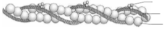
Label the different components of the actin filament in the diagram given below.
Solution
(N/A) The actin filament (thin filament) is composed of three main protein components:
$1$. $F$-actin: These are two filamentous actin helices that form the backbone of the filament.
$2$. Tropomyosin: These are two strands of tropomyosin protein that run close to the $F$-actin throughout its length.
$3$. Troponin: These are complex proteins distributed at regular intervals on the tropomyosin.
0 likes
View Solution210
MediumMCQ
It is known that red muscle fibres in animals can work for longer periods of time continuously. How is this possible?
A
They contain high amounts of myoglobin and mitochondria.
B
They contain high amounts of sarcoplasmic reticulum.
C
They contain high amounts of glycogen.
D
They contain high amounts of lactic acid.
Solution
(A) Generally,muscles are of two types: $(1)$ Red muscle,$(2)$ White muscle.
Red muscles can work for longer periods because:
$(i)$ They contain a red-coloured oxygen-binding protein called myoglobin. Myoglobin stores oxygen in the form of oxymyoglobin,which releases $O_{2}$ during muscle contraction.
$(ii)$ They have a high density of mitochondria,which allows for sustained aerobic respiration.
$(iii)$ They have a less developed sarcoplasmic reticulum compared to white muscles.
$(iv)$ They perform aerobic respiration for longer periods without accumulating significant amounts of lactic acid,thus delaying fatigue.
$(v)$ The rate of contraction is slower,allowing for sustained activity,for example,the back muscles in humans.
0 likes
View Solution211
Easy
Rahul exercises regularly by visiting a gymnasium. Of late,he is gaining weight. What could be the reason? Choose the correct answer and elaborate.
$A$. Rahul has gained weight due to accumulation of fats in the body.
$B$. Rahul has gained weight due to increased muscle mass and loss of fat.
$C$. Rahul has gained weight because his muscle shape has improved.
$D$. Rahul has gained weight because he is accumulating water in the body.
$A$. Rahul has gained weight due to accumulation of fats in the body.
$B$. Rahul has gained weight due to increased muscle mass and loss of fat.
$C$. Rahul has gained weight because his muscle shape has improved.
$D$. Rahul has gained weight because he is accumulating water in the body.
Solution
(B) The correct answer is $B$. Regular exercise,such as weight training,leads to hypertrophy of muscle fibers.
When an individual exercises regularly,the volume and size of the muscles increase due to an increase in the amount of sarcoplasm,the number of myofibrils,and the density of mitochondria within the muscle cells.
While fat mass is typically reduced through consistent physical activity,the overall body weight may increase because muscle tissue is denser and heavier than adipose (fat) tissue.
Therefore,the weight gain observed in a person who exercises regularly is primarily due to the development of lean muscle mass rather than fat accumulation.
0 likes
View Solution212
EasyMCQ
What is the source of energy for muscle contraction?
A
Glucose
B
$ATP$
C
Creatine phosphate
D
Lactic acid
Solution
(B) $ATP$ is the source of energy for muscle contraction.
In each myosin molecule,the $ATPase$ enzyme is present at the head region.
In the presence of this enzyme,along with $Ca^{2+}$ and $Mg^{2+}$ ions,the $ATP$ molecule dissociates into $ADP + Pi$.
Energy is released from the head of the myosin. With the energy obtained from $ATP$,the excited myosin forms a cross-bridge with actin,and muscle contraction is initiated.
0 likes
View Solution213
Easy
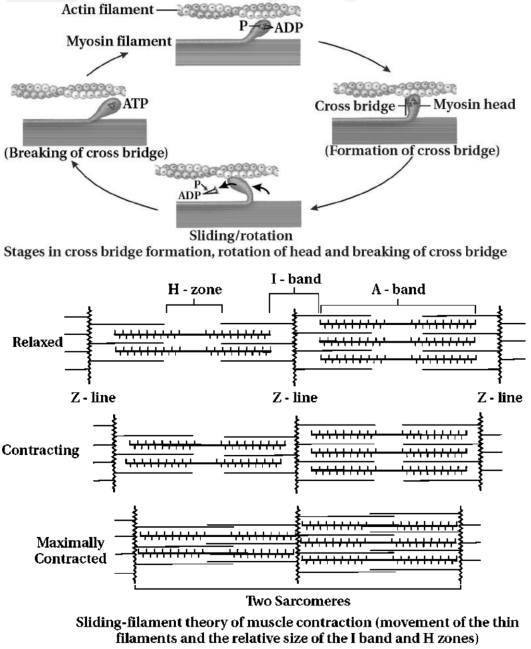
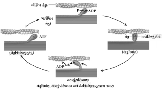
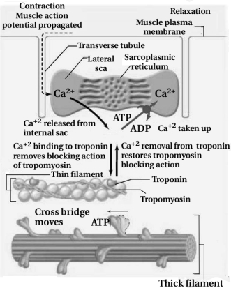
Explain the sliding filament theory of muscle contraction with neat sketches.
Solution
(N/A) The mechanism of muscle contraction is best explained by the sliding filament theory,which states that the contraction of a muscle fibre takes place by the sliding of the thin filaments over the thick filaments.
This theory was postulated by $A$.$F$. Huxley and $J$. Hanson.
Muscle contraction is initiated by a signal sent by the central nervous system $(CNS)$ via a motor neuron.
$A$ motor neuron along with the muscle fibres connected to it constitutes a motor unit. The junction between a motor neuron and the sarcolemma of the muscle fibre is called the neuromuscular junction.
$A$ neural signal reaching this junction releases a neurotransmitter (acetylcholine),which generates an action potential in the sarcolemma. This spreads through the muscle fibre and causes the release of $Ca^{++}$ (calcium ions) into the sarcoplasm.
An increase in $Ca^{++}$ level leads to the binding of $Ca^{++}$ with a subunit of troponin on actin filaments,thereby removing the masking of active sites for myosin.
Utilising the energy from $ATP$ hydrolysis,the myosin head now binds to exposed active sites on actin to form a cross-bridge.
This pulls the attached actin filaments towards the centre of the '$A$' band. The '$Z$' line attached to these actins is also pulled inwards,thereby causing a shortening of the sarcomere,i.e.,contraction.
It is clear from the above steps that during the shortening of the muscle (contraction),the '$I$' bands get reduced,whereas the '$A$' band retains its length.
The myosin,releasing the $ADP$ and $P_i$,goes back to its relaxed state. $A$ new $ATP$ binds,and the cross-bridge is broken.
The $ATP$ is again hydrolysed by the myosin head,and the cycle of cross-bridge formation and breakage is repeated,causing further sliding. The process continues until the $Ca^{++}$ ions are pumped back to the sarcoplasmic cisternae,resulting in the masking of actin filaments. This causes the return of '$Z$' lines back to their original position (i.e.,relaxation).
The reaction time of the fibres can vary in different muscles. Repeated activation of the muscles can lead to the accumulation of lactic acid due to the anaerobic breakdown of glycogen in them,causing fatigue.
0 likes
View Solution214
Easy
How does a muscle shorten during its contraction and return to its original form during relaxation?
Solution
(N/A) neural signal reaching the neuromuscular junction releases a neurotransmitter (Acetylcholine) which generates an action potential in the sarcolemma. This spreads through the muscle fibre and causes the release of $Ca^{++}$ (calcium ions) into the sarcoplasm.
Increase in $Ca^{++}$ level leads to the binding of $Ca^{++}$ with a subunit of troponin on actin filaments,thereby removing the masking of active sites for myosin.
Utilising the energy from $ATP$ hydrolysis,the myosin head now binds to exposed active sites on actin to form a cross-bridge.
This pulls the attached actin filaments towards the centre of the '$A$' band. The '$Z$' line attached to these actins are also pulled inwards,thereby causing a shortening of the sarcomere,i.e.,contraction.
It is clear from the above steps that during the shortening of the muscle (contraction),the '$I$' bands get reduced,whereas the '$A$' band retains its length.
The myosin,releasing the $ADP$ and $P_i$,goes back to its relaxed state. $A$ new $ATP$ binds and the cross-bridge is broken.
The $ATP$ is again hydrolysed by the myosin head and the cycle of cross-bridge formation and breakage is repeated,causing further sliding. The process continues until the $Ca^{++}$ ions are pumped back to the sarcoplasmic cisternae,resulting in the masking of actin filaments. This causes the return of '$Z$' lines back to their original position (i.e.,relaxation).
0 likes
View Solution215
Easy
Discuss the role of $Ca^{2+}$ ions in muscle contraction. Draw neat sketches to illustrate your answer.
Solution
(N/A) Calcium ions $(Ca^{2+})$ play a crucial regulatory role in muscle contraction.
$1$. Upon stimulation,$Ca^{2+}$ ions are released from the sarcoplasmic reticulum into the sarcoplasm.
$2$. These $Ca^{2+}$ ions bind to the troponin complex on the thin filaments ($F$-actin).
$3$. This binding induces a conformational change in the troponin-tropomyosin complex,which shifts the position of tropomyosin,thereby exposing the active binding sites on the actin filaments.
$4$. The myosin heads then bind to these exposed active sites on actin to form cross-bridges.
$5$. The myosin head,which possesses $ATPase$ activity,hydrolyzes $ATP$ into $ADP$ and inorganic phosphate $(Pi)$. The energy released from this hydrolysis allows the myosin head to pivot,pulling the actin filament towards the center of the sarcomere (power stroke),resulting in muscle contraction.
$6$. During relaxation,$Ca^{2+}$ ions are actively pumped back into the sarcoplasmic reticulum,causing the troponin-tropomyosin complex to return to its original position,blocking the active sites on actin and preventing further cross-bridge formation.
0 likes
View Solution216
EasyMCQ
Analogy type questions:
$(1)$ Red muscle fibers / White muscle fibers have fewer mitochondria but more sarcoplasmic reticulum.
$(2)$ When nerve impulses reach the neuromuscular junction,acetic acid / acetylcholine neurotransmitter is released.
$(1)$ Red muscle fibers / White muscle fibers have fewer mitochondria but more sarcoplasmic reticulum.
$(2)$ When nerve impulses reach the neuromuscular junction,acetic acid / acetylcholine neurotransmitter is released.
A
$(1)$ White muscle fibers,$(2)$ Acetylcholine
B
$(1)$ Red muscle fibers,$(2)$ Acetic acid
C
$(1)$ White muscle fibers,$(2)$ Acetic acid
D
$(1)$ Red muscle fibers,$(2)$ Acetylcholine
Solution
(A) $(1)$ White muscle fibers contain fewer mitochondria and a more extensive sarcoplasmic reticulum compared to red muscle fibers,which allows for faster contraction and anaerobic metabolism.
$(2)$ When a neural signal reaches the neuromuscular junction,it triggers the release of the neurotransmitter acetylcholine into the synaptic cleft,which initiates an action potential in the muscle fiber membrane.
$(2)$ When a neural signal reaches the neuromuscular junction,it triggers the release of the neurotransmitter acetylcholine into the synaptic cleft,which initiates an action potential in the muscle fiber membrane.
0 likes
View Solution217
EasyMCQ
Analogy type questions:
$(1)$ The portion between two successive $Z$-lines is the functional unit of contraction,which is called the sarcoplasmic reticulum / sarcomere.
$(2)$ The thick filaments in the $A$-band are held together in the middle of this band by a thin fibrous membrane. It is called the $Z$-line / $M$-line.
$(1)$ The portion between two successive $Z$-lines is the functional unit of contraction,which is called the sarcoplasmic reticulum / sarcomere.
$(2)$ The thick filaments in the $A$-band are held together in the middle of this band by a thin fibrous membrane. It is called the $Z$-line / $M$-line.
A
$(1)$ Sarcomere,$(2)$ $M$-line
B
$(1)$ Sarcoplasmic reticulum,$(2)$ $Z$-line
C
$(1)$ Sarcomere,$(2)$ $Z$-line
D
$(1)$ Sarcoplasmic reticulum,$(2)$ $M$-line
Solution
(A) $(1)$ The functional unit of muscle contraction is the sarcomere,which is defined as the segment of a myofibril between two successive $Z$-lines.
$(2)$ The thick filaments (myosin) in the center of the $A$-band are connected by a thin fibrous membrane known as the $M$-line.
$(2)$ The thick filaments (myosin) in the center of the $A$-band are connected by a thin fibrous membrane known as the $M$-line.
0 likes
View Solution218
EasyMCQ
Visceral muscles are involved in which functions?
A
Locomotion of the body
B
Transportation of food through the digestive tract
C
Contraction of the heart
D
Voluntary movement of limbs
Solution
(B) Visceral muscles,also known as smooth muscles,are involuntary muscles found in the walls of internal organs. They are involved in functions such as the transportation of food through the digestive tract and the movement of gametes through the genital tract.
0 likes
View Solution219
Medium
Explain sarcolemma.
Solution
(N/A) The sarcolemma is the specialized plasma membrane that surrounds each muscle fibre.
It encloses the sarcoplasm,which is the cytoplasm of the muscle cell.
Because the muscle fibre contains multiple nuclei,it is considered a syncytium.
It encloses the sarcoplasm,which is the cytoplasm of the muscle cell.
Because the muscle fibre contains multiple nuclei,it is considered a syncytium.
0 likes
View Solution220
MediumMCQ
Where are calcium ions stored in muscle cells?
A
Mitochondria
B
Sarcoplasmic reticulum
C
Golgi apparatus
D
Lysosomes
Solution
(B) In muscle cells,the specialized form of the endoplasmic reticulum is known as the sarcoplasmic reticulum.
It acts as the primary storage site for calcium ions $(Ca^{2+})$.
During muscle contraction,these calcium ions are released into the sarcoplasm to initiate the interaction between actin and myosin filaments.
It acts as the primary storage site for calcium ions $(Ca^{2+})$.
During muscle contraction,these calcium ions are released into the sarcoplasm to initiate the interaction between actin and myosin filaments.
0 likes
View Solution221
EasyMCQ
What is a sarcomere?
A
The structural unit of a muscle fiber.
B
The portion of the myofibril between two successive $Z$-lines.
C
The contractile protein found in muscles.
D
The junction between a nerve and a muscle.
Solution
(B) The portion of the myofibril between two successive $Z$-lines is considered as the functional unit of contraction and is called a sarcomere. It is the basic contractile unit of the striated muscle fiber.
0 likes
View Solution222
MediumMCQ
How is $G$-actin converted into $F$-actin?
A
By the addition of $Ca^{++}$ ions
B
By the polymerization of $G$-actin in the presence of $Mg^{++}$ ions
C
By the hydrolysis of $ATP$
D
By the breakdown of $F$-actin filaments
Solution
(B) $G$-actin (Globular actin) is a monomeric protein.
In the presence of $Mg^{++}$ ions and $ATP$,$G$-actin molecules undergo polymerization to form long,helical,polymeric filamentous structures known as $F$-actin (Filamentous actin).
This process is essential for the formation of thin filaments in muscle fibers.
In the presence of $Mg^{++}$ ions and $ATP$,$G$-actin molecules undergo polymerization to form long,helical,polymeric filamentous structures known as $F$-actin (Filamentous actin).
This process is essential for the formation of thin filaments in muscle fibers.
0 likes
View Solution223
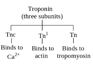
Medium
Give the name of the subunits of Troponin.
Solution
(N/A) Troponin is a regulatory protein complex associated with the actin filament in muscle fibers. It consists of three distinct subunits:
$(a)$ Troponin $I$ $(T_pI)$: This subunit inhibits the interaction between actin and myosin.
$(b)$ Troponin $T$ $(T_pT)$: This subunit binds to tropomyosin,anchoring the troponin complex to the tropomyosin strand.
$(c)$ Troponin $C$ $(T_pC)$: This subunit acts as a binding site for calcium ions $(Ca^{2+})$,which triggers the contraction process.
0 likes
View Solution224
MediumMCQ
How is the meromyosin formed?
A
By the polymerization of actin filaments.
B
By the polymerization of monomeric proteins called meromyosins.
C
By the breakdown of myosin filaments.
D
By the combination of actin and myosin filaments.
Solution
(B) Each myosin filament is a polymerised protein.
Many monomeric proteins called meromyosins constitute one thick filament.
Therefore,a thick filament (myosin) is formed by the polymerization of many meromyosin units.
Many monomeric proteins called meromyosins constitute one thick filament.
Therefore,a thick filament (myosin) is formed by the polymerization of many meromyosin units.
0 likes
View Solution225
Medium
What are $HMM$ and $LMM$?
Solution
(N/A) Each meromyosin molecule consists of two important parts: a globular head with a short arm and a tail. The globular head with the short arm is called the heavy meromyosin $(HMM)$,while the tail portion is called the light meromyosin $(LMM)$.
0 likes
View Solution226
EasyMCQ
Who put forward the sliding filament theory?
A
$A$.$F$. Huxley and $H$.$E$. Huxley
B
Andrew Huxley and Hugh Huxley
C
$A$.$F$. Huxley and $J$. Hanson
D
$H$.$E$. Huxley and $J$. Hanson
Solution
(C) The sliding filament theory was proposed by Andrew $F$. Huxley and Jean Hanson,along with Hugh $E$. Huxley and Rolf Niedergerke,in $1954$.
It explains the mechanism of muscle contraction where thin filaments slide over thick filaments,shortening the sarcomere without changing the length of the filaments themselves.
It explains the mechanism of muscle contraction where thin filaments slide over thick filaments,shortening the sarcomere without changing the length of the filaments themselves.
0 likes
View Solution227
MediumMCQ
What is muscle fatigue?
A
The accumulation of lactic acid in muscles due to anaerobic breakdown of glycogen.
B
The rapid contraction of muscles without rest.
C
The depletion of $ATP$ in the muscle fibres.
D
The relaxation of muscles after prolonged activity.
Solution
(A) Muscle fatigue is a condition where muscles lose their ability to contract effectively after prolonged or intense activity.
Repeated activation of the muscles leads to the accumulation of lactic acid due to the anaerobic breakdown of glycogen in the muscle fibres.
This accumulation of lactic acid causes the sensation of fatigue and muscle soreness.
Repeated activation of the muscles leads to the accumulation of lactic acid due to the anaerobic breakdown of glycogen in the muscle fibres.
This accumulation of lactic acid causes the sensation of fatigue and muscle soreness.
0 likes
View Solution228
Medium
Select the correct option for the following statements:
$(i)$ The most important respiratory organ in frogs is the skin / lungs.
(ii) Skeletal muscle / Cardiac muscle exhibits rhythmic contraction.
$(i)$ The most important respiratory organ in frogs is the skin / lungs.
(ii) Skeletal muscle / Cardiac muscle exhibits rhythmic contraction.
Solution
(SKIN, CARDIAC MUSCLE) $(i)$ Skin: Frogs respire through their skin (cutaneous respiration) both in water and on land,making it the most important respiratory organ.
(ii) Cardiac muscle: Cardiac muscles are involuntary and exhibit rhythmic,spontaneous contractions throughout life.
(ii) Cardiac muscle: Cardiac muscles are involuntary and exhibit rhythmic,spontaneous contractions throughout life.
0 likes
View Solution229
MediumMCQ
Myosin head separates from actin when:
A
$ATP$ hydrolysis
B
When $ATP$ releases from Actin
C
When $ATP$ releases from myosin head
D
When $ATP$ attached to myosin head
Solution
(D) During the muscle contraction cycle,the myosin head remains bound to the actin filament in a cross-bridge state. When a new molecule of $ATP$ binds to the myosin head,the affinity of the myosin head for actin decreases significantly. Consequently,the myosin head detaches from the actin filament. Therefore,the separation occurs when $ATP$ attaches to the myosin head.
0 likes
View Solution230
MediumMCQ
Which of the following muscles act involuntarily?
$(a)$ Striated muscles $(b)$ Smooth muscles
$(c)$ Cardiac muscles $(d)$ Skeletal muscles
$(a)$ Striated muscles $(b)$ Smooth muscles
$(c)$ Cardiac muscles $(d)$ Skeletal muscles
A
$(a) \text{ and } (b)$
B
$(b) \text{ and } (c)$
C
$(c) \text{ and } (d)$
D
$(a) \text{ and } (d)$
Solution
(B) Skeletal muscles (striated or striped muscles) are voluntary muscles,meaning they are under conscious control,$e.g.$,movement of arms and legs.
Smooth muscles (unstriped or non-striated muscles) are involuntary muscles,meaning they are not under conscious control,$e.g.$,muscles in the posterior part of the oesophagus,stomach,intestine,and blood vessels.
Cardiac muscles are specialized involuntary muscles found in the walls of the heart.
Therefore,both smooth muscles $(b)$ and cardiac muscles $(c)$ act involuntarily.
0 likes
View Solution231
EasyMCQ
In a skeletal muscle fibre,the nuclei are located:
A
Diffused
B
Centrally
C
Peripherally
D
Absent
Solution
(C) Skeletal or striated muscle fibers are long,cylindrical,and unbranched structures. These fibers are multinucleated,meaning they contain many nuclei. $A$ characteristic feature of these muscle fibers is that their nuclei are pushed towards the periphery,just beneath the sarcolemma.
0 likes
View Solution232
MediumMCQ
Cardiac muscle fibres are
A
Striated and involuntary
B
Striated and voluntary
C
Non-striated and involuntary
D
Non-striated and voluntary
Solution
(A) Cardiac muscle fibres exhibit characteristic transverse dark and light bands,which alternate with each other,giving them a striated appearance. Furthermore,these muscles are controlled by the autonomic nervous system and are not under conscious control,making them involuntary.
0 likes
View Solution233
MediumMCQ
Contractile proteins are found in
A
Bone
B
Blood
C
Cartilage
D
Muscles
Solution
(D) Muscles possess a contractile nature.
The contractile structures within muscle cells are known as myofibrils.
Myofibrils are composed of myofilaments.
Myofilaments are of two primary types: myosin and actin.
Muscle contraction occurs due to the sliding of actin filaments over myosin filaments.
The contractile structures within muscle cells are known as myofibrils.
Myofibrils are composed of myofilaments.
Myofilaments are of two primary types: myosin and actin.
Muscle contraction occurs due to the sliding of actin filaments over myosin filaments.
0 likes
View Solution234
MediumMCQ
Which of the following is not a characteristic feature of the biceps muscle?
A
We are usually able to make it contract merely by thinking about it
B
It has alternate light and dark bands
C
Its muscle fibres taper at both ends
D
Its muscle fibres are bundled together in a parallel fashion
Solution
(C) The biceps muscle is a type of skeletal muscle,which is also known as striated muscle.
Skeletal muscles are voluntary,meaning we can control their contraction through conscious thought.
They exhibit alternate light and dark bands (striations) due to the arrangement of actin and myosin filaments.
Their muscle fibres are bundled together in a parallel fashion.
In contrast,smooth muscle fibres are spindle-shaped (fusiform) and taper at both ends,which is not a characteristic of skeletal muscles like the biceps.
0 likes
View Solution235
MediumMCQ
An individual sarcomere of a myofibril consists of:
A
Overlapping actin and myosin
B
$A$ stack of actin fibres
C
$A$ stack of myosin units
D
Overlapping actin and relaxin
Solution
(A) sarcomere is the functional unit of a muscle contraction,defined as the segment between two successive $Z-$lines.
It consists of an arrangement of thick filaments (myosin) and thin filaments (actin).
In the resting state,the thin filaments partially overlap the thick filaments,which is the structural basis for the sliding filament theory.
Therefore,a sarcomere is characterized by the overlapping of actin and myosin filaments.
0 likes
View Solution236
MediumMCQ
Where is troponin found during muscle contraction?
A
Myosin filament
B
Meromyosin
C
Tropomyosin
D
$T$-tubule
Solution
(C) Troponin is a regulatory protein complex associated with the thin filament (actin). It consists of three subunits: $TnC$ (binds to $Ca^{2+}$),$TnI$ (binds to actin),and $TnT$ (binds to tropomyosin). During muscle contraction,troponin is found distributed at regular intervals on the tropomyosin protein,which in turn lies along the actin filament. Therefore,it is closely associated with tropomyosin.
0 likes
View Solution237
EasyMCQ
The largest muscle in the human body is:
A
Sartorius
B
Gluteus maximus
C
Stapedius
D
Masseter
Solution
(B) The $Gluteus$ $maximus$ (buttock muscle) is the largest and most powerful muscle in the human body. It is primarily responsible for the movement of the hip and thigh.
0 likes
View Solution238
MediumMCQ
Muscle is attached to bone by
A
Tendon
B
Ligament
C
Insertion
D
Cartilage
Solution
(A) Tendons are dense regular connective tissues that primarily consist of collagen fibers. They serve the specific function of attaching skeletal muscles to bones,allowing for the transmission of force generated by muscle contraction to the skeletal system,which facilitates movement.
0 likes
View Solution239
MediumMCQ
The mechanism of muscle contraction is best explained by:
A
Physical filament theory
B
Chemical filament theory
C
Sliding filament theory
D
Jumping filament theory
Solution
(C) The mechanism of muscle contraction is best explained by the $Sliding$ $Filament$ $Theory$.
This theory states that the contraction of a muscle fiber occurs due to the sliding of thin actin filaments over thick myosin filaments,which shortens the sarcomere without changing the length of the filaments themselves.
This theory states that the contraction of a muscle fiber occurs due to the sliding of thin actin filaments over thick myosin filaments,which shortens the sarcomere without changing the length of the filaments themselves.
0 likes
View Solution240
MediumMCQ
Both proteins,actin and myosin,are arranged in a rod-like structure in the muscles:
A
Radially
B
Parallely
C
Horizontally
D
Obliquely
Solution
(B) Both proteins,$i.e.$,actin and myosin,are arranged as rod-like structures,parallel to each other and also to the longitudinal axis of the myofibrils.
Actin filaments are thinner compared to myosin filaments; hence,they are commonly called thin and thick filaments,respectively.
Actin filaments are thinner compared to myosin filaments; hence,they are commonly called thin and thick filaments,respectively.
0 likes
View Solution241
MediumMCQ
Action potential in the sarcolemma of muscles causes the release of:
A
$Na^{+}$
B
$Cl^{-}$
C
$Ca^{2+}$
D
$HCO_{3}^{-}$
Solution
(C) The action potential in the sarcolemma travels through the $T$-tubules and triggers the release of $Ca^{2+}$ ions from the sarcoplasmic reticulum into the sarcoplasm.
An increase in the $Ca^{2+}$ concentration in the sarcoplasm leads to the binding of $Ca^{2+}$ with a subunit of troponin on the actin filaments.
This binding causes a conformational change in the troponin-tropomyosin complex,which removes the masking of the active sites on actin filaments for myosin heads,thereby initiating muscle contraction.
0 likes
View Solution242
MediumMCQ
The membrane sarcolemma is found over
A
Heart
B
Muscle fiber
C
Both $(a)$ and $(b)$
D
Nerve fiber
Solution
(B) Sarcolemma is the fine,transparent,and elastic plasma membrane that covers the muscle fiber (myocyte).
It encloses the sarcoplasm,which contains myofibrils,mitochondria,and other organelles.
Therefore,the correct answer is muscle fiber.
It encloses the sarcoplasm,which contains myofibrils,mitochondria,and other organelles.
Therefore,the correct answer is muscle fiber.
0 likes
View Solution243
MediumMCQ
Each myofibril of a muscle contains:
A
Regular dark bands
B
Regular light bands
C
Both $(a)$ and $(b)$
D
Alternate dark and light bands
Solution
(D) Each myofibril contains alternate dark and light bands. The light bands contain actin and are called $I$-bands or isotropic bands,whereas the dark bands are called $A$-bands or anisotropic bands,which contain myosin.
0 likes
View Solution244
MediumMCQ
The functional unit of skeletal muscle is called:
A
Sarcomere
B
Twitch
C
$Z$-band
D
None of these
Solution
(A) Skeletal muscles are responsible for voluntary movements under the conscious control of the brain,hence they are called voluntary muscles.
Each muscle fiber contains many parallel filaments called myofibrils.
The segment of a myofibril located between two adjacent $Z$-lines (or $Z$-bands) is known as a sarcomere.
The sarcomere is considered the functional unit of contraction in skeletal muscle because it contains the contractile proteins,actin and myosin,arranged in a specific pattern that allows for muscle shortening.
0 likes
View Solution245
MediumMCQ
The region between two successive $Z-$lines in a myofibril is
A
Sarcomere
B
Sarcosome
C
Fascia
D
Anisotropic band
Solution
(A) The sarcomere is the functional and structural unit of a muscle fiber.
It is defined as the region of a myofibril located between two successive $Z-$lines (or $Z-$discs).
These $Z-$lines anchor the actin filaments,and the contraction of the muscle occurs due to the shortening of the sarcomere.
It is defined as the region of a myofibril located between two successive $Z-$lines (or $Z-$discs).
These $Z-$lines anchor the actin filaments,and the contraction of the muscle occurs due to the shortening of the sarcomere.
0 likes
View Solution246
MediumMCQ
Which muscle component is the smallest among the given options?
A
Muscle fibre
B
Myofibril
C
Actin
D
Sarcomere
Solution
(C) The structural hierarchy of muscle tissue is as follows: $Actin$ and $Myosin$ filaments are the smallest contractile proteins.
$Actin$ and $Myosin$ filaments organize into $Sarcomeres$, which are the functional units of a muscle.
Many $Sarcomeres$ arranged in series form a $Myofibril$.
Many $Myofibrils$ bundled together form a $Muscle \text{ } fibre$ (muscle cell).
Therefore, among the given options, $Actin$ is the smallest component.
0 likes
View Solution247
MediumMCQ
The immediate energy source for muscle contraction is:
A
$ATP$
B
$ADP$
C
$Glucose$
D
$Lactic \; acid$
Solution
(A) Muscle contraction requires energy to facilitate the sliding of actin and myosin filaments.
$ATP$ (Adenosine Triphosphate) acts as the immediate source of chemical energy for this process.
When $ATP$ is hydrolyzed by the enzyme myosin ATPase,it releases the energy required for the power stroke of the myosin head.
$ATP$ (Adenosine Triphosphate) acts as the immediate source of chemical energy for this process.
When $ATP$ is hydrolyzed by the enzyme myosin ATPase,it releases the energy required for the power stroke of the myosin head.
0 likes
View Solution248
MediumMCQ
Which muscle band remains unchanged during the contraction and relaxation of the skeletal muscle?
A
$I$-band
B
$H$-zone
C
$A$-band
D
$Z$-line
Solution
(C) The $A$-band (anisotropic band),which is composed of thick filaments (myosin),remains unchanged in length during skeletal muscle contraction.
During contraction,the thin filaments (actin) slide over the thick filaments,which causes the $I$-band to shorten and the $H$-zone to disappear or narrow,but the $A$-band length remains constant.
During contraction,the thin filaments (actin) slide over the thick filaments,which causes the $I$-band to shorten and the $H$-zone to disappear or narrow,but the $A$-band length remains constant.
0 likes
View Solution249
MediumMCQ
$A$ motor unit is a
A
Neuron
B
Muscle fibre
C
Motor neuron with muscle fibre
D
All of the above
Solution
(C) motor unit consists of a motor neuron and the muscle fibres connected to it. Muscle contraction is initiated by a signal sent by the Central Nervous System $(CNS)$ via motor neurons. $A$ single motor neuron,along with all the muscle fibres it innervates,constitutes a motor unit.
0 likes
View Solution250
MediumMCQ
The thin filaments of a muscle fiber are made up of
A
Actin
B
Myoglobin
C
Myosin
D
Cellulose
Solution
(A) The thin filaments of a muscle fiber are primarily composed of two $F$-actin filaments, along with two regulatory proteins, $troponin$ and $tropomyosin$.
0 likes
View SolutionLocomotion and Movement — Muscles · Frequently Asked Questions
1Are these Locomotion and Movement questions useful for JEE and NEET?
Yes. All questions in this section are mapped to JEE Main and NEET exam patterns. Previous year questions from JEE Main, NEET, GUJCET and state-level exams are included with full solutions.
2Can I switch to Hindi or Gujarati for these questions?
Yes. Use the language tabs in the hero section or the sidebar to view the same questions and solutions in English, Hindi or Gujarati.
3How do I generate a question paper from this subtopic?
Use the Vedclass Exam Paper Generator — select the chapter and subtopic, set difficulty, and generate Sets A, B, C, D automatically. First 3 chapters of every subject are free.
Vedclass Products
For Students
Vedclass Test Series
Mock tests in real JEE/NEET style with performance analysis. 5-day free trial.
Start Free TrialFor Teachers
Exam Paper Generator
Generate Set A/B/C/D papers from this chapter in 2 minutes. 3 chapters free.
Try FreeFor Institutes
Online Exam Module
Live online exams with unlimited students, 360° analytics & white-label branding.
See DemoFor Teachers & Institutes
Generate a Locomotion and Movement Exam Paper in 2 Minutes
Select subtopic & difficulty — Sets A, B, C, D auto-generated with No Repeat logic.
First 3 chapters of every subject are free — no payment required.