
A English
Blood pressure, ECG Questions in English
Class 11 Biology · Body Fluids and Circulations · Blood pressure, ECG
87+
Questions
English
Language
100%
With Solutions
Showing 37 of 87 questions in English
51
MediumMCQ
What does a blood pressure instrument record?
A
Systolic pressure
B
Diastolic pressure
C
Both $(a)$ and $(b)$
D
None of these
Solution
(C) The lateral pressure exerted by the column of blood on the walls of the blood vessels in which it flows is called blood pressure.
It is usually measured in the brachial artery using an instrument called a sphygmomanometer.
$A$ sphygmomanometer measures both systolic blood pressure (the peak pressure during heart contraction) and diastolic blood pressure (the minimum pressure during heart relaxation).
It is usually measured in the brachial artery using an instrument called a sphygmomanometer.
$A$ sphygmomanometer measures both systolic blood pressure (the peak pressure during heart contraction) and diastolic blood pressure (the minimum pressure during heart relaxation).
0 likes
View Solution52
MediumMCQ
$ECG$ is a graphical representation of the electrical activity of the heart during
A
Cardiac systole
B
Cardiac diastole
C
Cardiac cycle
D
Ventricular and atrial diastole
Solution
(C) An $ECG$ (electrocardiogram) is a graphical representation of the electrical activity of the heart during a single cardiac cycle. It records the electrical impulses generated by the heart's conduction system,which triggers the contraction and relaxation of the atria and ventricles.
0 likes
View Solution53
MediumMCQ
Which one indicates hypertension or high blood pressure $(BP)$?
A
$120 / 80$
B
$110 / 70$
C
$140 / 90$
D
All of these
Solution
(C) Hypertension is the term for blood pressure that is higher than normal.
If repeated checks of blood pressure of an individual show $140 / 90$ ($140$ mmHg systolic and $90$ mmHg diastolic) or higher,it indicates hypertension.
High blood pressure leads to heart diseases and also affects vital organs like the brain and kidneys.
If repeated checks of blood pressure of an individual show $140 / 90$ ($140$ mmHg systolic and $90$ mmHg diastolic) or higher,it indicates hypertension.
High blood pressure leads to heart diseases and also affects vital organs like the brain and kidneys.
0 likes
View Solution54
MediumMCQ
Maximum blood pressure is experienced when blood enters from:
A
Right ventricle to aorta
B
Right auricle to aorta
C
Left ventricle to aorta
D
Left auricle to aorta
Solution
(C) Blood pressure is defined as the pressure exerted by the flow of blood on the elastic walls of the arteries.
Blood pressure is significantly higher during ventricular systole compared to diastole.
The maximum blood pressure is experienced when the left ventricle contracts,pumping oxygenated blood into the aorta,which then distributes it to the rest of the body.
Blood pressure is significantly higher during ventricular systole compared to diastole.
The maximum blood pressure is experienced when the left ventricle contracts,pumping oxygenated blood into the aorta,which then distributes it to the rest of the body.
0 likes
View Solution55
MediumMCQ
Which wave of the human heart $ECG$ $(PQRST)$ is used for determining the heart rate of an individual?
A
$P$
B
$QRS$
C
$T$
D
$RS$
Solution
(B) The $QRS$ complex represents the depolarization of the ventricles,which initiates ventricular contraction.
By counting the number of $QRS$ complexes that occur in a given time period,one can determine the heart rate of an individual.
Since $ECGs$ obtained from different individuals have roughly the same shape for a given lead configuration,any deviation from this shape indicates a possible abnormality or disease.
Therefore,the $QRS$ complex is of great clinical significance for determining heart rate.
0 likes
View Solution56
MediumMCQ
In an $ECG$,the depolarization of atria is indicated by
A
$P-$wave
B
$Q-$wave
C
$R-$wave
D
$S-$wave
Solution
(A) $ECG$ is the graphical recording of electrical changes that accompany the cardiac cycle.
It is represented by five waves: $P, Q, R, S,$ and $T$.
The $P-$wave indicates the depolarization of the atria.
The $QRS$ complex indicates ventricular depolarization.
The $T-$wave indicates ventricular repolarization.
It is represented by five waves: $P, Q, R, S,$ and $T$.
The $P-$wave indicates the depolarization of the atria.
The $QRS$ complex indicates ventricular depolarization.
The $T-$wave indicates ventricular repolarization.
0 likes
View Solution57
MediumMCQ
Which of the following sentences is correct?
$I$. $ECG$ is of a great clinical significance.
$II$. Electrocardiograph is the machine used to obtain an electrocardiogram.
$III$. To obtain a standard $ECG$,a patient is connected to the machine with $3$ electrical leads (one to each wrist and to the left ankle).
$IV$. Normal activities of the heart are regulated intrinsically (auto-regulated by nodal tissue).
$V$. Electrocardiogram is the graphical representation of the electrical activity of the heart during a cardiac cycle.
Which of the following options contains all the correct statements?
A
$I, II, III$ and $IV$
B
$I, II, III, IV$ and $V$
C
$II, III, IV$ and $V$
D
$I, III, IV$ and $V$
Solution
(B) $I$. $ECG$ is of great clinical significance as it helps in diagnosing various heart abnormalities. (Correct)
$II$. Electrocardiograph is the machine used to record the electrical activity of the heart,while the electrocardiogram is the actual recording (graph). The statement provided in the original question was technically imprecise regarding the definition. (Corrected context: $II$ is correct if defined as the machine).
$III$. $A$ standard $ECG$ is obtained by connecting the patient to the machine using $3$ electrical leads (one to each wrist and to the left ankle). (Correct)
$IV$. The heart is myogenic,meaning its normal activities are regulated intrinsically by specialized nodal tissue ($SA$ node and $AV$ node). (Correct)
$V$. Electrocardiogram is the graphical representation of the electrical activity of the heart during a cardiac cycle. (Correct)
Therefore,all statements $I, II, III, IV,$ and $V$ are correct.
0 likes
View Solution58
MediumMCQ
Repolarisation of the ventricles is represented by
A
$P$-wave
B
$QRS$-complex
C
$T$-wave
D
Both $P$ and $T$-wave
Solution
(C) $P$-wave: Represents the electrical excitation (or depolarisation) of the atria,which leads to the contraction of both the atria.
$QRS$-complex: Represents the depolarisation of the ventricles,which initiates the ventricular contraction. The contraction starts shortly after $Q$ and marks the beginning of the systole.
$T$-wave: Represents the return of the ventricles from the excited to the normal state (repolarisation). The end of the $T$-wave marks the end of the systole.
0 likes
View Solution59
EasyMCQ
To obtain a standard $ECG$,a patient is connected to the machine with three electrodes.
A
One to each wrist and to the left ankle.
B
One to each ankle and the left wrist.
C
One to each wrist and to the left chest region.
D
One to each ankle and to the left chest region.
Solution
(A) An $ECG$ (Electrocardiogram) is a graphical representation of the electrical activity of the heart during a cardiac cycle. To obtain a standard $ECG$,a patient is connected to the machine using three electrical leads (electrodes). These are typically attached to both wrists and the left ankle. This configuration allows the machine to measure the electrical potential differences generated by the heart muscles from different angles.
0 likes
View Solution60
EasyMCQ
What is represented by the $P$-wave in an $ECG$?
A
Ventricular depolarization
B
Atrial depolarization
C
Ventricular repolarization
D
Atrial repolarization
Solution
(B) The $P$-wave in an electrocardiogram $(ECG)$ represents the electrical excitation or depolarization of the atria.
This electrical activity leads to the contraction of both the atria.
This electrical activity leads to the contraction of both the atria.
0 likes
View Solution61
EasyMCQ
How can the rate of heart beating be obtained?
A
By counting the number of $P$-waves
B
By counting the number of $QRS$ complexes
C
By counting the number of $T$-waves
D
By measuring the duration of the $PR$ interval
Solution
(B) The heart beat rate of an individual can be determined by counting the number of $QRS$ complexes that occur in a given time period.
Each $QRS$ complex represents the depolarization of the ventricles,which initiates the ventricular contraction (systole).
Since one $QRS$ complex corresponds to one heartbeat,the total number of these complexes in a specific timeframe allows for the calculation of the heart rate.
Each $QRS$ complex represents the depolarization of the ventricles,which initiates the ventricular contraction (systole).
Since one $QRS$ complex corresponds to one heartbeat,the total number of these complexes in a specific timeframe allows for the calculation of the heart rate.
0 likes
View Solution62
EasyMCQ
What does the blood pressure reading $120/80 \ mmHg$ represent?
A
Systolic pressure of $120 \ mmHg$ and diastolic pressure of $80 \ mmHg$
B
Diastolic pressure of $120 \ mmHg$ and systolic pressure of $80 \ mmHg$
C
Average pressure of $120 \ mmHg$ and pulse pressure of $80 \ mmHg$
D
None of the above
Solution
(A) The blood pressure reading $120/80 \ mmHg$ is a standard measurement of arterial blood pressure.
In this value,$120 \ mmHg$ represents the systolic pressure,which is the pressure in the arteries during ventricular contraction (systole).
$80 \ mmHg$ represents the diastolic pressure,which is the pressure in the arteries during ventricular relaxation (diastole).
In this value,$120 \ mmHg$ represents the systolic pressure,which is the pressure in the arteries during ventricular contraction (systole).
$80 \ mmHg$ represents the diastolic pressure,which is the pressure in the arteries during ventricular relaxation (diastole).
0 likes
View Solution63
Medium
Complete the missing words in the statements given below:
$(a)$ Plasma without clotting factors is called serum.
$(b)$ $.........$ and monocytes are phagocytic cells.
$(c)$ Eosinophils are associated with $.........$ reactions.
$(d)$ $.........$ ions play a significant role in clotting.
$(e)$ One can determine the heart beat rate by counting the number of $.........$ in an $ECG$.
Solution
(N/A) Plasma without clotting factors is called serum.
$(b)$ Neutrophils and monocytes are phagocytic cells.
$(c)$ Eosinophils are associated with allergic reactions.
$(d)$ $Ca^{++}$ ions play a significant role in clotting.
$(e)$ One can determine the heart beat rate by counting the number of $QRS$ complexes in an $ECG$.
$(b)$ Neutrophils and monocytes are phagocytic cells.
$(c)$ Eosinophils are associated with allergic reactions.
$(d)$ $Ca^{++}$ ions play a significant role in clotting.
$(e)$ One can determine the heart beat rate by counting the number of $QRS$ complexes in an $ECG$.
0 likes
View Solution64
Medium
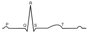
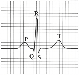
Given below is the diagrammatic representation of a standard $ECG$. Label its different peaks.
Solution
(N/A) The standard $ECG$ (Electrocardiogram) represents the electrical activity of the heart. The peaks are labeled as follows:
$1$. $P$-wave: Represents the electrical excitation (or depolarization) of the atria,which leads to the contraction of both the atria.
$2$. $QRS$-complex: Represents the depolarization of the ventricles,which initiates the ventricular contraction. The contraction starts shortly after $Q$ and marks the beginning of the systole.
$3$. $T$-wave: Represents the return of the ventricles from an excited to a normal state (repolarization). The end of the $T$-wave marks the end of the systole.
0 likes
View Solution65
MediumMCQ
How will you interpret an electrocardiogram $(ECG)$ in which the time taken in the $QRS$ complex is higher?
A
Normal heart function
B
Ventricular hypertrophy or conduction delay
C
Atrial fibrillation
D
Myocardial infarction
Solution
(B) The $QRS$ complex represents the depolarization of the ventricles,which initiates ventricular contraction.
In a normal $ECG$,the duration of the $QRS$ complex is typically less than $0.12$ seconds.
If the time taken for the $QRS$ complex is higher (prolonged),it indicates a delay in the conduction of electrical impulses through the ventricles.
This is often associated with conditions such as ventricular hypertrophy (enlargement of the heart muscle) or bundle branch blocks,where the electrical signal takes longer to spread through the ventricular myocardium.
0 likes
View Solution66
MediumMCQ
Select the correct option for $ECG$.
A
$P$-wave: Contraction of both atria; $QRS$-complex: Diastole of all chambers; $T$-wave: Contraction of both ventricles.
B
$P$-wave: Contraction of both atria; $QRS$-complex: Contraction of both ventricles; $T$-wave: Diastole of all chambers.
C
$P$-wave: Diastole of both atria; $QRS$-complex: Diastole of all chambers; $T$-wave: Diastole of both ventricles.
D
$P$-wave: Diastole of both atria; $QRS$-complex: Contraction of all chambers; $T$-wave: Diastole of both ventricles.
Solution
(B) The $ECG$ (Electrocardiogram) represents the electrical activity of the heart during a cardiac cycle:
$1$. The $P$-wave represents the electrical excitation (depolarization) of the atria,which leads to the contraction of both atria.
$2$. The $QRS$-complex represents the depolarization of the ventricles,which initiates ventricular contraction. This contraction starts shortly after the $Q$ and marks the beginning of the systole.
$3$. The $T$-wave represents the return of the ventricles from an excited to a normal state (repolarization). The end of the $T$-wave marks the end of systole and the beginning of ventricular diastole (relaxation of all chambers).
0 likes
View Solution67
MediumMCQ
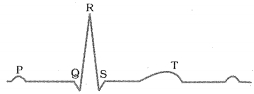
The following is a diagrammatic representation of a standard $ECG$. Which wave represents the contraction of both atria?
A
$P$
B
$QRS$
C
$R$
D
$T$
Solution
(A) In a standard $ECG$ (Electrocardiogram),the $P$-wave represents the electrical excitation (depolarization) of the atria,which leads to the contraction of both atria.
The $QRS$ complex represents the depolarization of the ventricles,which initiates ventricular contraction.
The $T$-wave represents the return of the ventricles from an excited to a normal state (repolarization).
Therefore,the $P$-wave is the correct answer.
The $QRS$ complex represents the depolarization of the ventricles,which initiates ventricular contraction.
The $T$-wave represents the return of the ventricles from an excited to a normal state (repolarization).
Therefore,the $P$-wave is the correct answer.
0 likes
View Solution68
MediumMCQ
Which wave is produced when the action potential is conducted from the $SA$ node to the $AV$ node?
A
$P$ wave
B
$QRS$ complex
C
$T$ wave
D
None of the above
Solution
(A) The $P$ wave in an $ECG$ represents the electrical excitation (or depolarization) of the atria,which leads to the contraction of both the atria.
This electrical impulse originates from the $SA$ node and travels to the $AV$ node,causing atrial depolarization.
Therefore,the conduction of the action potential from the $SA$ node to the $AV$ node is represented by the $P$ wave.
This electrical impulse originates from the $SA$ node and travels to the $AV$ node,causing atrial depolarization.
Therefore,the conduction of the action potential from the $SA$ node to the $AV$ node is represented by the $P$ wave.
0 likes
View Solution69
MediumMCQ
Which wave is produced when the action potential is conducted from the $AV$ node to the Purkinje fibers?
A
$P$ wave
B
$QRS$ complex
C
$T$ wave
D
None of the above
Solution
(B) The $ECG$ (Electrocardiogram) represents the electrical activity of the heart.
$1$. The $P$ wave represents the electrical excitation (or depolarization) of the atria,which leads to the contraction of both the atria.
$2$. The $QRS$ complex represents the depolarization of the ventricles,which initiates the ventricular contraction. This process occurs as the action potential is conducted from the $AV$ node through the Bundle of His and into the Purkinje fibers,causing the ventricles to contract.
$3$. The $T$ wave represents the return of the ventricles from an excited to a normal state (repolarization). The end of the $T$ wave marks the end of systole.
Therefore,the conduction of the action potential from the $AV$ node to the Purkinje fibers corresponds to the $QRS$ complex.
0 likes
View Solution70
MediumMCQ
What is the systolic and diastolic blood pressure of a healthy individual?
Systolic Pressure $\quad$ Diastolic Pressure
$(mm\, Hg) \quad (mm\, Hg)$
Systolic Pressure $\quad$ Diastolic Pressure
$(mm\, Hg) \quad (mm\, Hg)$
A
$80 \quad 120$
B
$120 \quad 80$
C
$90 \quad 140$
D
$140 \quad 90$
Solution
(B) In a healthy adult human,the standard blood pressure is measured as $120/80 \, mm\, Hg$.
Systolic pressure refers to the pressure exerted on the arterial walls during ventricular contraction,which is typically $120 \, mm\, Hg$.
Diastolic pressure refers to the pressure during ventricular relaxation,which is typically $80 \, mm\, Hg$.
Therefore,the correct sequence is $120 \, mm\, Hg$ (systolic) and $80 \, mm\, Hg$ (diastolic).
0 likes
View Solution71
MediumMCQ
Match List $I$ with List $II$.
Choose the correct answer from the options given below:
| List $I$ | List $II$ |
|---|---|
| $A$. $P$-wave | $I$. Beginning of systole |
| $B$. $Q$-wave | $II$. Repolarisation of ventricles |
| $C$. $QRS$ complex | $III$. Depolarisation of atria |
| $D$. $T$-wave | $IV$. Depolarisation of ventricles |
Choose the correct answer from the options given below:
A
$A-I, B-II, C-III, D-IV$
B
$A-III, B-I, C-IV, D-II$
C
$A-IV, B-III, C-II, D-I$
D
$A-II, B-IV, C-I, D-III$
Solution
(B) In a standard $ECG$:
- The $P$-wave represents the electrical excitation (or depolarisation) of the atria,which leads to the contraction of both atria $(A-III)$.
- The $Q$-wave marks the beginning of ventricular systole $(B-I)$.
- The $QRS$ complex represents the depolarisation of the ventricles,which initiates ventricular contraction $(C-IV)$.
- The $T$-wave represents the return of the ventricles from an excited to a normal state (repolarisation) $(D-II)$.
Therefore,the correct matching is $A-III, B-I, C-IV, D-II$.
0 likes
View Solution72
MediumMCQ
Match List $I$ with List $II$ :
Choose the correct answer from the options given below:
| List $I$ | List $II$ |
| $A$. $P$ wave | $I$. Heart muscles are electrically silent. |
| $B$. $QRS$ complex | $II$. Depolarisation of ventricles. |
| $C$. $T$ wave | $III$. Depolarisation of atria. |
| $D$. $T-P$ gap | $IV$. Repolarisation of ventricles. |
Choose the correct answer from the options given below:
A
$A-III, B-II, C-IV, D-I$
B
$A-II, B-III, C-I, D-IV$
C
$A-IV, B-II, C-I, D-III$
D
$A-I, B-III, C-IV, D-II$
Solution
$(A)$ The correct matching is as follows:
$A$. $P$ wave represents the electrical excitation (or depolarisation) of the atria, which leads to the contraction of both the atria. Thus, $A-III$.
$B$. $QRS$ complex represents the depolarisation of the ventricles, which initiates the ventricular contraction. Thus, $B-II$.
$C$. $T$ wave represents the return of the ventricles from an excited to a normal state (repolarisation). The end of the $T$ wave marks the end of systole. Thus, $C-IV$.
$D$. $T-P$ gap represents the period when the heart muscles are electrically silent, occurring between the end of the $T$ wave and the beginning of the next $P$ wave. Thus, $D-I$.
Therefore, the correct sequence is $A-III, B-II, C-IV, D-I$.
0 likes
View Solution73
MediumMCQ
In $\text{ECG}$,the $\text{QRS}$ complex represents:
A
Atrial depolarisation
B
Atrial repolarisation
C
Ventricular depolarisation
D
Ventricular repolarisation
Solution
(C) The $\text{ECG}$ (Electrocardiograph) is a graphical representation of the electrical activity of the heart during a cardiac cycle.
- The $P$-wave represents the electrical excitation (or depolarisation) of the atria,which leads to the contraction of both the atria.
- The $\text{QRS}$ complex represents the depolarisation of the ventricles,which initiates the ventricular contraction.
- The $T$-wave represents the return of the ventricles from an excited to a normal state (repolarisation).
Therefore,the $\text{QRS}$ complex corresponds to ventricular depolarisation.
0 likes
View Solution74
MediumMCQ
$ECG$ depicts the depolarization and repolarization processes during the cardiac cycle. In the $ECG$ of a normal healthy individual,one of the following waves is not represented?
A
Depolarization of atria
B
Repolarization of atria
C
Depolarization of ventricles
D
Repolarization of ventricles
Solution
(B) The $ECG$ (Electrocardiogram) consists of the $P$-wave,$QRS$ complex,and $T$-wave.
$1$. The $P$-wave represents the electrical excitation (or depolarization) of the atria,which leads to the contraction of both the atria.
$2$. The $QRS$ complex represents the depolarization of the ventricles,which initiates the ventricular contraction.
$3$. The $T$-wave represents the return of the ventricles from an excited to a normal state (repolarization).
$4$. The repolarization of the atria occurs during the ventricular depolarization ($QRS$ complex) and is masked by it; therefore,it is not represented as a distinct wave in the $ECG$ of a normal healthy individual.
0 likes
View Solution75
EasyMCQ
$ECG$ helps in the diagnosis of the following abnormalities in conducting pathways $EXCEPT$ . . . . . . .
A
enlargement of heart chambers
B
damage to cardiac muscles
C
reduction in blood supply to cardiac muscles
D
leucopenia
Solution
(D) An Electrocardiogram $(ECG)$ is a graphical representation of the electrical activity of the heart during a cardiac cycle. It is used to diagnose various heart conditions such as enlargement of heart chambers,damage to cardiac muscles,and reduction in blood supply to cardiac muscles (ischemia). Leucopenia,on the other hand,refers to a decrease in the number of white blood cells $(WBCs)$ in the blood,which is a hematological condition and cannot be diagnosed using an $ECG$.
0 likes
View Solution76
EasyMCQ
Given below are two statements.
Statement $I$ - Graphical recording of electrical variations detected at the surface of body during their propagation through the wall of heart is electrocardiograph.
Statement $II$ - The instrument used for the graphical recording of electrical variations detected at the surface of body during their propagation through wall of heart is electrocardiogram.
In the light of above statements,select the correct option given below:
A
Both statement $I$ and statement $II$ are correct.
B
Both statement $I$ and statement $II$ are incorrect.
C
Statement $I$ is correct but statement $II$ is incorrect.
D
Statement $I$ is incorrect but statement $II$ is correct.
Solution
(B) Statement $I$ is incorrect because the graphical recording of electrical activity of the heart is called an electrocardiogram $(ECG)$,not an electrocardiograph.
Statement $II$ is incorrect because the machine or instrument used to obtain this recording is called an electrocardiograph,not an electrocardiogram.
Therefore,both statements are incorrect.
Statement $II$ is incorrect because the machine or instrument used to obtain this recording is called an electrocardiograph,not an electrocardiogram.
Therefore,both statements are incorrect.
0 likes
View Solution77
EasyMCQ
Rise in blood pressure is directly related to the following $EXCEPT$ . . . . . . .
A
length of the blood vessel
B
venous return
C
vasoconstriction
D
vasodilation
Solution
(D) $i.$ $\text{Vasoconstriction:}$ narrows blood vessels $\rightarrow$ increases $BP$.
$ii.$ $\text{Venous return:}$ more blood returning to the heart $\rightarrow$ raises cardiac output and $BP$.
$iii.$ $\text{Longer vessel length:}$ increases peripheral resistance $\rightarrow$ higher $BP$.
$iv.$ $\text{Vasodilation:}$ widens blood vessels,which reduces resistance and lowers $BP$.
Therefore,vasodilation is the factor that is not related to a rise in blood pressure.
0 likes
View Solution78
EasyMCQ
In a normal $ECG$,a duration of $0.16$ seconds is called:
A
$P-R$ interval
B
$R-R$ interval
C
$Q-T$ interval
D
$S-T$ segment
Solution
(A) In a standard $ECG$ tracing,the $P-R$ interval represents the time taken for the electrical impulse to travel from the $SA$ node through the atria and the $AV$ node to the ventricles.
This interval typically ranges from $0.12$ to $0.20$ seconds.
Therefore,a duration of $0.16$ seconds falls within the normal range for the $P-R$ interval.
This interval typically ranges from $0.12$ to $0.20$ seconds.
Therefore,a duration of $0.16$ seconds falls within the normal range for the $P-R$ interval.
0 likes
View Solution79
EasyMCQ
Arrange and select the following events in the correct sequence during the measurement of blood pressure by a sphygmomanometer.
$i.$ Pulsatile sound disappears.
$ii.$ Cuff is inflated to block the brachial artery by external pressure.
$iii.$ First pulsatile sound is heard indicating systolic pressure.
$iv.$ Pressure in the cuff is slowly lowered.
$v.$ Cuff is wrapped tightly around the upper arm.
$i.$ Pulsatile sound disappears.
$ii.$ Cuff is inflated to block the brachial artery by external pressure.
$iii.$ First pulsatile sound is heard indicating systolic pressure.
$iv.$ Pressure in the cuff is slowly lowered.
$v.$ Cuff is wrapped tightly around the upper arm.
A
$v \rightarrow ii \rightarrow iv \rightarrow iii \rightarrow i$
B
$iv \rightarrow ii \rightarrow i \rightarrow iii \rightarrow v$
C
$iii \rightarrow v \rightarrow i \rightarrow ii \rightarrow iv$
D
$i \rightarrow v \rightarrow ii \rightarrow iii \rightarrow iv$
Solution
(A) The correct sequence for measuring blood pressure using a sphygmomanometer is as follows:
$1$. First,the cuff is wrapped tightly around the upper arm $(v)$.
$2$. Then,the cuff is inflated to block the brachial artery by applying external pressure $(ii)$.
$3$. Next,the pressure in the cuff is slowly lowered $(iv)$.
$4$. As the pressure drops,the first pulsatile sound is heard,which indicates the systolic pressure $(iii)$.
$5$. Finally,the pulsatile sound disappears,which indicates the diastolic pressure $(i)$.
Therefore,the correct sequence is $v \rightarrow ii \rightarrow iv \rightarrow iii \rightarrow i$.
0 likes
View Solution80
EasyMCQ
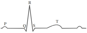
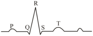
The given diagram represents a normal $ECG$ record of a human heart. Select the $CORRECT$ match from the options given below:
A
$P$ wave - Atrial repolarization,$QRS$ wave - Ventricular repolarization,$T$ wave - Atrial depolarization
B
$P$ wave - Atrial depolarization,$QRS$ wave - Ventricular depolarization,$T$ wave - Ventricular repolarization
C
$P$ wave - Ventricular depolarization,$QRS$ wave - Atrial repolarization,$T$ wave - Ventricular repolarization
D
$P$ wave - Ventricular polarization,$QRS$ wave - Ventricular depolarization,$T$ wave - Atrial repolarization
Solution
(B) The $P$-wave represents the electrical excitation (or depolarization) of the atria,which leads to the contraction of both the atria.
The $QRS$ complex represents the depolarization of the ventricles,which initiates the ventricular contraction. The contraction starts shortly after $Q$ and marks the beginning of the systole.
The $T$-wave represents the return of the ventricles from an excited to a normal state (repolarization). The end of the $T$-wave marks the end of systole.
Therefore,the correct match is: $P$ wave - Atrial depolarization,$QRS$ wave - Ventricular depolarization,$T$ wave - Ventricular repolarization.
0 likes
View Solution81
EasyMCQ
Blood pressure is directly proportional to the following $EXCEPT$ . . . . . . .
A
Length of blood vessel
B
Increased level of $ANF$
C
Vasoconstriction
D
Venous return
Solution
(B) Blood pressure is influenced by various physiological factors.
$1$. Length of blood vessel: Increased length increases resistance,which increases blood pressure.
$2$. Vasoconstriction: Narrowing of blood vessels increases resistance and thus increases blood pressure.
$3$. Venous return: Increased venous return increases stroke volume and cardiac output,leading to higher blood pressure.
$4$. Atrial Natriuretic Factor $(ANF)$: $ANF$ is released by the atrial wall in response to increased blood pressure. It causes vasodilation and promotes sodium excretion by the kidneys,which reduces blood volume and consequently lowers blood pressure.
Therefore,blood pressure is inversely related to the level of $ANF$.
0 likes
View Solution82
EasyMCQ
The $T$-wave in a normal $ECG$ represents . . . . . . .
A
atrial depolarization.
B
ventricular depolarization.
C
atrial repolarization.
D
ventricular repolarization.
Solution
(D) In a normal $ECG$:
$1$. The $P$-wave represents the electrical excitation (depolarization) of the atria.
$2$. The $QRS$ complex represents the depolarization of the ventricles.
$3$. The $T$-wave represents the return of the ventricles from an excited state to a normal state,which is known as ventricular repolarization.
Therefore,the correct option is $D$.
0 likes
View Solution83
EasyMCQ
In $ECG$,$P$-wave represents
A
Ventricular repolarisation
B
Ventricular depolarisation
C
Atrial depolarisation
D
Atrial repolarisation
Solution
(C) The $ECG$ (Electrocardiogram) is a graphical representation of the electrical activity of the heart during a cardiac cycle.
- The $P$-wave represents the electrical excitation (or depolarisation) of the atria,which leads to the contraction of both the atria.
- The $QRS$ complex represents the depolarisation of the ventricles,which initiates the ventricular contraction.
- The $T$-wave represents the return of the ventricles from an excited to a normal state (repolarisation).
Therefore,the $P$-wave represents atrial depolarisation.
0 likes
View Solution84
EasyMCQ
Systolic pressure in an adult human is
A
$120 \text{ mm Hg}$
B
$120/80 \text{ mm Hg}$
C
$150/120 \text{ mm Hg}$
D
$80 \text{ mm Hg}$
Solution
(A) Systolic blood pressure refers to the pressure exerted on the arterial walls during the contraction of the ventricles (systole).
In a healthy adult human,the normal systolic blood pressure is approximately $120 \text{ mm Hg}$.
The value $120/80 \text{ mm Hg}$ represents the entire blood pressure reading,where $120 \text{ mm Hg}$ is the systolic pressure and $80 \text{ mm Hg}$ is the diastolic pressure.
In a healthy adult human,the normal systolic blood pressure is approximately $120 \text{ mm Hg}$.
The value $120/80 \text{ mm Hg}$ represents the entire blood pressure reading,where $120 \text{ mm Hg}$ is the systolic pressure and $80 \text{ mm Hg}$ is the diastolic pressure.
0 likes
View Solution85
EasyMCQ
In a standard $ECG$,one of the following functions of its components is not correctly interpreted.
A
$P$ is the contraction of only left atria
B
$QRS$ complex represents ventricular contraction
C
$T$ is the end of systole
D
$P$ is the contraction of both atria
Solution
(A) $P$ is the contraction of only left atria is incorrect because the $P$-wave represents the electrical excitation (depolarisation) of both atria,which leads to the contraction of both atria simultaneously.
$ECG$ or Electrocardiogram records the electrical activity of the heart to diagnose various cardiac conditions.
$1$. The $P$-wave represents atrial depolarisation,which leads to the contraction of both atria.
$2$. The $QRS$ complex represents ventricular depolarisation,which initiates ventricular contraction (systole).
$3$. The $T$-wave represents ventricular repolarisation,which marks the end of ventricular systole as the ventricles begin to relax.
0 likes
View Solution86
EasyMCQ
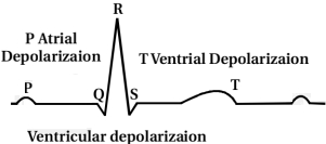
In the following diagrammatic representation of a standard $ECG$,the '$T$' wave represents:
A
Depolarisation of atria
B
Depolarisation of ventricles
C
Repolarisation of atria
D
Repolarisation of ventricles
Solution
(D) The correct answer is $(D)$.
In a standard $ECG$,the '$P$' wave represents the electrical excitation or depolarisation of the atria,which leads to the contraction of both the atria.
The '$QRS$' complex represents the depolarisation of the ventricles,which initiates the ventricular contraction.
The '$T$' wave represents the return of the ventricles from an excited to a normal state,i.e.,the repolarisation of the ventricles. The end of the '$T$' wave marks the end of systole.
0 likes
View Solution87
EasyMCQ
The $T$-wave in an $ECG$ represents
A
depolarisation of ventricles
B
electrical excitation of atria
C
beginning of systole
D
return of the ventricles from excited state
Solution
(D) The correct answer is $D$.
In an $ECG$,the $P$-wave represents the electrical excitation (or depolarisation) of the atria.
The $QRS$ complex represents the depolarisation of the ventricles,which initiates the ventricular contraction (systole).
The $T$-wave represents the return of the ventricles from an excited state to a normal state,which is known as ventricular repolarisation.
0 likes
View SolutionBody Fluids and Circulations — Blood pressure, ECG · Frequently Asked Questions
1Are these Body Fluids and Circulations questions useful for JEE and NEET?
Yes. All questions in this section are mapped to JEE Main and NEET exam patterns. Previous year questions from JEE Main, NEET, GUJCET and state-level exams are included with full solutions.
2Can I switch to Hindi or Gujarati for these questions?
Yes. Use the language tabs in the hero section or the sidebar to view the same questions and solutions in English, Hindi or Gujarati.
3How do I generate a question paper from this subtopic?
Use the Vedclass Exam Paper Generator — select the chapter and subtopic, set difficulty, and generate Sets A, B, C, D automatically. First 3 chapters of every subject are free.
Vedclass Products
For Students
Vedclass Test Series
Mock tests in real JEE/NEET style with performance analysis. 5-day free trial.
Start Free TrialFor Teachers
Exam Paper Generator
Generate Set A/B/C/D papers from this chapter in 2 minutes. 3 chapters free.
Try FreeFor Institutes
Online Exam Module
Live online exams with unlimited students, 360° analytics & white-label branding.
See DemoFor Teachers & Institutes
Generate a Body Fluids and Circulations Exam Paper in 2 Minutes
Select subtopic & difficulty — Sets A, B, C, D auto-generated with No Repeat logic.
First 3 chapters of every subject are free — no payment required.