
A English
Digestive system Questions in English
Class 11 Biology · Digestion and Absorption · Digestive system
272+
Questions
English
Language
100%
With Solutions
Showing 50 of 272 questions in English
201
EasyMCQ
In the wall of the alimentary canal, what is the actual sequence from outer to inner?
A
Serosa, longitudinal muscle, mucosa, submucosa
B
Mucosa, serosa, longitudinal muscle
C
Serosa, longitudinal muscle, circular muscle, submucosa, mucosa
D
Serosa, longitudinal muscle, submucosa, mucosa
Solution
(C) The wall of the alimentary canal is composed of four main layers from the outside to the inside:
$1$. $Serosa$: The outermost layer made of a thin mesothelium with some connective tissues.
$2$. $Muscularis$: This layer consists of an outer layer of longitudinal muscles and an inner layer of circular muscles.
$3$. $Submucosa$: This layer is formed of loose connective tissues containing nerves, blood, and lymph vessels.
$4$. $Mucosa$: The innermost lining of the lumen of the alimentary canal.
Therefore, the correct sequence from outer to inner is $Serosa \rightarrow \text{longitudinal muscle} \rightarrow \text{circular muscle} \rightarrow \text{submucosa} \rightarrow \text{mucosa}$.
0 likes
View Solution202
MediumMCQ
The gastrointestinal functions like secretion and motility are controlled by which system?
A
Intrinsic neural system
B
Extrinsic neural system
C
Both $(a)$ and $(b)$
D
None of the above
Solution
(C) The activities of the gastrointestinal tract are regulated by both the neural system and hormones.
Neural control is mediated by the intrinsic neural system (enteric nervous system) located within the wall of the gastrointestinal tract and the extrinsic neural system (autonomic nervous system) which connects the gut to the central nervous system.
Therefore,both systems play a role in controlling functions like secretion and motility.
0 likes
View Solution203
MediumMCQ
Which one is correct regarding the number of teeth and dental formula with reference to a child of age between $4$ to $6$ years?
A
$2102 / 2102$
B
$2103 / 2103$
C
$2123 / 2123$
D
$2122 / 2122$
Solution
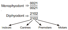
(A) Human dentition is diphyodont,heterodont,and thecodont. Diphyodont means teeth appear in two sets during a lifetime. The first set is the deciduous or milk teeth,which are replaced by permanent teeth between $6$ and $12$ years of age. $A$ child between $4$ and $6$ years of age possesses $20$ milk teeth. These include $8$ incisors,$4$ canines,and $8$ molars. Premolars $(PM)$ are absent in milk teeth. The dental formula is represented as $I \frac{2}{2}, C \frac{1}{1}, PM \frac{0}{0}, M \frac{2}{2}$. Thus,the total number of teeth is $(2+1+0+2) \times 2 = 20$.
0 likes
View Solution204
EasyMCQ
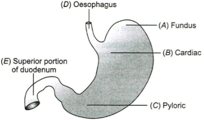
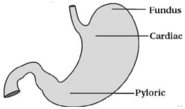
What is the correct labelling of the diagram given below? Choose the correct option accordingly.
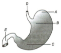
A
$A-Fundic portion, B-Cardiac region, C-Pyloric region, D-Food pipe, E-Wind pipe$
B
$A-Fundus, B-Pyloric region, C-Cardiac region, D-Oesophagus, E-Duodenum$
C
$A-Fundic region, B-Cardiac region, C-Pyloric region, D-Oesophagus, E-Duodenum$
D
$A-Cardiac region, B-Pyloric region, C-Fundic region, D-Oesophagus, E-Duodenum$
Solution
(C) Based on the anatomical structure of the human stomach:
$A$ represents the Fundic region (the upper dome-shaped part).
$B$ represents the Cardiac region (the part into which the oesophagus opens).
$C$ represents the Pyloric region (the lower part that opens into the duodenum).
$D$ represents the Oesophagus (the food pipe).
$E$ represents the Duodenum (the first part of the small intestine).
Therefore,the correct labelling is $A-Fundic region, B-Cardiac region, C-Pyloric region, D-Oesophagus, E-Duodenum$.
0 likes
View Solution205
MediumMCQ
In which layer of the wall of the alimentary canal are secretory glands present?
A
Serosa
B
Mucosa
C
Muscularis
D
Submucosa
Solution
(B) The wall of the alimentary canal consists of four layers: Serosa, Muscularis, Submucosa, and Mucosa.
$1$. The $Mucosa$ is the innermost layer lining the lumen of the alimentary canal.
$2$. It contains the $Lamina \, propria$, which consists of loose connective tissue, blood vessels, and glands.
$3$. Most secretory glands of the alimentary canal (such as gastric glands and intestinal crypts) are located within the $Mucosa$.
$4$. Note: $Brunner's \, glands$ are a notable exception, as they are located in the $Submucosa$ of the duodenum.
0 likes
View Solution206
EasyMCQ
In which type of dentition,each tooth in the buccal cavity is embedded in a socket of jaw bone?
A
Heterodont
B
Thecodont
C
Diphyodont
D
Monophyodont
Solution
(B) Thecodont is a condition in which each tooth is embedded in a socket of the jaw bone and has well-developed roots.
Monophyodont: Teeth that appear only once in a lifetime.
Diphyodont: Teeth that appear twice in a lifetime (e.g.,milk teeth and permanent teeth).
Heterodont: Humans (adults) have $32$ permanent teeth of four different types: incisors,canines,premolars,and molars. This type of dentition is called Heterodont.
0 likes
View Solution207
EasyMCQ
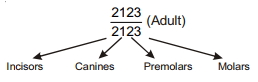
The dental formula of human beings is
A
$\frac{2123}{2123}$
B
$\frac{2021}{2021}$
C
$\frac{2321}{2321}$
D
$\frac{2133}{2133}$
Solution
(A) The dental formula represents the arrangement of teeth in one half of each jaw in the order of Incisors $(I)$,Canines $(C)$,Premolars $(PM)$,and Molars $(M)$.
For an adult human,the dental formula is $\frac{2123}{2123}$.
This means in each half of the upper and lower jaw,there are $2$ incisors,$1$ canine,$2$ premolars,and $3$ molars.
Therefore,the correct option is $A$.
For an adult human,the dental formula is $\frac{2123}{2123}$.
This means in each half of the upper and lower jaw,there are $2$ incisors,$1$ canine,$2$ premolars,and $3$ molars.
Therefore,the correct option is $A$.
0 likes
View Solution208
EasyMCQ
All of the following are the parts of the large intestine,except:
A
Caecum
B
Colon
C
Ileum
D
Rectum
Solution
(C) The large intestine consists of the caecum,colon,and rectum. The ileum is the final section of the small intestine,not the large intestine. Therefore,the correct answer is $C$.
0 likes
View Solution209
EasyMCQ
The oesophagus opens into which part of the stomach?
A
Pyloric
B
Cardiac
C
Fundic
D
Caecum
Solution
(B) The human stomach has four major parts: the cardiac,fundic,body,and pyloric regions.
The oesophagus is a muscular tube that transports food to the stomach.
It opens into the cardiac portion of the stomach,which is the part into which the oesophagus opens.
The oesophagus is a muscular tube that transports food to the stomach.
It opens into the cardiac portion of the stomach,which is the part into which the oesophagus opens.
0 likes
View Solution210
EasyMCQ
Which of the following organs is present in the abdominal cavity,just below the diaphragm?
A
Pharynx
B
Pancreas
C
Liver
D
Tongue
Solution
(C) The liver is the largest gland of the body,which is situated in the upper right portion of the abdominal cavity,just below the diaphragm.
It consists of two main lobes,namely the right and left lobes.
It consists of two main lobes,namely the right and left lobes.
0 likes
View Solution211
EasyMCQ
The wall of the alimentary canal from the oesophagus to the rectum possesses . . . . . . layers.
A
$2$ layers
B
$3$ layers
C
$4$ layers
D
$5$ layers
Solution
(C) The wall of the human alimentary canal from the oesophagus to the rectum is composed of four distinct layers.
These layers, from the outermost to the innermost, are:
$1$. Serosa: The outermost layer made of a thin mesothelium with some connective tissues.
$2$. Muscularis: Formed by smooth muscles, usually arranged into an inner circular and an outer longitudinal layer.
$3$. Sub-mucosa: Formed of loose connective tissues containing nerves, blood, and lymph vessels.
$4$. Mucosa: The innermost lining of the lumen of the alimentary canal.
0 likes
View Solution212
EasyMCQ
The major functions of the buccal cavity is/are:
A
Mastication of food
B
Facilitation of swallowing
C
Help in the secretion of glucagon
D
Both $(a)$ & $(b)$
Solution
(D) The buccal cavity performs the following functions:
$1$. Mastication of food: Teeth and tongue help in breaking down food into smaller particles.
$2$. Facilitation of swallowing: The tongue helps in mixing saliva with food to form a bolus,which is then swallowed.
$3$. Glucagon is a hormone secreted by the $\alpha$-cells of the pancreas,not the buccal cavity.
Therefore,both $(a)$ and $(b)$ are correct functions of the buccal cavity.
0 likes
View Solution213
EasyMCQ
Crypts of Lieberkuhn are present in
A
Stomach
B
Pharynx
C
Oesophagus
D
Intestine
Solution
(D) Crypts of Lieberkuhn,also known as intestinal glands,are tubular glands found between the villi of the small intestine. They secrete intestinal juice,which contains various enzymes that help in the final stages of digestion.
0 likes
View Solution214
EasyMCQ
The structural and functional unit of the liver is:
A
Cystic duct
B
Hepatic lobule
C
Hepatopancreatic duct
D
Sphincter of Oddi
Solution
(B) Hepatic lobules are the structural and functional units of the liver.
They consist of hepatic cells arranged in the form of cords.
They consist of hepatic cells arranged in the form of cords.
0 likes
View Solution215
EasyMCQ
Bile can be prevented from being released into the duodenum by:
A
Sphincter of Oddi
B
Cardiac sphincter
C
Pyloric sphincter
D
Ileo-caecal valve
Solution
(A) The $Sphincter$ $of$ $Oddi$ guards the opening of the common hepatopancreatic duct into the duodenum. It regulates the flow of bile and pancreatic juice into the duodenum,thereby preventing their release when not required.
0 likes
View Solution216
MediumMCQ
Which of the following is a modification of the mucosa of the alimentary canal?
A
Villi
B
Microvilli
C
Rugae
D
All of these
Solution
(D) The mucosa of the alimentary canal exhibits several modifications to increase surface area and facilitate function.
$1$. Villi are finger-like projections of the mucosa in the small intestine.
$2$. Microvilli are microscopic projections on the surface of the epithelial cells of the mucosa.
$3$. Rugae are irregular folds of the mucosa in the stomach.
Since all these structures are modifications of the mucosa,the correct answer is $D$.
0 likes
View Solution217
MediumMCQ
The dental formula for the monophyodont teeth of humans is:
A
$\frac{0021}{0021}$
B
$\frac{0003}{0003}$
C
$\frac{2120}{2120}$
D
$\frac{2102}{2102}$
Solution
(A) In humans,teeth are diphyodont,meaning they erupt twice in a lifetime. The permanent teeth include both diphyodont teeth (which replace milk teeth) and monophyodont teeth (which erupt only once).
The dental formula for the permanent teeth in humans is $\frac{2123}{2123}$.
The diphyodont teeth (those that replace deciduous teeth) are the incisors,canines,and the first two premolars,represented by the formula $\frac{2102}{2102}$.
The monophyodont teeth are the three molars on each side of each jaw,which erupt only once. Their dental formula is $\frac{0021}{0021}$ (representing $0$ incisors,$0$ canines,$0$ premolars,and $3$ molars per quadrant).
0 likes
View Solution218
EasyMCQ
Upper molars in human dentition have
A
Four roots
B
Three roots
C
Two roots
D
Single root
Solution
(B) In human dentition,the root structure varies based on the tooth type.
Upper molars (specifically the first and second molars) typically possess $3$ roots.
Lower molars typically possess $2$ roots.
Therefore,the correct answer is $3$ roots.
Upper molars (specifically the first and second molars) typically possess $3$ roots.
Lower molars typically possess $2$ roots.
Therefore,the correct answer is $3$ roots.
0 likes
View Solution219
EasyMCQ
Which of the following can be taken as the true stomach in ruminants?
A
Rumen
B
Reticulum
C
Omasum
D
Abomasum
Solution
(D) In ruminants,the stomach is divided into four chambers: $Rumen$,$Reticulum$,$Omasum$,and $Abomasum$.
The $Abomasum$ is considered the true stomach because it is the only chamber that contains gastric glands,which secrete gastric juices containing hydrochloric acid and digestive enzymes for chemical digestion.
The $Abomasum$ is considered the true stomach because it is the only chamber that contains gastric glands,which secrete gastric juices containing hydrochloric acid and digestive enzymes for chemical digestion.
0 likes
View Solution220
EasyMCQ
Oblique muscle layer is present in
A
Stomach
B
Duodenum
C
Colon
D
All of these
Solution
(A) The wall of the alimentary canal is made up of four layers: serosa,muscularis,submucosa,and mucosa.
The muscularis layer is usually made up of an inner circular and an outer longitudinal muscle layer.
However,an additional oblique muscle layer is present in the stomach,which helps in the churning of food.
The muscularis layer is usually made up of an inner circular and an outer longitudinal muscle layer.
However,an additional oblique muscle layer is present in the stomach,which helps in the churning of food.
0 likes
View Solution221
EasyMCQ
Pernicious anaemia is caused by the deficiency of . . . . . . vitamin.
A
$B_{1}$
B
$B_{12}$
C
$C$
D
$D$
Solution
(B) Deficiency of vitamin $B_{12}$ causes pernicious anaemia.
It is a severe form of anaemia because this vitamin is essential for the maturation of $RBCs$,$DNA$ synthesis,and the maintenance of the myelin sheath in the nervous system.
It is a severe form of anaemia because this vitamin is essential for the maturation of $RBCs$,$DNA$ synthesis,and the maintenance of the myelin sheath in the nervous system.
0 likes
View Solution222
EasyMCQ
Which of the following papillae are without taste buds in human tongue?
A
Vallate
B
Fungiform
C
Fusiform
D
Filiform
Solution
(D) The human tongue has four types of papillae: Vallate,Fungiform,Foliate,and Filiform. Among these,the Filiform papillae are the most numerous but do not contain taste buds. They are primarily involved in providing friction for food manipulation.
0 likes
View Solution223
MediumMCQ
The $Sphincter$ of $Boyden$,which helps in the filling of the gallbladder,is present in:
A
Ductus choledochus
B
Duct of Wirsung
C
Ampulla of Vater
D
Duct of Santorini
Solution
(A) The $Sphincter$ of $Boyden$ is located at the junction of the common bile duct ($Ductus$ $choledochus$) and the cystic duct.
It regulates the flow of bile into the gallbladder.
When this sphincter contracts,bile is diverted into the gallbladder for storage and concentration.
Therefore,it is present in the $Ductus$ $choledochus$.
It regulates the flow of bile into the gallbladder.
When this sphincter contracts,bile is diverted into the gallbladder for storage and concentration.
Therefore,it is present in the $Ductus$ $choledochus$.
0 likes
View Solution224
EasyMCQ
Cholecystitis refers to inflammation of which organ?
A
Gall bladder
B
Stomach
C
Spleen
D
Lungs
Solution
(A) Cholecystitis is a medical term derived from 'cholecyst' (gall bladder) and '-itis' (inflammation).
Therefore,it refers to the inflammation of the gall bladder,which is often caused by gallstones blocking the cystic duct.
Therefore,it refers to the inflammation of the gall bladder,which is often caused by gallstones blocking the cystic duct.
0 likes
View Solution225
EasyMCQ
Sphincter of Oddi is present at:
A
Ileo-caecal junction
B
Junction of hepato-pancreatic duct and duodenum
C
Gastro-oesophageal junction
D
Junction of jejunum and duodenum
Solution
(B) The Sphincter of Oddi (also known as the hepato-pancreatic sphincter) is a muscular valve that controls the flow of digestive juices (bile and pancreatic juice) through the ampulla of Vater into the duodenum.
It is located at the junction where the hepato-pancreatic duct opens into the duodenum.
Therefore,the correct option is $B$.
It is located at the junction where the hepato-pancreatic duct opens into the duodenum.
Therefore,the correct option is $B$.
0 likes
View Solution226
EasyMCQ
What is called the digestive system?
A
The process of breaking down food into simpler substances.
B
The group of organs that work together to convert food into energy and basic nutrients.
C
The network of nerves that controls the movement of food through the gut.
D
The process of eliminating waste products from the body.
Solution
(B) The organs involved in the ingestion of food,digestion,absorption,and egestion are called digestive organs.
Examples include the mouth,tongue,teeth,pharynx,alimentary canal,stomach,small intestine,large intestine,rectum,and anus.
Digestive organs also include accessory glands such as salivary glands,liver,and pancreas.
The system formed by the coordination of all these organs is called the digestive system.
0 likes
View Solution227
Medium
Explain the thecodont and diphyodont type of dental arrangement.
Solution
(N/A) Thecodont: Each tooth is embedded in a socket of the jaw bone. This type of attachment is known as thecodont.
Diphyodont: In humans,a set of temporary milk teeth or deciduous teeth is replaced by a set of permanent or adult teeth. This type of dentition,where two sets of teeth are formed during a lifetime,is called diphyodont.
Diphyodont: In humans,a set of temporary milk teeth or deciduous teeth is replaced by a set of permanent or adult teeth. This type of dentition,where two sets of teeth are formed during a lifetime,is called diphyodont.
0 likes
View Solution228
EasyMCQ
What do we call the type of teeth attachment to jaw bones in which each tooth is embedded in a socket of jaw bones?
A
Thecodont
B
Pleurodont
C
Acrodont
D
Heterodont
Solution
(A) In humans,each tooth is embedded in a socket of the jaw bone. This type of attachment is known as thecodont.
0 likes
View Solution229
Easy
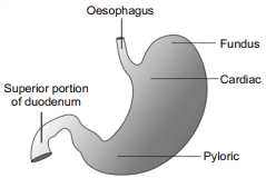
The stomach is located in the upper left portion of the abdominal cavity and has three major parts. Name these three parts.
Solution
(N/A) The human stomach is a $J$-shaped muscular organ located in the upper left portion of the abdominal cavity. It consists of three major parts:
$1$. Cardiac portion: Into which the oesophagus opens.
$2$. Fundus region: Which is typically filled with air or gas.
$3$. Pyloric portion: Which opens into the first part of the small intestine (duodenum).
$1$. Cardiac portion: Into which the oesophagus opens.
$2$. Fundus region: Which is typically filled with air or gas.
$3$. Pyloric portion: Which opens into the first part of the small intestine (duodenum).
0 likes
View Solution230
EasyMCQ
How many types of teeth are found in humans?
A
$2$
B
$3$
C
$4$
D
$8$
Solution
(C) Humans possess four distinct types of teeth,which are categorized based on their shape and function:
$1$. Incisors $(I)$: Used for cutting food.
$2$. Canines $(C)$: Used for tearing food.
$3$. Premolars $(PM)$: Used for crushing and grinding food.
$4$. Molars $(M)$: Used for grinding food.
Therefore,there are $4$ types of teeth in humans.
$1$. Incisors $(I)$: Used for cutting food.
$2$. Canines $(C)$: Used for tearing food.
$3$. Premolars $(PM)$: Used for crushing and grinding food.
$4$. Molars $(M)$: Used for grinding food.
Therefore,there are $4$ types of teeth in humans.
0 likes
View Solution231
MediumMCQ
Identify the incorrect statement from the following.
A
Digestion is the process of converting complex food substances into simple absorbable forms.
B
Digestion does not occur in poriferans.
C
The chemical form of food changes during digestion.
D
The physical form of food changes during digestion.
Solution
(B) Digestion is defined as the process of converting complex food substances into simple absorbable forms. This process involves both physical and chemical methods. During digestion,the physical state (e.g.,mastication) and the chemical composition (e.g.,enzymatic hydrolysis) of food are altered. Regarding option $B$,poriferans (sponges) do perform digestion,but it is strictly intracellular,occurring within the cells after the food is engulfed via phagocytosis. Therefore,stating that digestion does not occur in poriferans is incorrect.
0 likes
View Solution232
EasyMCQ
Select the correct sequence for the human digestive tract.
A
Mouth $\rightarrow$ Oral cavity $\rightarrow$ Pharynx $\rightarrow$ Oesophagus $\rightarrow$ Stomach $\rightarrow$ Small intestine $\rightarrow$ Large intestine $\rightarrow$ Anus
B
Mouth $\rightarrow$ Oral cavity $\rightarrow$ Oesophagus $\rightarrow$ Pharynx $\rightarrow$ Stomach $\rightarrow$ Small intestine $\rightarrow$ Large intestine $\rightarrow$ Anus
C
Mouth $\rightarrow$ Oral cavity $\rightarrow$ Pharynx $\rightarrow$ Oesophagus $\rightarrow$ Stomach $\rightarrow$ Large intestine $\rightarrow$ Small intestine $\rightarrow$ Anus
D
None of these
Solution
(A) The human digestive system consists of the alimentary canal and associated glands. The alimentary canal begins with an anterior opening,the mouth,and it opens out posteriorly through the anus. The correct sequence of the passage of food through the alimentary canal is: Mouth $\rightarrow$ Oral cavity (Buccal cavity) $\rightarrow$ Pharynx $\rightarrow$ Oesophagus $\rightarrow$ Stomach $\rightarrow$ Small intestine $\rightarrow$ Large intestine $\rightarrow$ Anus. Therefore,option $A$ is the correct sequence.
0 likes
View Solution233
EasyMCQ
The human digestive system is shown below. What are $P, Q$ and $R$?
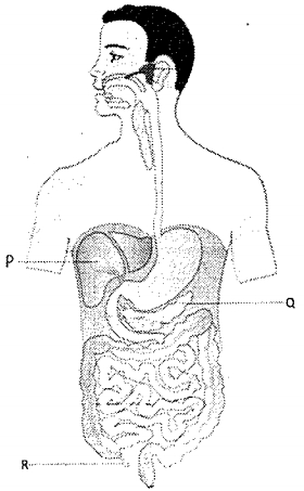
A
Gall bladder,Pancreas,Appendix
B
Gall bladder,Pancreas,Caecum
C
Pancreas,Gall bladder,Appendix
D
Pancreas,Gall bladder,Caecum
Solution
(A) Based on the anatomical diagram of the human digestive system:
$1$. $P$ points to the Gall bladder,which is a small pouch-like structure located under the liver.
$2$. $Q$ points to the Pancreas,which is a leaf-shaped gland located behind the stomach.
$3$. $R$ points to the Appendix (vermiform appendix),which is a small,finger-like projection arising from the caecum.
Therefore,the correct sequence is Gall bladder,Pancreas,and Appendix.
0 likes
View Solution234
MediumMCQ
Match the following columns:
| Column-$I$ | Column-$II$ |
|---|---|
| $P$. Thecodont | $I$. Human possesses four types of teeth |
| $Q$. Diphyodont | $II$. Permanent teeth replace milk teeth |
| $R$. Heterodont | $III$. Human teeth are embedded in the jaw bone |
A
$(P-II), (Q-I), (R-III)$
B
$(P-I), (Q-III), (R-II)$
C
$(P-I), (Q-II), (R-III)$
D
$(P-III), (Q-II), (R-I)$
Solution
(D) $1$. Thecodont $(P)$: Human teeth are embedded in the jaw bone,which is described in statement $III$.
$2$. Diphyodont $(Q)$: Humans have two sets of teeth during their lifetime,where permanent teeth replace milk teeth,which is described in statement $II$.
$3$. Heterodont $(R)$: Humans possess four different types of teeth (incisors,canines,premolars,and molars),which is described in statement $I$.
Therefore,the correct matching is $(P-III), (Q-II), (R-I)$.
0 likes
View Solution235
EasyMCQ
Which teeth are most numerous in the human jaw?
A
Incisors
B
Canines
C
Premolars
D
Molars
Solution
(D) The dental formula for an adult human is $2123/2123$.
This means in each half of the upper and lower jaw,there are $2$ incisors,$1$ canine,$2$ premolars,and $3$ molars.
Total number of teeth per jaw half is $8$,making $32$ teeth in total.
Molars are the most numerous teeth,with $3$ in each quadrant,totaling $12$ molars in the entire mouth $(3 \times 4 = 12)$.
0 likes
View Solution236
EasyMCQ
What is the dental formula of an adult human?
A
$\frac{2120}{2120}$
B
$\frac{2123}{2123}$
C
$\frac{2102}{2102}$
D
None of these
Solution
(B) The dental formula represents the arrangement of teeth in one half of each jaw,in the order of Incisors $(I)$,Canines $(C)$,Premolars $(PM)$,and Molars $(M)$.
For an adult human,the dental formula is $\frac{2123}{2123}$.
This means in each half of the upper and lower jaw,there are $2$ Incisors,$1$ Canine,$2$ Premolars,and $3$ Molars.
Total number of teeth = $2 \times (2+1+2+3) = 2 \times 8 = 16$ per jaw,resulting in $32$ teeth in total.
For an adult human,the dental formula is $\frac{2123}{2123}$.
This means in each half of the upper and lower jaw,there are $2$ Incisors,$1$ Canine,$2$ Premolars,and $3$ Molars.
Total number of teeth = $2 \times (2+1+2+3) = 2 \times 8 = 16$ per jaw,resulting in $32$ teeth in total.
0 likes
View Solution237
EasyMCQ
The structure of a tooth is given below. What are $P$ and $Q$?
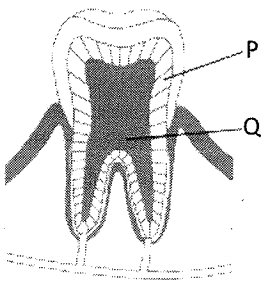
A
Dentine,Enamel
B
Enamel,Pulp cavity
C
Dentine,Pulp cavity
D
Enamel,Dentine
Solution
(C) In the given diagram of a tooth:
$P$ represents the Dentine,which is the hard,calcified tissue forming the bulk of the tooth.
$Q$ represents the Pulp cavity,which is the central part of the tooth containing nerves and blood vessels.
Therefore,the correct option is $C$.
$P$ represents the Dentine,which is the hard,calcified tissue forming the bulk of the tooth.
$Q$ represents the Pulp cavity,which is the central part of the tooth containing nerves and blood vessels.
Therefore,the correct option is $C$.
0 likes
View Solution238
EasyMCQ
What is the number of canines in each jaw of a human adult?
A
$1$
B
$2$
C
$4$
D
$8$
Solution
(B) The human dental formula is represented as $2123/2123$.
In this formula,the numbers represent the number of incisors,canines,premolars,and molars in one half of each jaw.
The canine is represented by the number $1$ in the formula.
Since there are two halves in each jaw,the total number of canines per jaw is $1 \times 2 = 2$.
In this formula,the numbers represent the number of incisors,canines,premolars,and molars in one half of each jaw.
The canine is represented by the number $1$ in the formula.
Since there are two halves in each jaw,the total number of canines per jaw is $1 \times 2 = 2$.
0 likes
View Solution239
EasyMCQ
What is the number of premolars in each half of each jaw?
A
$1$
B
$2$
C
$4$
D
$3$
Solution
(B) The human dental formula is represented as $I: 2/2, C: 1/1, PM: 2/2, M: 3/3$.
Here,$I$ stands for Incisors,$C$ for Canines,$PM$ for Premolars,and $M$ for Molars.
Each half of each jaw contains $2$ premolars.
Therefore,the correct option is $B$.
Here,$I$ stands for Incisors,$C$ for Canines,$PM$ for Premolars,and $M$ for Molars.
Each half of each jaw contains $2$ premolars.
Therefore,the correct option is $B$.
0 likes
View Solution240
EasyMCQ
In the jaw of an adult human,which teeth are equal in number?
A
Premolars and Incisors
B
Molars and Premolars
C
Canines and Incisors
D
Premolars and Canines
Solution
(A) The dental formula of an adult human is $2123/2123$.
This represents:
Incisors $(I)$: $2$ per quadrant
Canines $(C)$: $1$ per quadrant
Premolars $(PM)$: $2$ per quadrant
Molars $(M)$: $3$ per quadrant
Comparing the options:
$A$: Premolars $(2)$ and Incisors $(2)$ - These are equal in number per quadrant.
$B$: Molars $(3)$ and Premolars $(2)$ - Not equal.
$C$: Canines $(1)$ and Incisors $(2)$ - Not equal.
$D$: Premolars $(2)$ and Canines $(1)$ - Not equal.
Therefore,the number of Premolars and Incisors is equal in each quadrant of the jaw.
0 likes
View Solution241
EasyMCQ
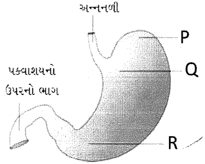
Identify the parts of the stomach labeled $P$,$Q$,and $R$ in the given diagram.
A
$P$: Fundus,$Q$: Cardiac,$R$: Pyloric
B
$P$: Cardiac,$Q$: Fundus,$R$: Pyloric
C
$P$: Pyloric,$Q$: Fundus,$R$: Cardiac
D
$P$: Pyloric,$Q$: Cardiac,$R$: Fundus
Solution
(A) The human stomach is divided into four major parts:
$1$. Cardiac portion: Into which the esophagus opens.
$2$. Fundic region: The upper dome-shaped portion.
$3$. Body: The main central region.
$4$. Pyloric portion: The lower part which opens into the first part of the small intestine (duodenum).
Based on the provided diagram:
- $P$ points to the upper dome-shaped region,which is the Fundus.
- $Q$ points to the region near the esophageal opening,which is the Cardiac part.
- $R$ points to the lower region leading towards the duodenum,which is the Pyloric part.
Therefore,the correct sequence is $P$: Fundus,$Q$: Cardiac,$R$: Pyloric.
0 likes
View Solution242
EasyMCQ
What is the correct sequence of the parts of the small intestine?
A
Duodenum $\rightarrow$ Jejunum $\rightarrow$ Ileum
B
Duodenum $\rightarrow$ Ileum $\rightarrow$ Jejunum
C
Ileum $\rightarrow$ Jejunum $\rightarrow$ Duodenum
D
Jejunum $\rightarrow$ Ileum $\rightarrow$ Duodenum
Solution
(A) The small intestine is a distinguishable region of the alimentary canal and is divided into three parts in a specific sequence:
$1$. Duodenum: The $C$-shaped first part of the small intestine.
$2$. Jejunum: The long coiled middle portion.
$3$. Ileum: The highly coiled final part that opens into the large intestine.
Therefore, the correct sequence is $Duodenum \rightarrow Jejunum \rightarrow Ileum$.
$1$. Duodenum: The $C$-shaped first part of the small intestine.
$2$. Jejunum: The long coiled middle portion.
$3$. Ileum: The highly coiled final part that opens into the large intestine.
Therefore, the correct sequence is $Duodenum \rightarrow Jejunum \rightarrow Ileum$.
0 likes
View Solution243
EasyMCQ
Which part of our body serves as a habitat for symbiotic microorganisms?
A
Ileum
B
Appendix
C
Caecum
D
Duodenum
Solution
(C) The $Caecum$ is a small blind sac which hosts some symbiotic microorganisms. It is a part of the large intestine where the small intestine opens. In humans, it is small and vestigial, but it still serves as a site for certain symbiotic bacteria.
0 likes
View Solution244
MediumMCQ
$A$ sphincter muscle is $NOT$ present between which of the following?
A
Oesophagus and stomach
B
Stomach and duodenum
C
Duodenum and hepato-pancreatic duct
D
Pharynx and oesophagus
Solution
(D) The gastro-oesophageal sphincter regulates the opening of the oesophagus into the stomach.
The pyloric sphincter regulates the opening of the stomach into the duodenum.
The sphincter of Oddi guards the opening of the hepato-pancreatic duct into the duodenum.
There is no specific sphincter muscle present between the pharynx and the oesophagus; the upper oesophageal sphincter is a physiological structure,but it is not typically classified as a distinct anatomical sphincter muscle in the same manner as the others listed.
0 likes
View Solution245
MediumMCQ
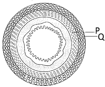
The given figure shows a cross-section of the alimentary canal. Identify the layers labeled $P$ and $Q$.
A
Longitudinal muscle layer $\quad$ Circular muscle layer
B
Circular muscle layer $\quad$ Longitudinal muscle layer
C
Oblique muscle layer $\quad$ Longitudinal muscle layer
D
Circular muscle layer $\quad$ Oblique muscle layer
Solution
(B) The wall of the alimentary canal consists of four layers: serosa,muscularis,submucosa,and mucosa.
In the muscularis layer,the arrangement of smooth muscles is typically organized into an inner circular layer and an outer longitudinal layer.
In the provided cross-section diagram,the inner layer labeled $P$ consists of fibers arranged in a circular pattern,representing the circular muscle layer.
The outer layer labeled $Q$ consists of fibers arranged longitudinally (appearing as dots in cross-section),representing the longitudinal muscle layer.
Therefore,$P$ is the circular muscle layer and $Q$ is the longitudinal muscle layer.
0 likes
View Solution246
EasyMCQ
The layers of the alimentary canal are given from the inside to the outside.
A
Mucosa $\rightarrow$ Submucosa $\rightarrow$ Muscularis $\rightarrow$ Serosa
B
Serosa $\rightarrow$ Muscularis $\rightarrow$ Submucosa $\rightarrow$ Mucosa
C
Submucosa $\rightarrow$ Mucosa $\rightarrow$ Muscularis $\rightarrow$ Serosa
D
Serosa $\rightarrow$ Muscularis $\rightarrow$ Mucosa $\rightarrow$ Submucosa
Solution
(A) The wall of the human alimentary canal from the esophagus to the rectum possesses four layers:
$1$. Serosa (outermost layer)
$2$. Muscularis
$3$. Submucosa
$4$. Mucosa (innermost layer)
Since the question asks for the sequence from the inside to the outside,the order is: Mucosa $\rightarrow$ Submucosa $\rightarrow$ Muscularis $\rightarrow$ Serosa.
$1$. Serosa (outermost layer)
$2$. Muscularis
$3$. Submucosa
$4$. Mucosa (innermost layer)
Since the question asks for the sequence from the inside to the outside,the order is: Mucosa $\rightarrow$ Submucosa $\rightarrow$ Muscularis $\rightarrow$ Serosa.
0 likes
View Solution247
EasyMCQ
In which organ,besides longitudinal and circular muscles,are oblique muscles also present?
A
Oesophagus
B
Stomach
C
Small intestine
D
Large intestine
Solution
(B) The wall of the alimentary canal is made up of four layers: serosa,muscularis,submucosa,and mucosa.
The muscularis layer is usually formed by an inner circular and an outer longitudinal muscle layer.
However,the stomach is unique because it possesses an additional third layer of muscles known as the oblique muscle layer,which is located internal to the circular layer.
This extra layer of muscle helps in the churning and mixing of food with gastric juices.
0 likes
View Solution248
MediumMCQ
Which of the four layers of the wall of the alimentary canal shows the most variation?
A
Serosa
B
Muscularis
C
Submucosa
D
Mucosa
Solution
(D) The wall of the alimentary canal consists of four layers: $Serosa$,$Muscularis$,$Submucosa$,and $Mucosa$.
$1$. $Serosa$ is the outermost layer and is made up of a thin mesothelium with some connective tissues.
$2$. $Muscularis$ is formed by smooth muscles usually arranged into an inner circular and an outer longitudinal layer.
$3$. $Submucosa$ consists of loose connective tissues containing nerves,blood,and lymph vessels.
$4$. $Mucosa$ is the innermost lining of the lumen of the alimentary canal. It shows significant modifications and variations in different parts of the digestive tract,such as the formation of rugae in the stomach and villi in the small intestine,to increase surface area and facilitate secretion and absorption.
0 likes
View Solution249
EasyMCQ
Identify the location of gastric glands in the stomach.
A
Serosa
B
Muscularis
C
Submucosa
D
Mucosa
Solution
(D) The wall of the human alimentary canal consists of four layers: Serosa, Muscularis, Submucosa, and Mucosa.
$1$. The $Serosa$ is the outermost layer.
$2$. The $Muscularis$ is formed by smooth muscles.
$3$. The $Submucosa$ consists of loose connective tissue containing nerves, blood, and lymph vessels.
$4$. The $Mucosa$ is the innermost lining of the lumen of the alimentary canal.
Gastric glands are located in the $Mucosa$ layer of the stomach wall, specifically within the gastric pits of the gastric mucosa.
0 likes
View Solution250
MediumMCQ
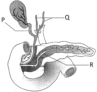
The following diagram shows the duct system of the liver,gallbladder,and pancreas. Identify the ducts labeled $P, Q$,and $R$.
A
$P$: Common bile duct,$Q$: Hepatic duct,$R$: Pancreatic duct
B
$P$: Bile duct,$Q$: Hepatic duct,$R$: Pancreatic duct
C
$P$: Common bile duct,$Q$: Hepatic duct,$R$: Hepato-pancreatic duct
D
$P$: Bile duct,$Q$: Pancreatic duct,$R$: Hepato-pancreatic duct
Solution
(C) Based on the anatomical structure of the human digestive system:
$1$. The duct emerging from the gallbladder is the common bile duct $(P)$.
$2$. The ducts coming from the liver lobes are the hepatic ducts $(Q)$.
$3$. The duct running through the pancreas is the pancreatic duct,which joins the common bile duct to form the common hepato-pancreatic duct $(R)$ before entering the duodenum.
Therefore,the correct identification is $P$: Common bile duct,$Q$: Hepatic duct,$R$: Hepato-pancreatic duct.
0 likes
View SolutionDigestion and Absorption — Digestive system · Frequently Asked Questions
1Are these Digestion and Absorption questions useful for JEE and NEET?
Yes. All questions in this section are mapped to JEE Main and NEET exam patterns. Previous year questions from JEE Main, NEET, GUJCET and state-level exams are included with full solutions.
2Can I switch to Hindi or Gujarati for these questions?
Yes. Use the language tabs in the hero section or the sidebar to view the same questions and solutions in English, Hindi or Gujarati.
3How do I generate a question paper from this subtopic?
Use the Vedclass Exam Paper Generator — select the chapter and subtopic, set difficulty, and generate Sets A, B, C, D automatically. First 3 chapters of every subject are free.
Vedclass Products
For Students
Vedclass Test Series
Mock tests in real JEE/NEET style with performance analysis. 5-day free trial.
Start Free TrialFor Teachers
Exam Paper Generator
Generate Set A/B/C/D papers from this chapter in 2 minutes. 3 chapters free.
Try FreeFor Institutes
Online Exam Module
Live online exams with unlimited students, 360° analytics & white-label branding.
See DemoFor Teachers & Institutes
Generate a Digestion and Absorption Exam Paper in 2 Minutes
Select subtopic & difficulty — Sets A, B, C, D auto-generated with No Repeat logic.
First 3 chapters of every subject are free — no payment required.