
A English
Digestive system Questions in English
Class 11 Biology · Digestion and Absorption · Digestive system
272+
Questions
English
Language
100%
With Solutions
Showing 50 of 272 questions in English
151
EasyMCQ
What is the dental formula of an adult human being?
A
$2123$/$2123$
B
$2102$/$2102$
C
$2123$/$2123$ * $2$
D
$2102$/$2102$ * $2$
Solution
(C) The dental formula expresses the arrangement of teeth in each half of the upper and lower jaw.
The dental formula for permanent teeth in an adult human is represented as: $\frac{2123}{2123} \times 2 = 32$.
This indicates that in each half of the upper and lower jaw,there are $2$ incisors,$1$ canine,$2$ premolars,and $3$ molars.
Therefore,the total number of teeth in an adult human is $32$.
The dental formula for permanent teeth in an adult human is represented as: $\frac{2123}{2123} \times 2 = 32$.
This indicates that in each half of the upper and lower jaw,there are $2$ incisors,$1$ canine,$2$ premolars,and $3$ molars.
Therefore,the total number of teeth in an adult human is $32$.
0 likes
View Solution152
Medium
Explain the terms thecodont and diphyodont.
Solution
(N/A) Thecodont is a type of dentition in which each tooth is embedded in a socket of the jaw bone. This is a characteristic feature of mammals.
Diphyodont is a type of dentition in which two successive sets of teeth are developed during the lifetime of an organism. In humans,a temporary milk or deciduous set of teeth is replaced by a set of permanent or adult teeth.
Diphyodont is a type of dentition in which two successive sets of teeth are developed during the lifetime of an organism. In humans,a temporary milk or deciduous set of teeth is replaced by a set of permanent or adult teeth.
0 likes
View Solution153
Easy
Name different types of teeth and their number in an adult human.
Solution
(N/A) There are four different types of teeth in an adult human. They are as follows:
$(i)$ Incisors: The eight teeth in the front are incisors. There are four incisors each in the upper jaw and the lower jaw. They are meant for cutting.
$(ii)$ Canines: The pointy teeth on either side of the incisors are canines. They are four in number,two each placed in the upper jaw and the lower jaw. They are meant for tearing.
$(iii)$ Premolars: They are present next to the canines. They are eight in number,four each placed in the upper jaw and the lower jaw. They are meant for grinding.
$(iv)$ Molars: They are present at the end of the jaw,next to the premolars. There are twelve molars,six each placed in the upper jaw and the lower jaw.
Hence,the dental formula in humans is $\frac{2123}{2123} \times 2 = 32$.
This means each half of the upper jaw and the lower jaw has $2$ incisors,$1$ canine,$2$ premolars,and $3$ molars. Thus,an adult human has $32$ permanent teeth.
0 likes
View Solution154
Easy
Answer briefly:
What are the basic layers of the wall of alimentary canal?
What are the basic layers of the wall of alimentary canal?
Solution
(N/A) The walls of the alimentary canal are made up of four layers. These are as follows:
$(i)$ Serosa is the outermost layer of the human alimentary canal. It is made up of a thin layer of secretory epithelial cells,with some connective tissues underneath.
$(ii)$ Muscularis is a thin layer of smooth muscles arranged into an outer longitudinal layer and an inner circular layer.
$(iii)$ Sub-mucosa is a layer of loose connective tissues,containing nerves,blood,and lymph vessels. It supports the mucosa.
$(iv)$ Mucosa is the innermost lining of the lumen of the alimentary canal. It is mainly involved in absorption and secretion.
0 likes
View Solution155
Medium
What is included in the components of food? What is their contribution to metabolic processes?
Solution
(N/A) Food is the basic requirement of all living organisms.
- The major components of our food are carbohydrates,proteins,and fats.
- Minerals and vitamins are also required in small quantities.
- Food provides energy for various activities.
- Food provides organic materials for the growth and repair of tissues.
- Water plays a crucial role in metabolic processes and prevents dehydration of the body.
- Bio-macromolecules in food cannot be utilized by our body in their original form.
- They must be broken down and converted into simple substances in the digestive system.
- The process of conversion of complex food substances into simple absorbable forms is called digestion,which is carried out by our digestive system through mechanical and biochemical methods.
0 likes
View Solution156
Medium
What are digestive organs? Describe the various parts of the human digestive system.
Solution
(N/A) Digestive organs are the structures involved in the ingestion of food,digestion,absorption,and egestion.
These include the alimentary canal and associated digestive glands.
$1$. Alimentary Canal: It starts with the mouth and ends with the anus. Key parts include the mouth,oral cavity,pharynx,oesophagus,stomach,small intestine (duodenum,jejunum,ileum),and large intestine (caecum,colon,rectum,anus).
$2$. Associated Digestive Glands: These include the salivary glands (parotid,submaxillary,and sublingual glands),liver (with gall bladder),and pancreas.
The human digestive system is formed by the coordination of these organs to process food into absorbable nutrients.

0 likes
View Solution157
Medium
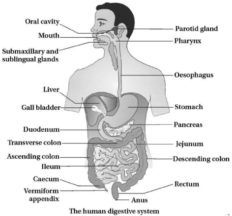
Draw a labelled diagram of the human digestive system.
Solution
(N/A) The human digestive system consists of the alimentary canal and associated digestive glands.
The alimentary canal begins with an anterior opening—the mouth,and it opens out posteriorly through the anus.
The mouth leads to the oral cavity or buccal cavity. The oral cavity has a number of teeth and a muscular tongue.
The pharynx serves as a common passage for food and air. The oesophagus and the trachea (wind pipe) open into the pharynx.
$A$ cartilaginous flap called epiglottis prevents the entry of food into the glottis—opening of the wind pipe—during swallowing.
The oesophagus is a thin,long tube which extends posteriorly passing through the neck,thorax and diaphragm and leads to a '$J$' shaped bag-like structure called stomach.
The stomach leads to the small intestine,which is distinguishable into three regions—a '$C$' shaped duodenum,a long coiled middle portion jejunum and a highly coiled ileum.
The opening of the stomach into the duodenum is guarded by the pyloric sphincter.
Ileum opens into the large intestine. It consists of caecum,colon and rectum. The caecum is a small blind sac which hosts some symbiotic micro-organisms. $A$ narrow finger-like tubular projection,the vermiform appendix which is a vestigial organ,arises from the caecum.
The caecum opens into the colon. The colon is divided into three parts—an ascending,a transverse and a descending part. The descending part opens into the rectum which opens out through the anus.
The digestive glands associated with the alimentary canal include the salivary glands,the liver and the pancreas.
0 likes
View Solution158
Medium
Explain the structure of the mouth.
Solution
(N/A) The mouth is the anterior opening of the alimentary canal.
It leads into the oral or buccal cavity.
The oral cavity contains a number of teeth and a muscular tongue.
The roof of the oral cavity is formed by the palate,which consists of an anterior hard palate and a posterior soft palate.
The uvula is a small,fleshy projection hanging from the middle of the soft palate.
The sides of the oral cavity are formed by muscular cheeks,which are connected to the lips.
The lips are muscular folds covered by skin on the outer surface and a mucous membrane on the inner surface.
The lower jaw contains the hyoid bone,to which the tongue is attached.
0 likes
View Solution159
Medium
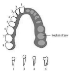
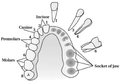
Give types of teeth and describe the arrangement of teeth in the mouth with a diagram.
Solution
(N/A) Thecodont: Each tooth is embedded in a socket of the jaw bone. This type of attachment is called thecodont.
Diphyodont: At the age of $6$ years,a set of temporary milk teeth or deciduous teeth are replaced by a set of permanent or adult teeth. This type of dentition is called diphyodont.
Types of Teeth: The majority of mammals,including human beings,form two sets of teeth during their life:
$(i)$ Deciduous or milk teeth
(ii) Permanent or adult teeth
The number of milk teeth is $20$. The dental formula of a child is $\frac{2102}{2102}$.
An adult human has $32$ permanent teeth which are of four different types. There are $16$ teeth in each jaw. The arrangement of teeth in each half of the upper and lower jaw in the order of Incisors $(I)$,Canines $(C)$,Premolars $(PM)$,and Molars $(M)$ is represented by a dental formula,which in humans is $\frac{2123}{2123}$.
The hard chewing surface of the teeth is made up of enamel.
0 likes
View Solution160
Medium
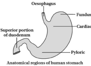
Describe the pharynx,oesophagus,and external structure of the stomach in the human digestive tract.
Solution
(N/A) Pharynx: The oesophagus and the trachea open into the pharynx.
The pharynx is located behind the nasal cavity,oral cavity,and larynx. It is divided into three parts:
$(i)$ Nasopharynx
$(ii)$ Oropharynx
$(iii)$ Laryngopharynx
Function: It serves as a common passage for food and air. $A$ cartilaginous flap called the epiglottis prevents the entry of food into the glottis during swallowing.
$(b)$ Oesophagus: The oesophagus is a thin,long tube. It is situated behind the trachea and in front of the vertebral column. It extends posteriorly,passing through the neck,thorax,and diaphragm,and leads to a '$J$'-shaped,bag-like structure called the stomach.
$A$ muscular gastro-oesophageal sphincter regulates the opening of the oesophagus into the stomach.
Function: It transports food into the stomach via peristaltic movements.
$(c)$ Stomach: The stomach is the most enlarged part of the digestive tract. It is located in the upper left portion of the abdominal cavity. It has three major parts:
$(i)$ Cardiac portion: The part into which the oesophagus opens.
$(ii)$ Fundic region: The main,dome-shaped part of the stomach.
$(iii)$ Pyloric portion: The distal part that opens into the first part of the small intestine (duodenum).
Function: It stores food temporarily,churns it,and mixes it with gastric juices to convert it into chyme.
0 likes
View Solution161
Medium
Describe the external structure of the small intestine and large intestine.
Solution
(N/A) Small Intestine: It is a tube approximately $6.25 \,m$ long, beginning at the pyloric sphincter of the stomach. It is divided into three parts:
$(i)$ Duodenum: The initial $'U'$-shaped part, approximately $25 \,cm$ long.
$(ii)$ Jejunum: The long, coiled middle portion, approximately $1 \,m$ long.
$(iii)$ Ileum: The final, highly coiled part of the small intestine.
Function: Primary site for digestion of food and absorption of nutrients.
$(b)$ Large Intestine: It is approximately $1.5 \,m$ long and consists of the caecum, colon, and rectum.
$(i)$ Caecum: $A$ small blind sac that hosts symbiotic microorganisms. $A$ narrow, finger-like tubular projection, the vermiform appendix (a vestigial organ), arises from it.
$(ii)$ Colon: Divided into three parts: Ascending, Transverse, and Descending colon.
$(iii)$ Rectum: The descending colon opens into the rectum, which stores feces until it is eliminated through the anus.
Function: Absorption of water, minerals, and certain drugs.
0 likes
View Solution162
Medium
Draw a labelled diagram of the transverse section $(T.S.)$ of the intestine.
Solution
(N/A) The wall of the alimentary canal from the oesophagus to the rectum possesses four layers:
$(a)$ Serosa: Serosa is the outermost layer and is made up of a thin mesothelium with some connective tissues.
$(b)$ Muscularis: It is formed by smooth muscles usually arranged into an inner circular and an outer longitudinal layer.
$(c)$ Sub-mucosa: The sub-mucosal layer is formed of loose connective tissues containing nerves,blood,and lymph vessels. In the duodenum,glands are also present in the sub-mucosa.
$(d)$ Mucosa: The innermost layer lining the lumen of the alimentary canal is the mucosa. This layer forms irregular folds in the stomach and small finger-like projections called villi in the small intestine.
The cells lining the villi produce numerous microscopic projections called microvilli,giving a brush-border appearance. These modifications increase the surface area enormously.
Villi are supplied with a network of capillaries and a large lymph vessel called a lacteal.
Mucosal epithelium has goblet cells which secrete mucus that helps in lubrication.
Mucosa also forms glands in the stomach (gastric glands) and crypts in between the bases of villi in the intestine (Crypts of Lieberkuhn).
All the four layers show modifications in different parts of the alimentary canal.

0 likes
View Solution163
Medium
Describe the tissue structure of the wall of the digestive tract.
Solution
(N/A) The wall of the alimentary canal from the oesophagus to the rectum possesses four layers:
$(a)$ Serosa: Serosa is the outermost layer and is made up of a thin mesothelium (epithelium of visceral organs) with some connective tissues.
$(b)$ Muscularis: It is formed by smooth muscles,usually arranged into an inner circular and an outer longitudinal layer.
$(c)$ Sub-mucosa: The sub-mucosal layer is formed of loose connective tissues containing nerves,blood,and lymph vessels. In the duodenum,glands are also present in the sub-mucosa.
$(d)$ Mucosa: The innermost layer lining the lumen of the alimentary canal is the mucosa. This layer forms irregular folds (rugae) in the stomach and small finger-like projections called villi in the small intestine.
The cells lining the villi produce numerous microscopic projections called microvilli,giving a brush-border appearance. These modifications increase the surface area enormously.
Villi are supplied with a network of capillaries and a large lymph vessel called a lacteal.
Mucosal epithelium has goblet cells which secrete mucus that helps in lubrication.
Mucosa also forms glands in the stomach (gastric glands) and crypts in between the bases of villi in the intestine (Crypts of Lieberkuhn).
All the four layers show modifications in different parts of the alimentary canal.

0 likes
View Solution164
EasyMCQ
Give the dental formula of an adult human.
A
$2123$/$2123$
B
$2102$/$2102$
C
$2122$/$2122$
D
$2124$/$2124$
Solution
(A) The arrangement of teeth in each half of the upper and lower jaw in the order of Incisors $(I)$,Canines $(C)$,Premolars $(PM)$,and Molars $(M)$ is represented by a dental formula.
For an adult human,the dental formula is $\frac{2123}{2123}$,which means there are $2$ incisors,$1$ canine,$2$ premolars,and $3$ molars in each half of both the upper and lower jaws.
This results in a total of $32$ teeth $(8 \times 4 = 32)$.
For an adult human,the dental formula is $\frac{2123}{2123}$,which means there are $2$ incisors,$1$ canine,$2$ premolars,and $3$ molars in each half of both the upper and lower jaws.
This results in a total of $32$ teeth $(8 \times 4 = 32)$.
0 likes
View Solution165
MediumMCQ
Give scientific reason:
$(1)$ Heterodontic dentition is seen in human beings.
$(1)$ Heterodontic dentition is seen in human beings.
A
Humans have only one type of tooth.
B
Humans have different types of teeth specialized for different functions.
C
Humans have teeth that are all identical in shape.
D
Humans have teeth that are replaced only once in a lifetime.
Solution
(B) Human beings possess four different types of teeth,namely incisors,canines,premolars,and molars. This arrangement of different types of teeth is known as heterodont dentition.
Incisors are chisel-shaped and are used for cutting and biting.
Canines are pointed and are used for tearing food.
Premolars and molars have broad,flat surfaces and are specialized for chewing and grinding food.
Incisors are chisel-shaped and are used for cutting and biting.
Canines are pointed and are used for tearing food.
Premolars and molars have broad,flat surfaces and are specialized for chewing and grinding food.
0 likes
View Solution166
Medium
Define / Explain the following terms:
$(1)$ Diphyodont
$(2)$ Thecodont
$(1)$ Diphyodont
$(2)$ Thecodont
Solution
(N/A) $(1)$ Diphyodont: Human beings and the majority of mammals form two sets of teeth during their life: a set of temporary milk or deciduous teeth replaced by a set of permanent or adult teeth. This type of dentition is called diphyodont.
$(2)$ Thecodont: In mammals,each tooth is embedded in a socket of the jaw bone. This type of attachment is called thecodont.
$(2)$ Thecodont: In mammals,each tooth is embedded in a socket of the jaw bone. This type of attachment is called thecodont.
0 likes
View Solution167
Medium
Provide definitions/explanations for the following terms:
$(1)$ Heterodont
$(2)$ Appendix
$(1)$ Heterodont
$(2)$ Appendix
Solution
(N/A) $(1)$ Heterodont: In humans and many mammals,teeth are of different types based on their shape and function. These are categorized into four types: Incisors,Canines,Premolars,and Molars. This condition of having different types of teeth is known as heterodont dentition.
$(2)$ Appendix: The caecum is a small blind sac which hosts some symbiotic micro-organisms and is located at the junction of the small and large intestine. $A$ narrow,finger-like tubular projection,known as the vermiform appendix,arises from the caecum.
$(2)$ Appendix: The caecum is a small blind sac which hosts some symbiotic micro-organisms and is located at the junction of the small and large intestine. $A$ narrow,finger-like tubular projection,known as the vermiform appendix,arises from the caecum.
0 likes
View Solution168
Medium
Identify the location and function of the following:
$(1)$ Villi
$(2)$ Oxyntic cells
$(1)$ Villi
$(2)$ Oxyntic cells
Solution
(N/A) $(1)$ Villi: Located in the mucosa of the small intestine. They provide a large surface area for the absorption of digested food components.
$(2)$ Oxyntic cells (Parietal cells): Located in the gastric glands of the stomach. They secrete dilute $HCl$ and intrinsic factor,which is essential for the absorption of vitamin $B_{12}$.
$(2)$ Oxyntic cells (Parietal cells): Located in the gastric glands of the stomach. They secrete dilute $HCl$ and intrinsic factor,which is essential for the absorption of vitamin $B_{12}$.
0 likes
View Solution169
Medium
List the organs of the human alimentary canal and name the major digestive glands with their location.
Solution
(N/A) The human alimentary canal consists of the following organs:
$(i)$ Mouth,$(ii)$ Pharynx,$(iii)$ Oesophagus,$(iv)$ Stomach,$(v)$ Small intestine,$(vi)$ Large intestine,$(vii)$ Rectum,$(viii)$ Anus.
The major digestive glands and their locations are:
$(i)$ Salivary glands: Located in the oral cavity; they secrete saliva.
$(ii)$ Liver: The largest gland of the body,located in the upper right quadrant of the abdominal cavity,just below the diaphragm. It secretes bile.
$(iii)$ Pancreas: Located in the '$U$' shaped curve of the duodenum. It functions as both an endocrine and an exocrine gland.
0 likes
View Solution170
EasyMCQ
Analogy type question:
$(1)$ Teeth embedded in the socket of the jaw bone : Thecodont : Teeth that replace milk teeth : ...........
$(1)$ Teeth embedded in the socket of the jaw bone : Thecodont : Teeth that replace milk teeth : ...........
A
Diphyodont
B
Heterodont
C
Monophyodont
D
Polyphyodont
Solution
(A) The term 'Thecodont' describes the arrangement where teeth are embedded in the jaw bone socket.
Similarly,the term 'Diphyodont' describes the condition where teeth are formed twice in a lifetime,i.e.,a set of temporary milk or deciduous teeth replaced by a set of permanent or adult teeth.
Similarly,the term 'Diphyodont' describes the condition where teeth are formed twice in a lifetime,i.e.,a set of temporary milk or deciduous teeth replaced by a set of permanent or adult teeth.
0 likes
View Solution171
EasyMCQ
Select the correct option:
$(1)$ The non-enzymatic digestive juice is Saliva / Bile.
$(2)$ The Caecum / Colon is a vestigial organ in humans.
$(1)$ The non-enzymatic digestive juice is Saliva / Bile.
$(2)$ The Caecum / Colon is a vestigial organ in humans.
A
$(1)$ Saliva,$(2)$ Caecum
B
$(1)$ Bile,$(2)$ Caecum
C
$(1)$ Saliva,$(2)$ Colon
D
$(1)$ Bile,$(2)$ Colon
Solution
(B) $(1)$ Bile is the correct answer because it does not contain any digestive enzymes. It helps in the emulsification of fats.
$(2)$ The Caecum is a vestigial organ in humans,often associated with the vermiform appendix,which is a narrow finger-like tubular projection arising from it.
$(2)$ The Caecum is a vestigial organ in humans,often associated with the vermiform appendix,which is a narrow finger-like tubular projection arising from it.
0 likes
View Solution172
EasyMCQ
Name the main parts of the human stomach.
A
Cardiac,Fundus,Pyloric
B
Cardiac,Body,Pyloric
C
Fundus,Body,Pyloric
D
Cardiac,Fundus,Body
Solution
(A) The human stomach is anatomically divided into three major parts:
$(i)$ Cardiac portion: Into which the oesophagus opens.
$(ii)$ Fundic region: Which is the upper,dome-shaped portion.
$(iii)$ Pyloric portion: Which opens into the first part of the small intestine (duodenum).
$(i)$ Cardiac portion: Into which the oesophagus opens.
$(ii)$ Fundic region: Which is the upper,dome-shaped portion.
$(iii)$ Pyloric portion: Which opens into the first part of the small intestine (duodenum).
0 likes
View Solution173
EasyMCQ
Into how many parts is the large intestine distributed?
A
Two
B
Three
C
Four
D
Five
Solution
(B) The large intestine is distributed into three parts: $caecum$, $colon$, and $rectum$.
0 likes
View Solution174
Medium
Define the following terms:
$(i)$ Chyme
$(ii)$ Lymph
$(i)$ Chyme
$(ii)$ Lymph
Solution
(N/A) $(i)$ Chyme: In the stomach,food is mixed thoroughly with the acidic gastric juice (containing $HCl$ and enzymes). This partially digested,acidic,semi-solid food mass is known as chyme.
$(ii)$ Lymph: Lymph is a colourless fluid containing specialized lymphocytes,which are responsible for the immune responses of the body. It is essentially a filtered blood plasma that lacks red blood cells and large protein molecules.
$(ii)$ Lymph: Lymph is a colourless fluid containing specialized lymphocytes,which are responsible for the immune responses of the body. It is essentially a filtered blood plasma that lacks red blood cells and large protein molecules.
0 likes
View Solution175
EasyMCQ
Goblet cells of the alimentary canal are modified from:
A
Compound epithelial cells
B
Squamous epithelial cells
C
Columnar epithelial cells
D
Chondrocytes
Solution
(C) Goblet cells are specialized unicellular glands found in the epithelial lining of the alimentary canal. These cells are modified from columnar epithelial cells. They are responsible for the secretion of mucus,which helps in the lubrication and protection of the intestinal lining.
0 likes
View Solution176
EasyMCQ
Identify the correct statement with reference to the human digestive system.
A
Vermiform appendix arises from the duodenum.
B
Ileum opens into the small intestine.
C
Serosa is the innermost layer of the alimentary canal.
D
Ileum is a highly coiled part.
Solution
(D) The correct statement is that the ileum is a highly coiled part of the small intestine.
- The vermiform appendix arises from the caecum,not the duodenum.
- The ileum opens into the large intestine,specifically the caecum,not the small intestine.
- The serosa is the outermost layer of the alimentary canal,not the innermost (the innermost is the mucosa).
- The vermiform appendix arises from the caecum,not the duodenum.
- The ileum opens into the large intestine,specifically the caecum,not the small intestine.
- The serosa is the outermost layer of the alimentary canal,not the innermost (the innermost is the mucosa).
0 likes
View Solution177
MediumMCQ
Identify the correct statement regarding the human digestive system.
A
The ileum opens into the small intestine.
B
The mucosa is the innermost layer of the alimentary canal.
C
The ileum is a highly coiled part.
D
The vermiform appendix arises from the duodenum.
Solution
(B) The wall of the alimentary canal is made up of $4$ layers: mucosa,submucosa,muscularis,and serosa. The mucosa is the innermost layer of the alimentary canal. Therefore,option $B$ is correct.
- The ileum is the final part of the small intestine itself,so it does not open into the small intestine.
- While the ileum is a coiled part,it opens into the caecum of the large intestine.
- The vermiform appendix arises from the caecum,not the duodenum.
- The ileum is the final part of the small intestine itself,so it does not open into the small intestine.
- While the ileum is a coiled part,it opens into the caecum of the large intestine.
- The vermiform appendix arises from the caecum,not the duodenum.
0 likes
View Solution178
MediumMCQ
From which of the following was Vitamin $B_{12}$ first isolated?
A
Liver
B
Intestine
C
Bacteria
D
Plants
Solution
(A) Vitamin $B_{12}$,also known as cobalamin,was first isolated from the liver. It is a water-soluble vitamin that plays a crucial role in the functioning of the brain and nervous system,and in the formation of red blood cells. While it is now produced industrially using specific bacteria,its initial discovery and isolation were achieved from animal liver tissue.
0 likes
View Solution179
EasyMCQ
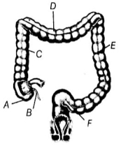
The diagram of the large intestine is given below. Identify the parts $A, B, C, D, E$ and $F$.
A
$A$ - Sigmoid colon,$B$ - Vermiform appendix,$C$ - Ascending colon,$D$ - Transverse colon,$E$ - Descending colon,$F$ - Caecum
B
$A$ - Caecum,$B$ - Vermiform appendix,$C$ - Sigmoid colon,$D$ - Ascending colon,$E$ - Transverse colon,$F$ - Descending colon
C
$A$ - Caecum,$B$ - Vermiform appendix,$C$ - Ascending colon,$D$ - Transverse colon,$E$ - Descending colon,$F$ - Sigmoid colon
D
$A$ - Sigmoid colon,$B$ - Vermiform appendix,$C$ - Descending colon,$D$ - Transverse colon,$E$ - Ascending colon,$F$ - Caecum
Solution
(C) In the given figure:
$A$ represents the caecum,which is a small blind sac that hosts some symbiotic microorganisms.
$B$ is the vermiform appendix,a narrow finger-like tubular projection arising from the caecum.
$C$ is the ascending colon,which is the first part of the colon moving upwards.
$D$ is the transverse colon,which runs across the abdomen.
$E$ is the descending colon,which moves downwards.
$F$ is the sigmoid colon,which is the $S$-shaped part leading to the rectum.
Therefore,the correct identification is $A$ - Caecum,$B$ - Vermiform appendix,$C$ - Ascending colon,$D$ - Transverse colon,$E$ - Descending colon,$F$ - Sigmoid colon.
0 likes
View Solution180
EasyMCQ
Goblet cells secrete
A
$Mucus$
B
$Enzymes$
C
$HCl$
D
$Hormones$
Solution
(A) Goblet cells are specialized simple columnar epithelial cells that are responsible for the production and secretion of mucus.
These cells are named 'goblet' cells because their shape resembles a goblet or a wine cup,which is caused by the accumulation of mucin granules in the apical portion of the cell during the secretion process.
Mucus acts as a lubricant and protects the lining of various organs,such as the digestive and respiratory tracts.
0 likes
View Solution181
EasyMCQ
The backflow of faecal matter in the large intestine is prevented by the presence of
A
Epiglottis
B
Sphincter of Oddi
C
Ileo-caecal valve
D
Pyloric sphincter
Solution
(C) The $Ileo-caecal$ valve is a sphincter muscle situated at the junction of the small intestine (ileum) and the large intestine (caecum).
Its primary function is to allow the passage of digested food (chyme) from the ileum into the caecum.
Crucially,it prevents the backflow of faecal matter from the large intestine into the small intestine,thereby maintaining the unidirectional flow of the digestive process.
Its primary function is to allow the passage of digested food (chyme) from the ileum into the caecum.
Crucially,it prevents the backflow of faecal matter from the large intestine into the small intestine,thereby maintaining the unidirectional flow of the digestive process.
0 likes
View Solution182
EasyMCQ
Cud chewing animals are known as
A
Frugivorous
B
Sanguivorous
C
Ruminants
D
Cannibals
Solution
(C) Cud chewing animals are called ruminants. These animals swallow their grazed food into the rumen,which is a specialized chamber of the stomach. In the rumen,the food is mixed with mucus and undergoes partial anaerobic digestion of cellulose by symbiotic bacteria.
0 likes
View Solution183
EasyMCQ
Which part of the small intestine opens into the large intestine?
A
Colon
B
Jejunum
C
Ileum
D
Duodenum
Solution
(C) The small intestine is divisible into three regions: a $C$-shaped duodenum,a long coiled middle portion jejunum,and a highly coiled ileum.
The ileum is the distal part of the small intestine.
It opens into the large intestine,specifically into the first part known as the caecum.
The ileum is the distal part of the small intestine.
It opens into the large intestine,specifically into the first part known as the caecum.
0 likes
View Solution184
EasyMCQ
Crypts of Lieberkühn are present in
A
Small intestine
B
Liver
C
Stomach
D
Colon
Solution
(A) Crypts of Lieberkühn,also known as intestinal glands,are found in the mucosa of the small intestine between the bases of the villi.
These glands are responsible for the secretion of $succus$ $entericus$,which is the intestinal juice containing various enzymes for digestion.
Therefore,the correct location is the small intestine.
These glands are responsible for the secretion of $succus$ $entericus$,which is the intestinal juice containing various enzymes for digestion.
Therefore,the correct location is the small intestine.
0 likes
View Solution185
EasyMCQ
In rabbits, the reverse flow of food from the stomach into the esophagus is prevented by:
A
Pyloric sphincter
B
Ileo-caecal valve
C
Cardiac sphincter
D
Uvula
Solution
(C) In rabbits (as in other mammals), the reverse flow of food from the stomach back into the esophagus is prevented by the $Cardiac$ $\text{sphincter}$.
This sphincter acts as a valve at the junction of the esophagus and the stomach.
The $Pyloric$ $\text{sphincter}$ is located at the junction of the stomach and the small intestine, which regulates the passage of chyme into the intestine.
This sphincter acts as a valve at the junction of the esophagus and the stomach.
The $Pyloric$ $\text{sphincter}$ is located at the junction of the stomach and the small intestine, which regulates the passage of chyme into the intestine.
0 likes
View Solution186
MediumMCQ
Vitamin-$B_{12}$ is available to ruminants by
A
Plants
B
Microorganisms in caecum
C
Animals
D
All of the above
Solution
(B) Vitamin-$B_{12}$ is not found in plants. While some sources suggest Spirulina (an alga) may contain $B_{12}$,it is primarily synthesized by microorganisms. In ruminant mammals,the rumen (a specialized stomach compartment) contains a dense population of symbiotic bacteria that synthesize large quantities of Vitamin-$B_{12}$,which the animal then absorbs.
0 likes
View Solution187
EasyMCQ
What is the another name of gastro-oesophageal sphincter?
A
Pyloric sphincter
B
Gastro-duodenal sphincter
C
Cardiac sphincter
D
Sphincter of Oddi
Solution
(C) The gastro-oesophageal sphincter is also known as the cardiac sphincter.
It is located at the junction of the oesophagus and the cardiac part of the stomach.
It is named 'cardiac' because it is situated near the heart.
It functions as a sphincter to prevent the backflow of gastric contents into the oesophagus.
It is located at the junction of the oesophagus and the cardiac part of the stomach.
It is named 'cardiac' because it is situated near the heart.
It functions as a sphincter to prevent the backflow of gastric contents into the oesophagus.
0 likes
View Solution188
EasyMCQ
The type of dentition found in human beings is
A
Polyphyodont,thecodont
B
Diphyodont and thecodont
C
Diphyodont and acrodont
D
Diphyodont and homodont
Solution
(B) The oral cavity consists of a number of teeth and a muscular tongue.
Teeth are embedded in a socket of the jaw bone; this type of attachment is called $thecodont$.
Most mammals have two sets of teeth during their life,which is known as $diphyodont$.
Temporary or deciduous teeth ($20$ in number) are replaced by permanent teeth.
An adult human has $32$ teeth,i.e.,$16$ in each jaw.
These are of four different types $(heterodont)$,namely incisors $(I)$,canines $(C)$,premolars $(PM)$,and molars $(M)$.
Therefore,the dentition in human beings is $diphyodont$,$heterodont$,and $thecodont$.
0 likes
View Solution189
EasyMCQ
The human dental formula is:
A
$I \frac{2}{2} C \frac{1}{1} PM \frac{2}{2} M \frac{3}{3}$
B
$I \frac{2}{1} C \frac{1}{2} PM \frac{2}{2} M \frac{3}{3}$
C
$I \frac{1}{2} C \frac{2}{1} PM \frac{2}{2} M \frac{3}{3}$
D
$I \frac{1}{1} C \frac{2}{2} PM \frac{2}{2} M \frac{3}{3}$
Solution
(A) The dental formula represents the number of teeth in one half of the upper jaw divided by the teeth in one half of the lower jaw.
For an adult human,the arrangement of teeth is $2$ incisors,$1$ canine,$2$ premolars,and $3$ molars in each half of both jaws.
Therefore,the human dental formula is $I \frac{2}{2} C \frac{1}{1} PM \frac{2}{2} M \frac{3}{3}$.
For an adult human,the arrangement of teeth is $2$ incisors,$1$ canine,$2$ premolars,and $3$ molars in each half of both jaws.
Therefore,the human dental formula is $I \frac{2}{2} C \frac{1}{1} PM \frac{2}{2} M \frac{3}{3}$.
0 likes
View Solution190
EasyMCQ
Diastema refers to
A
Gap between the teeth
B
Gap between tongue and teeth
C
Ciliary cells on alimentary wall
D
Cell lining along pharynx
Solution
(A) Diastema is the gap in the teeth along the jawbone.
In herbivores,the diastema separates the incisors from the premolars,resulting in an elongation of the jaw and aiding in feeding.
In herbivores,the diastema separates the incisors from the premolars,resulting in an elongation of the jaw and aiding in feeding.
0 likes
View Solution191
EasyMCQ
The layer of cells that secretes enamel of tooth is
A
Dentoblast
B
Ameloblast
C
Osteoblast
D
Odontoblast
Solution
(B) Enamel is the hardest substance in the human body. It covers the dentine in the crown of the tooth.
There are two primary types of cells involved in tooth formation:
$1$. $Odontoblasts$: These cells are responsible for the formation of dentine.
$2$. $Ameloblasts$: These cells are responsible for the secretion and formation of enamel.
There are two primary types of cells involved in tooth formation:
$1$. $Odontoblasts$: These cells are responsible for the formation of dentine.
$2$. $Ameloblasts$: These cells are responsible for the secretion and formation of enamel.
0 likes
View Solution192
MediumMCQ
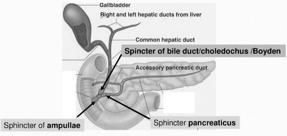
The opening of the common bile duct is guarded by which sphincter?
A
Pyloric Sphincter
B
Sphincter of Boyden
C
Sphincter of Oddi
D
Ileo-caecal Sphincter
Solution
(B) The common bile duct and the pancreatic duct open together into the duodenum as the common hepato-pancreatic duct.
This duct is guarded by a sphincter called the Sphincter of Oddi.
However,the specific opening of the common bile duct (ductus choledochus) before it joins the pancreatic duct is guarded by the Sphincter of Boyden.
In many standard contexts,the Sphincter of Oddi is the primary answer for the hepato-pancreatic opening,but based on the provided diagram,the sphincter guarding the bile duct specifically is the Sphincter of Boyden.
0 likes
View Solution193
EasyMCQ
Choose the incorrect statement regarding the human digestive system with reference to a normal person.
A
Human saliva is slightly acidic.
B
In human beings,four pairs of salivary glands secrete saliva.
C
The quantity of saliva in an adult man may be $1$ to $1.5 \; L$ per day.
D
Enzyme amylase present in saliva is responsible for the breakdown of starch into simple sugar.
Solution
(B) There are three pairs of salivary glands in human beings,namely,parotid glands,sublingual glands,and submaxillary (submandibular) glands.
All three pairs of glands secrete saliva into the buccal cavity through their respective ducts.
Therefore,the statement that there are four pairs of salivary glands is incorrect.
An adult person secretes about $1000-1500 \; mL$ $(1-1.5 \; L)$ of saliva per day.
All three pairs of glands secrete saliva into the buccal cavity through their respective ducts.
Therefore,the statement that there are four pairs of salivary glands is incorrect.
An adult person secretes about $1000-1500 \; mL$ $(1-1.5 \; L)$ of saliva per day.
0 likes
View Solution194
MediumMCQ
Which one of the following sequences is in the correct order?
A
Descending part of colon $\rightarrow$ Rectum $\rightarrow$ Anus
B
Colon $\rightarrow$ Anus $\rightarrow$ Rectum
C
Stomach $\rightarrow$ Jejunum $\rightarrow$ Duodenum
D
Ileum $\rightarrow$ Colon $\rightarrow$ Caecum
Solution
(A) The human digestive system consists of the alimentary canal and associated digestive glands. The sequence of the alimentary canal is: Mouth $\rightarrow$ Buccal cavity $\rightarrow$ Pharynx $\rightarrow$ Oesophagus $\rightarrow$ Stomach $\rightarrow$ Small intestine (Duodenum $\rightarrow$ Jejunum $\rightarrow$ Ileum) $\rightarrow$ Large intestine (Caecum $\rightarrow$ Colon $\rightarrow$ Rectum). The large intestine terminates into a $2-3 \; cm$ long anal canal,which opens to the exterior through the anus. Therefore,the sequence Descending part of colon $\rightarrow$ Rectum $\rightarrow$ Anus is the correct order.
0 likes
View Solution195
EasyMCQ
Aggregates of lymphoid tissue present in the distal portion of the small intestine are known as
A
Villi
B
Peyer's patches
C
Rugae
D
Choroid plexus
Solution
(B) In the wall of the small intestine,specifically in the distal portion known as the ileum,there are aggregates of lymphoid tissue called Peyer's patches.
These patches are groups of lymph nodules that play a crucial role in the immune system by producing lymphocytes.
Rugae are prominent folds found in the lining of an empty stomach.
Villi are finger-like projections of the mucosa in the small intestine that increase the surface area for absorption.
Choroid plexus is a network of blood vessels in the ventricles of the brain that produces cerebrospinal fluid.
0 likes
View Solution196
EasyMCQ
Food is masticated with the help of which part of teeth?
A
Enamel
B
Root
C
Dentine
D
None of these
Solution
(A) Enamel is the hardest substance in the human body and covers the crown of the tooth. It provides the necessary hardness and durability required for the mastication (chewing) of food. The bulk of the tooth is composed of a hard substance called dentine,which is secreted by odontoblast cells. Since enamel is the specific outer layer that directly contacts and grinds food,it is the primary part involved in mastication.
0 likes
View Solution197
EasyMCQ
Which is the hardest material of the human body?
A
Dentine
B
Enamel
C
Teeth
D
Bone
Solution
(B) tooth consists of three regions,i.e.,crown,neck,and root.
The exposed part,the crown,is covered by the hardest material of the human body.
This hardest material is enamel,which is the secretion of $ameloblast$ cells.
The exposed part,the crown,is covered by the hardest material of the human body.
This hardest material is enamel,which is the secretion of $ameloblast$ cells.
0 likes
View Solution198
EasyMCQ
Brunner's glands are located in
A
Oesophagus
B
Intestine
C
Stomach
D
Duodenum
Solution
(D) Brunner's glands are simple,branched tubular glands present in the submucosal layer of the duodenum.
These glands open into the crypts of Lieberkühn.
The goblet cells of Brunner's glands secrete mucus,which helps in lubricating the food and protecting the intestinal wall from acidic chyme.
These glands open into the crypts of Lieberkühn.
The goblet cells of Brunner's glands secrete mucus,which helps in lubricating the food and protecting the intestinal wall from acidic chyme.
0 likes
View Solution199
MediumMCQ
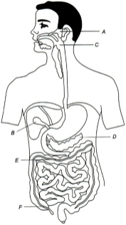
The diagram given below depicts the human digestive system. Label it from $A$ to $F$ and choose the correct option accordingly.
A
$A$-Parotid gland,$B$-Liver,$C$-Larynx,$D$-Pancreas,$E$-Transverse colon,$F$-Caecum
B
$A$-Parotid gland,$B$-Gall bladder,$C$-Pharynx,$D$-Pancreas,$E$-Transverse colon,$F$-Caecum
C
$A$-Parotid gland,$B$-Liver,$C$-Pharynx,$D$-Pancreas,$E$-Ascending colon,$F$-Caecum
D
$A$-Parotid gland,$B$-Gall bladder,$C$-Thymus,$D$-Pancreas,$E$-Ascending colon,$F$-Caecum
Solution
(B) By observing the provided diagram of the human digestive system:
$A$ points to the Parotid gland,which is a major salivary gland.
$B$ points to the Gall bladder,which stores bile.
$C$ points to the Pharynx,the common passage for food and air.
$D$ points to the Pancreas,a glandular organ.
$E$ points to the Transverse colon,a part of the large intestine.
$F$ points to the Caecum,the blind sac at the beginning of the large intestine.
Therefore,the correct sequence is $A$-Parotid gland,$B$-Gall bladder,$C$-Pharynx,$D$-Pancreas,$E$-Transverse colon,$F$-Caecum.
0 likes
View Solution200
EasyMCQ
Symbiotic bacteria present in the colon of the large intestine of humans produce:
A
Cyanocobalamin
B
Riboflavin
C
Thiamine
D
All of these
Solution
(A) The symbiotic bacteria present in the colon of the human large intestine play a vital role in synthesizing certain vitamins.
Specifically,these bacteria produce Vitamin $B_{12}$ (Cyanocobalamin) and Vitamin $K$ (naphthoquinone).
Among the given options,Cyanocobalamin is the correct product synthesized by these bacteria.
Specifically,these bacteria produce Vitamin $B_{12}$ (Cyanocobalamin) and Vitamin $K$ (naphthoquinone).
Among the given options,Cyanocobalamin is the correct product synthesized by these bacteria.
0 likes
View SolutionDigestion and Absorption — Digestive system · Frequently Asked Questions
1Are these Digestion and Absorption questions useful for JEE and NEET?
Yes. All questions in this section are mapped to JEE Main and NEET exam patterns. Previous year questions from JEE Main, NEET, GUJCET and state-level exams are included with full solutions.
2Can I switch to Hindi or Gujarati for these questions?
Yes. Use the language tabs in the hero section or the sidebar to view the same questions and solutions in English, Hindi or Gujarati.
3How do I generate a question paper from this subtopic?
Use the Vedclass Exam Paper Generator — select the chapter and subtopic, set difficulty, and generate Sets A, B, C, D automatically. First 3 chapters of every subject are free.
Vedclass Products
For Students
Vedclass Test Series
Mock tests in real JEE/NEET style with performance analysis. 5-day free trial.
Start Free TrialFor Teachers
Exam Paper Generator
Generate Set A/B/C/D papers from this chapter in 2 minutes. 3 chapters free.
Try FreeFor Institutes
Online Exam Module
Live online exams with unlimited students, 360° analytics & white-label branding.
See DemoFor Teachers & Institutes
Generate a Digestion and Absorption Exam Paper in 2 Minutes
Select subtopic & difficulty — Sets A, B, C, D auto-generated with No Repeat logic.
First 3 chapters of every subject are free — no payment required.