
A English
Centrosomes , CentrioleCilia Questions in English
Class 11 Biology · Cell: The Unit of Life · Centrosomes , CentrioleCilia
121+
Questions
English
Language
100%
With Solutions
Showing 50 of 121 questions in English
51
MediumMCQ
Which organelle is present in all animal cells?
A
Cell wall
B
Vacuole
C
Flagella
D
Centrosome
Solution
(D) $1$. $\text{Cell wall}$ is absent in animal cells; it is a characteristic feature of plant cells, fungi, and bacteria.
$2$. $\text{Vacuoles}$ are present in animal cells but are typically small and temporary, unlike the large central vacuole in plant cells.
$3$. $\text{Flagella}$ are not present in all animal cells; they are specific to certain cell types like sperm cells.
$4$. $\text{Centrosomes}$ (containing centrioles) are organelles that are present in almost all animal cells and play a crucial role in cell division by organizing microtubules.
0 likes
View Solution52
EasyMCQ
In which of the following organisms are centrioles found?
A
Angiosperms
B
Algae
C
Fungi
D
Both $(B)$ and $(C)$
Solution
(D) Centrioles are membrane-less organelles typically found in animal cells and some lower plant groups.
In the plant kingdom,centrioles are generally absent in higher plants like Angiosperms and Gymnosperms.
However,they are present in the motile cells of certain lower plants,such as some species of Algae and Fungi.
Therefore,both Algae and Fungi can contain centrioles in their motile stages.
Thus,the correct option is $(D)$.
0 likes
View Solution53
EasyMCQ
How many triplets are present in the structure of a centriole,and what is the angle between them?
A
$7, 30^{\circ}$
B
$8, 40^{\circ}$
C
$9, 40^{\circ}$
D
$9, 90^{\circ}$
Solution
(C) centriole is a cylindrical structure composed of $9$ sets of microtubule triplets arranged in a ring.
These microtubule triplets are arranged at an angle of $40^{\circ}$ relative to each other.
This arrangement is characteristic of the $9+0$ organization found in centrioles and basal bodies.
These microtubule triplets are arranged at an angle of $40^{\circ}$ relative to each other.
This arrangement is characteristic of the $9+0$ organization found in centrioles and basal bodies.
0 likes
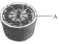
View Solution54
EasyMCQ
The central part of the centriole is known as:
A
Hub
B
Axoneme
C
Triplet
D
Basal body
Solution
(A) The centriole is a cylindrical structure composed of $9$ sets of peripheral triplet microtubules.
At the center of the centriole,there is a proteinaceous,rod-like structure called the $Hub$.
The peripheral triplets are connected to this central $Hub$ by radial spokes made of proteins.
Therefore,the central part of the centriole is the $Hub$.
At the center of the centriole,there is a proteinaceous,rod-like structure called the $Hub$.
The peripheral triplets are connected to this central $Hub$ by radial spokes made of proteins.
Therefore,the central part of the centriole is the $Hub$.
0 likes
View Solution55
EasyMCQ
The cytoplasm surrounding the centrosome is known as:
A
Centrosphere
B
Centriole
C
Cytosphere
D
Cytoplasm
Solution
(A) The centrosome consists of two centrioles surrounded by a specialized,dense,and clear region of cytoplasm called the centrosphere or kinoplasm. This region is devoid of organelles and is responsible for the organization of microtubules during cell division.
0 likes
View Solution56
MediumMCQ
Which organelle directs the formation of the bipolar spindle during cell division?
A
Flagella
B
Mitochondria
C
Microtubule
D
Centriole
Solution
(D) During cell division in animal cells, the $\text{Centriole}$ (part of the centrosome) plays a crucial role in organizing the cytoskeleton.
It acts as the microtubule-organizing center $(MTOC)$ and directs the formation of the bipolar spindle apparatus, which is essential for the segregation of chromosomes into daughter cells.
It acts as the microtubule-organizing center $(MTOC)$ and directs the formation of the bipolar spindle apparatus, which is essential for the segregation of chromosomes into daughter cells.
0 likes
View Solution57
EasyMCQ
Which of the following is $NOT$ associated with the centrosome?
A
Organization of bipolar spindle formation
B
Formation of basal bodies
C
Formation of cilia and flagella
D
Movement of chromosomes
Solution
(D) The centrosome is an organelle that serves as the main microtubule organizing center $(MTOC)$ of the animal cell.
$1$. It is responsible for the organization of the bipolar spindle during cell division.
$2$. Centrioles,which are components of the centrosome,give rise to the basal bodies that form the cilia and flagella.
$3$. While the centrosome organizes the spindle fibers that attach to chromosomes,the actual movement of chromosomes is mediated by the motor proteins and the dynamic instability of the spindle microtubules themselves,not by the centrosome directly. Therefore,'Movement of chromosomes' is the most appropriate answer as it is a process facilitated by the spindle apparatus rather than a direct function of the centrosome structure.
0 likes
View Solution58
MediumMCQ
$S$ - Statement: Basal bodies are associated with the formation of cilia and flagella.
$R$ - Reason: Nucleoli contain the nucleus and chromatin material.
$R$ - Reason: Nucleoli contain the nucleus and chromatin material.
A
$S$ and $R$ are both true,and $R$ is the correct explanation of $S$.
B
$S$ and $R$ are both true,but $R$ is not the correct explanation of $S$.
C
$S$ is true and $R$ is false.
D
$S$ is false and $R$ is true.
Solution
(C) $S$ - Statement: Basal bodies (also known as kinetosomes) are derived from centrioles and are responsible for the formation of the axoneme in cilia and flagella. This statement is true.
$R$ - Reason: Nucleoli are spherical structures present inside the nucleus. They are the sites of ribosomal $RNA$ $(rRNA)$ synthesis. They do not contain the nucleus or chromatin material; rather,they are located within the nucleoplasm of the nucleus. Thus,the reason is false.
$R$ - Reason: Nucleoli are spherical structures present inside the nucleus. They are the sites of ribosomal $RNA$ $(rRNA)$ synthesis. They do not contain the nucleus or chromatin material; rather,they are located within the nucleoplasm of the nucleus. Thus,the reason is false.
0 likes
View Solution59
EasyMCQ
Cilia and flagella are responsible for which of the following processes?
A
Respiration
B
Locomotion
C
Lipid synthesis
D
Protein synthesis
Solution
(B) Cilia and flagella are hair-like outgrowths of the cell membrane.
They are responsible for the movement of the cell or the movement of surrounding fluid.
In unicellular organisms,they facilitate locomotion.
In multicellular organisms,they help in moving fluids or mucus over the surface of cells.
Therefore,the primary function associated with them is locomotion or movement.
0 likes
View Solution60
MediumMCQ
Statement $X$ : Basal bodies are involved in the formation of cilia and flagella.
Statement $Y$ : Nucleoli contain the nucleus and chromatin material.
Statement $Y$ : Nucleoli contain the nucleus and chromatin material.
A
Both statements $X$ and $Y$ are false.
B
Statement $X$ is true and statement $Y$ is false.
C
Both statements $X$ and $Y$ are true,but statement $Y$ is not the explanation of $X$.
D
Both statements $X$ and $Y$ are true,and statement $Y$ is the explanation of $X$.
Solution
(B) Statement $X$ is true because basal bodies (also known as kinetosomes) are derived from centrioles and act as the base for the growth of cilia and flagella.
Statement $Y$ is false because the nucleolus is a spherical structure present inside the nucleus. It is the site of ribosomal $RNA$ $(rRNA)$ synthesis. It does not contain the nucleus or chromatin material; rather,it is a component within the nucleus.
Statement $Y$ is false because the nucleolus is a spherical structure present inside the nucleus. It is the site of ribosomal $RNA$ $(rRNA)$ synthesis. It does not contain the nucleus or chromatin material; rather,it is a component within the nucleus.
0 likes
View Solution61
MediumMCQ
Which of the following is the function of the centrosome?
A
Storage and secretion of various substances
B
Involved in cellular movement and intracellular transport
C
Involved in the formation of basal bodies,cilia,and flagella
D
Regulation of various activities within the cell
Solution
(C) The centrosome is an organelle usually containing two cylindrical structures called centrioles.
Its primary function is to form the basal bodies that give rise to cilia and flagella.
It also plays a crucial role in organizing the spindle fibers during cell division.
Its primary function is to form the basal bodies that give rise to cilia and flagella.
It also plays a crucial role in organizing the spindle fibers during cell division.
0 likes
View Solution62
EasyMCQ
Which of the following organisms contains cilia?
A
Paramecium
B
Yeast
C
Euglena
D
Amoeba
Solution
(A) Cilia are hair-like outgrowths of the cell membrane that help in movement and feeding.
$Paramecium$ is a ciliate protozoan that uses cilia for locomotion and to direct food into its cytostome.
$Yeast$ is a fungus and does not possess cilia.
$Euglena$ uses flagella for movement.
$Amoeba$ uses pseudopodia for movement and feeding.
Therefore,the correct option is $A$.
$Paramecium$ is a ciliate protozoan that uses cilia for locomotion and to direct food into its cytostome.
$Yeast$ is a fungus and does not possess cilia.
$Euglena$ uses flagella for movement.
$Amoeba$ uses pseudopodia for movement and feeding.
Therefore,the correct option is $A$.
0 likes
View Solution63
MediumMCQ
It possesses two central and nine peripheral microtubule doublets.
A
Cilia
B
Flagella
C
Centriole
D
Both $(A)$ and $(B)$
Solution
(D) The axoneme of both cilia and flagella consists of a core called the axoneme,which possesses a number of microtubules running parallel to the long axis. The axoneme usually has nine pairs of doublets of radially arranged peripheral microtubules and a pair of centrally located microtubules. This arrangement is known as the $9+2$ array. Therefore,both cilia and flagella share this structural organization.
0 likes
View Solution64
EasyMCQ
Which organelle is associated with the formation of the bipolar spindle during cell division?
A
Cilia
B
Centriole
C
Golgi apparatus
D
Ribosome
Solution
(B) The $Centriole$ is a cylindrical structure composed of microtubules.
During cell division in animal cells, centrioles organize the formation of the bipolar spindle apparatus, which is essential for the segregation of chromosomes.
These structures are typically found in the centrosome, which acts as the microtubule-organizing center.
During cell division in animal cells, centrioles organize the formation of the bipolar spindle apparatus, which is essential for the segregation of chromosomes.
These structures are typically found in the centrosome, which acts as the microtubule-organizing center.
0 likes
View Solution65
EasyMCQ
What type of arrangement is shown by the cilia/flagella of a eukaryotic cell?
A
$9 + 0$ arrangement
B
$9 + 2$ arrangement
C
$7 + 2$ arrangement
D
$2 + 9$ arrangement
Solution
(B) The axoneme of a eukaryotic cilium or flagellum typically possesses a core called the axoneme,which is composed of microtubules.
These microtubules are arranged in a $9 + 2$ pattern.
This consists of $9$ doublets of radially arranged peripheral microtubules and a pair of centrally located microtubules.
These microtubules are arranged in a $9 + 2$ pattern.
This consists of $9$ doublets of radially arranged peripheral microtubules and a pair of centrally located microtubules.
0 likes
View Solution66
MediumMCQ
How many total microtubules are present in the structure of a centrosome?
A
$20$
B
$54$
C
$27$
D
$56$
Solution
(B) centrosome consists of two centrioles arranged at right angles to each other.
Each centriole is composed of $9$ peripheral triplets of microtubules.
Therefore,the number of microtubules in one centriole is $9 \times 3 = 27$.
Since a centrosome contains two centrioles,the total number of microtubules is $27 \times 2 = 54$.
Each centriole is composed of $9$ peripheral triplets of microtubules.
Therefore,the number of microtubules in one centriole is $9 \times 3 = 27$.
Since a centrosome contains two centrioles,the total number of microtubules is $27 \times 2 = 54$.
0 likes
View Solution67
EasyMCQ
The centrosome is not involved in the formation of which of the following?
A
Cilia
B
Flagella
C
Basal body
D
Ribosomes
Solution
(D) The centrosome is an organelle that serves as the main microtubule organizing center $(MTOC)$ of the animal cell.
Centrioles,which are components of the centrosome,are responsible for the formation of the basal body,which gives rise to cilia and flagella.
Ribosomes are complex molecular machines composed of ribosomal $RNA$ (rRNA) and proteins,and their assembly occurs in the nucleolus,not the centrosome.
Centrioles,which are components of the centrosome,are responsible for the formation of the basal body,which gives rise to cilia and flagella.
Ribosomes are complex molecular machines composed of ribosomal $RNA$ (rRNA) and proteins,and their assembly occurs in the nucleolus,not the centrosome.
0 likes
View Solution68
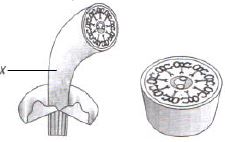
MediumMCQ
Which cell structure is shown in the given figure?
A
Cilia
B
Flagella
C
Centriole
D
Both $(A)$ and $(B)$
Solution
(D) The figure shows the cross-section of a cilium or flagellum,which exhibits a $9+2$ arrangement of microtubules. Both cilia and flagella share this internal structure,known as the axoneme. Therefore,the figure represents both cilia and flagella.
0 likes
View Solution69
EasyMCQ
What does $A$ indicate in the given figure?
A
Axoneme
B
Plasma membrane
C
Central sheath
D
Radial spoke
Solution
(B) The given figure represents the cross-section of a cilium or flagellum.
The structure labeled $A$ points to the outer boundary of the cilium/flagellum,which is the plasma membrane.
The axoneme is the core structure,the central sheath surrounds the central pair of microtubules,and radial spokes connect the outer doublets to the central sheath.
The structure labeled $A$ points to the outer boundary of the cilium/flagellum,which is the plasma membrane.
The axoneme is the core structure,the central sheath surrounds the central pair of microtubules,and radial spokes connect the outer doublets to the central sheath.
0 likes
View Solution70
EasyMCQ
Which cell structure is shown in the given figure?
A
Aster
B
Centrosphere
C
Centriole
D
Nucleus
Solution
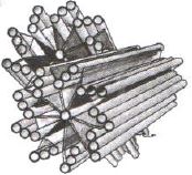
(C) The figure shows the structure of a centriole. $A$ centriole is a cylindrical structure composed mainly of the protein tubulin. It consists of nine sets of microtubule triplets arranged in a ring. Two centrioles lie perpendicular to each other and are surrounded by amorphous pericentriolar material,collectively forming the centrosome.
0 likes
View Solution71
EasyMCQ
What is the other name for the structure shown in the given figure?
A
$9 + 2$
B
$9 + 0$
C
$9 + 1$
D
All of the above $(A), (B)$ and $(C)$
Solution
(B) The figure shows a centriole. $A$ centriole is made up of nine sets of peripheral microtubule triplets,arranged in a circular pattern. There are no microtubules in the center. This arrangement is known as the $9 + 0$ arrangement. Therefore,the correct option is $B$.
0 likes
View Solution72
EasyMCQ
At what angle are the nine triplets arranged in the structure shown in the given figure (in $^\circ$)?
A
$45$
B
$50$
C
$60$
D
$40$
Solution
(D) The figure shows the structure of a centriole,which is composed of nine peripheral triplets of microtubules arranged in a cartwheel pattern.
These nine triplets are arranged at an angle of $40^\circ$ relative to each other in the cylindrical structure.
These nine triplets are arranged at an angle of $40^\circ$ relative to each other in the cylindrical structure.
0 likes
View Solution73
MediumMCQ
The tubules in each triplet of the structure shown in the figure are made of which protein?
A
Tubulin
B
Flagellin
C
Actin
D
Myosin
Solution
(A) The figure shows the structure of a centriole,which consists of nine sets of microtubule triplets arranged in a ring.
Each microtubule is composed of the protein tubulin.
These triplets are arranged in a $9+0$ pattern,which is characteristic of centrioles and basal bodies.
Therefore,the tubules in each triplet are made of tubulin protein.
Each microtubule is composed of the protein tubulin.
These triplets are arranged in a $9+0$ pattern,which is characteristic of centrioles and basal bodies.
Therefore,the tubules in each triplet are made of tubulin protein.
0 likes
View Solution74
EasyMCQ
What is $X$ in the given figure?
A
Intermediate filament
B
Central sheath
C
Axoneme
D
Central microtubules
Solution
(C) The given figure represents the structure of a cilium or flagellum.
$X$ points to the core structure of the cilium/flagellum,which is known as the axoneme.
The axoneme possesses a number of microtubules running parallel to the long axis.
It is usually composed of nine doublets of radially arranged peripheral microtubules and a pair of centrally located microtubules,often referred to as the $9+2$ arrangement.
0 likes
View Solution75
EasyMCQ
Which organelle,involved in cell division,is absent in plant cells?
A
Golgi apparatus
B
Centriole
C
Mitochondria
D
Ribosome
Solution
(B) In animal cells,the centrosome is an organelle that contains two cylindrical structures called centrioles. These centrioles play a crucial role in organizing the spindle fibers during cell division (mitosis and meiosis). Plant cells lack centrosomes and centrioles,yet they are still able to undergo cell division by forming a cell plate instead of using spindle poles organized by centrioles.
0 likes
View Solution76
MediumMCQ
What do the centrosomes form during cell division?
A
Proteins
B
Microtubules
C
Bipolar spindle
D
Enzymes
Solution
(C) During cell division,the centrosomes act as the primary microtubule-organizing centers (MTOCs) in animal cells.
As the cell enters the $M$ phase,the centrosomes move to opposite poles of the cell.
They organize the assembly of microtubules to form the $Bipolar$ $spindle$ apparatus,which is essential for the segregation of chromosomes during mitosis and meiosis.
As the cell enters the $M$ phase,the centrosomes move to opposite poles of the cell.
They organize the assembly of microtubules to form the $Bipolar$ $spindle$ apparatus,which is essential for the segregation of chromosomes during mitosis and meiosis.
0 likes
View Solution77
EasyMCQ
Which cytoplasmic organelle plays an important role during cell division?
A
Mitochondria
B
Centrosome
C
Golgi apparatus
D
None of these
Solution
(B) During cell division,the $Centrosome$ (which contains $Centrioles$) plays a crucial role in the formation of the spindle apparatus.
In animal cells,the centrosome duplicates during the $S$-phase of the cell cycle.
During prophase,these centrosomes move to opposite poles of the cell and initiate the formation of spindle fibers,which are essential for the separation of chromosomes during mitosis and meiosis.
In animal cells,the centrosome duplicates during the $S$-phase of the cell cycle.
During prophase,these centrosomes move to opposite poles of the cell and initiate the formation of spindle fibers,which are essential for the separation of chromosomes during mitosis and meiosis.
0 likes
View Solution78
MediumMCQ
During cell division,where are the spindle fibers formed from?
A
Centromere
B
Nucleus
C
Centriole
D
Mitochondria
Solution
(C) During cell division,the spindle fibers are formed from the centrosome,which contains a pair of centrioles.
In animal cells,the centrioles act as the microtubule-organizing center $(MTOC)$ and are responsible for the formation of the spindle apparatus.
Therefore,the correct option is $C$.
In animal cells,the centrioles act as the microtubule-organizing center $(MTOC)$ and are responsible for the formation of the spindle apparatus.
Therefore,the correct option is $C$.
0 likes
View Solution79
EasyMCQ
Which cytoplasmic structures are essential during cell division?
A
Mitochondria
B
Centrioles
C
Golgi apparatus
D
Nuclear envelope
Solution
(B) Centrioles are essential cytoplasmic structures during cell division in animal cells. They organize the spindle fibers,which are responsible for the segregation of chromosomes into daughter cells during mitosis and meiosis. Therefore,the correct option is $B$.
0 likes
View Solution80
MediumMCQ
The centrosome is involved in:
A
Only present in dividing animal cells.
B
Absent in dividing animal cells.
C
Participates in the formation of spindle fibers in dividing animal cells.
D
Participates in the formation of spindle fibers in both dividing animal and plant cells.
Solution
(C) The centrosome consists of two cylindrical structures called centrioles.
It plays a crucial role in the formation of spindle fibers during animal cell division.
Centrosomes are absent in plant cells; therefore,they are specific to animal cells.
It plays a crucial role in the formation of spindle fibers during animal cell division.
Centrosomes are absent in plant cells; therefore,they are specific to animal cells.
0 likes
View Solution81
EasyMCQ
Which of the following is the odd one out?
A
Centromere
B
Primary constriction
C
Kinetochore
D
Centriole
Solution
(D) The $Centromere$, $Primary \text{ } constriction$, and $Kinetochore$ are all structural components associated with the chromosome.
$Centromere$ is the region where two sister chromatids are held together.
$Primary \text{ } constriction$ is another name for the $Centromere$.
$Kinetochore$ is a disc-shaped structure present on the surface of the $Centromere$ to which spindle fibers attach during cell division.
$Centriole$, on the other hand, is a cylindrical organelle involved in the formation of the spindle apparatus during cell division, but it is not a part of the chromosome structure.
Therefore, $Centriole$ is the odd one out.
0 likes
View Solution82
EasyMCQ
Statement $A$: Plant cells lack centrioles.
Statement $R$: Plant cells form a bipolar spindle during cell division.
Statement $R$: Plant cells form a bipolar spindle during cell division.
A
$A$ and $R$ are both true,$R$ is not the correct explanation of $A$.
B
$A$ and $R$ are both false.
C
$A$ and $R$ are both true,$R$ is the correct explanation of $A$.
D
$A$ is false and $R$ is true.
Solution
(A) Statement $A$ is true because plant cells do not possess centrioles,which are typically found in animal cells.
Statement $R$ is also true because,despite the absence of centrioles,plant cells still form a bipolar spindle during mitosis using microtubule organizing centers (MTOCs) located in the cytoplasm.
Since the formation of the bipolar spindle in plants occurs independently of centrioles,statement $R$ does not explain why plant cells lack centrioles. Therefore,both statements are true,but $R$ is not the correct explanation of $A$.
0 likes
View Solution83
EasyMCQ
Which protein is present in cilia and flagella?
A
Actin
B
Myosin
C
Tubulin
D
Myoglobin
Solution
(C) Cilia and flagella are hair-like outgrowths of the cell membrane. Their core,called the axoneme,possesses a number of microtubules running parallel to the long axis. The primary protein component of these microtubules is $Tubulin$.
0 likes
View Solution84
MediumMCQ
Which of the following statements regarding cilia is not correct?
A
Cilia contain an outer ring of nine doublet microtubules surrounding two singlet microtubules.
B
The organized beating of cilia is controlled by fluxes of $Ca^{2+}$ across the membrane.
C
Cilia are hair-like cellular appendages.
D
Microtubules of cilia are composed of tubulin.
Solution
(B) The correct answer is $B$.
Cilia are fine,hair-like,vibratile cytoplasmic processes found on certain cell types.
Their movement either propels the organism or moves the surrounding medium past a fixed cell.
Cilia are enclosed by a unit membrane,which is an extension of the plasma membrane.
Within the membrane is a fluid matrix containing a supporting axial shaft called the axoneme.
The axoneme typically shows a $9+2$ arrangement,where nine doublets of microtubules are arranged in a ring around two central singlet microtubules.
These microtubules are composed of globular units of the protein tubulin.
The arms of the $A$ microtubules contain the protein dynein,which acts as an $ATPase$ enzyme to catalyze the hydrolysis of $ATP$ to $ADP$,providing energy for ciliary movement.
Statement $B$ is incorrect because the beating of cilia is primarily driven by the sliding of microtubule doublets powered by dynein-mediated $ATP$ hydrolysis,not by $Ca^{2+}$ fluxes across the membrane.
0 likes
View Solution85
MediumMCQ
Study the diagram properly and select the correct option for labelled parts $A, B, C$ and $D$.
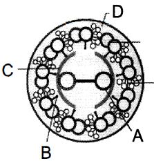
A
$A-$ Central microtubule,$B-$ Radial spoke,$C-$ Central sheath,$D-$ Plasma membrane
B
$A-$ Radial spoke,$B-$ Central sheath,$C-$ Plasma membrane,$D-$ Central microtubule
C
$A-$ Plasma membrane,$B-$ Radial spoke,$C-$ Plasma membrane,$D-$ Central microtubule
D
$A-$ Central sheath,$B-$ Plasma membrane,$C-$ Central microtubule,$D-$ Radial spoke
Solution
(A) The provided diagram represents the cross-section of a cilium or flagellum,which exhibits a $9+2$ arrangement of microtubules.
$1$. Part $A$ points to the central pair of microtubules.
$2$. Part $B$ points to the radial spoke,which connects the peripheral doublets to the central sheath.
$3$. Part $C$ points to the central sheath,which surrounds the central microtubules.
$4$. Part $D$ points to the plasma membrane,which encloses the entire structure.
Therefore,the correct identification is $A-$ Central microtubule,$B-$ Radial spoke,$C-$ Central sheath,$D-$ Plasma membrane.
0 likes
View Solution86
MediumMCQ
How many microtubule triplets are arranged in the peripheral area of a centriole?
A
$2$
B
$9$
C
$5$
D
$7$
Solution
(B) The centriole is a cylindrical structure composed of $9$ evenly spaced peripheral fibrils of tubulin protein.
Each of these peripheral fibrils is a triplet,meaning it consists of three microtubules fused together.
These $9$ triplets are arranged in a circular pattern in the peripheral area of the centriole.
Therefore,the correct number of triplets is $9$.
Each of these peripheral fibrils is a triplet,meaning it consists of three microtubules fused together.
These $9$ triplets are arranged in a circular pattern in the peripheral area of the centriole.
Therefore,the correct number of triplets is $9$.
0 likes
View Solution87
MediumMCQ
Which is correct for centriole function in animal cell?
$(I)$ Formation of bipolar spindle
$(II)$ Formation of lysosome
$(III)$ Formation of mesosome
$(IV)$ Formation of ribosome
$(I)$ Formation of bipolar spindle
$(II)$ Formation of lysosome
$(III)$ Formation of mesosome
$(IV)$ Formation of ribosome
A
$I$ and $II$
B
$I$ and $III$
C
$III$ and $IV$
D
Only $I$
Solution
(D) Centrioles are cylindrical structures found in animal cells that play a crucial role in cell division.
$(I)$ Formation of bipolar spindle: During cell division,centrioles organize the microtubules to form the spindle apparatus,which is essential for chromosome segregation.
$(II)$ Formation of lysosome: Lysosomes are formed by the Golgi apparatus,not by centrioles.
$(III)$ Formation of mesosome: Mesosomes are specialized structures found in prokaryotic cells,not in animal cells.
$(IV)$ Formation of ribosome: Ribosomes are synthesized in the nucleolus of the cell.
Therefore,only statement $(I)$ is correct regarding the function of centrioles.
0 likes
View Solution88
EasyMCQ
Which of the following organelles is helpful in cell division?
A
Chloroplast
B
Mitochondria
C
Centrosome
D
Lysosome
Solution
(C) The $Centrosome$ is a non-membrane-bound organelle found in animal cells that plays a crucial role in cell division.
It consists of two cylindrical structures called $Centrioles$.
During cell division, the $Centrosome$ organizes the $Microtubules$ to form the $Spindle$ apparatus, which is essential for the separation of chromosomes into daughter cells.
Note: The original option $Centromere$ is a region on a chromosome, not an organelle, so $Centrosome$ is the correct biological organelle involved in this process.
0 likes
View Solution89
MediumMCQ
$Cilia$ and $Flagella$ are hair-like outgrowths of the cell membrane. $Cilia$ are small structures which work like oars,causing the movement of either the cell or the surrounding fluid.
A
Cilia
B
Flagella
C
Pili
D
Fimbriae
Solution
(A) The correct answer is $A$. $Cilia$ are small,hair-like projections from the cell surface that function like oars. Their coordinated rhythmic beating causes the movement of either the cell itself (in unicellular organisms) or the movement of fluid and particles across the cell surface (in multicellular organisms). $Flagella$ are generally longer and responsible for cell movement,while $Pili$ and $Fimbriae$ are surface structures in bacteria primarily involved in attachment and conjugation,not locomotion.
0 likes
View Solution90
MediumMCQ
What is the correct description for the internal structure of cilia and flagella?
A
$A$ pair of centrally located microtubules and $9$ pairs of radially arranged peripheral doublets.
B
$A$ middle hub and $9$ peripherally arranged triplets.
C
$A$ central sheath and $9$ pairs of peripherally arranged doublets.
D
One pair of centrally located microtubules and $9$ peripherally arranged triplets.
Solution
(A) The internal structure of cilia and flagella is known as the $9+2$ arrangement.
It consists of a core called the axoneme,which possesses a number of microtubules running parallel to the long axis.
The axoneme usually has $9$ pairs of doublets of radially arranged peripheral microtubules and a pair of centrally located microtubules.
Thus,the arrangement is described as $9+2$.
It consists of a core called the axoneme,which possesses a number of microtubules running parallel to the long axis.
The axoneme usually has $9$ pairs of doublets of radially arranged peripheral microtubules and a pair of centrally located microtubules.
Thus,the arrangement is described as $9+2$.
0 likes
View Solution91
EasyMCQ
Animal cells contain non-membrane-bound organelles called $..........$ which help in cell division.
A
nucleus
B
centriole
C
mitochondria
D
vacuoles
Solution
(B) The correct answer is $B$.
Centrioles are non-membrane-bound organelles found in animal cells.
They are composed of microtubules arranged in a specific pattern.
During cell division, centrioles play a crucial role in the formation of the spindle apparatus, which helps in the separation of chromosomes.
Centrioles are non-membrane-bound organelles found in animal cells.
They are composed of microtubules arranged in a specific pattern.
During cell division, centrioles play a crucial role in the formation of the spindle apparatus, which helps in the separation of chromosomes.
0 likes
View Solution92
MediumMCQ
Microtubules are found in the structure of which of the following?
A
Spindle fibers, centrioles, and cilia
B
Centrioles, spindle fibers, and chromatin
C
Centrosome, nucleosomes, and centrioles
D
Cilia, flagella, and peroxisomes
Solution
(A) Microtubules are hollow, cylindrical structures composed of tubulin proteins. They form the structural components of the cytoskeleton.
Specifically, they are the primary building blocks of:
$1$. $Spindle fibers$: Essential for chromosome movement during cell division.
$2$. $Centrioles$: Cylindrical structures that organize the spindle apparatus.
$3$. $Cilia and flagella$: Hair-like appendages involved in cell movement, which possess a $9+2$ arrangement of microtubules.
Chromatin, nucleosomes, and peroxisomes do not contain microtubules as their structural components.
0 likes
View Solution93
MediumMCQ
Assertion : Centrosomes and centrioles are related to each other.
Reason : Centrosome usually contains two cylindrical structures called centrioles.
Reason : Centrosome usually contains two cylindrical structures called centrioles.
A
If both Assertion and Reason are correct and the Reason is a correct explanation of the Assertion.
B
If both Assertion and Reason are correct but Reason is not a correct explanation of the Assertion.
C
If the Assertion is correct but Reason is incorrect.
D
If both the Assertion and Reason are incorrect.
Solution
(A) The centrosome is an organelle that serves as the main microtubule organizing center $(MTOC)$ of the animal cell.
It typically contains two cylindrical structures known as centrioles,which are oriented at right angles to each other.
These centrioles are composed of microtubules arranged in a $9 + 0$ pattern (nine triplets of microtubules).
Since the centrosome is defined by the presence of these centrioles,the Reason correctly explains the Assertion.
0 likes
View Solution94
Easy
Describe the microstructure of cilia and flagella.
Solution
(N/A) Cilia and flagella are hair-like outgrowths of the cell membrane.
Cilia are small structures that work like oars,causing the movement of either the cell or the surrounding fluid.
Flagella are comparatively longer and are responsible for cell movement. Prokaryotic bacteria also possess flagella,but these are structurally different from eukaryotic flagella.
The electron microscopic study of a cilium or flagellum shows that they are covered with a plasma membrane. Their core,called the axoneme,possesses a number of microtubules running parallel to the long axis.
The axoneme usually has nine pairs of doublets of radially arranged peripheral microtubules and a pair of centrally located microtubules. Such an arrangement of axonemal microtubules is referred to as the $9+2$ array.
The central tubules are connected by a central sheath and are attached to one of the tubules of each peripheral doublet by a radial spoke. Thus,there are nine radial spokes.
The peripheral doublets are also interconnected by linkers.
0 likes
View Solution95
Easy
Explain Centrosome and Centrioles.
Solution
(N/A) $\Rightarrow$ Centrosome is an organelle usually containing two cylindrical structures called centrioles. They are surrounded by amorphous pericentriolar materials.
$\Rightarrow$ Both the centrioles in a centrosome lie perpendicular to each other,in which each has an organization like the cartwheel.
$\Rightarrow$ They are made up of nine evenly spaced peripheral fibrils of tubulin. Each of the peripheral fibril is a triplet. The adjacent triplets are also linked.
$\Rightarrow$ The central part of the proximal region of the centriole is also proteinaceous and called the hub,which is connected with tubules of the peripheral triplets by radial spokes made of protein.
$\Rightarrow$ The centrioles form the basal body of cilia or flagella,and spindle fibres that give rise to spindle apparatus during cell division in animal cells.
0 likes
View Solution96
Medium
State the role of centrioles other than spindle formation.
Solution
(N/A) The centrioles play a crucial role in the formation of the basal body of cilia and flagella.
Additionally,they are involved in the organization of microtubules and the formation of the tail (axoneme) in sperm cells.
Additionally,they are involved in the organization of microtubules and the formation of the tail (axoneme) in sperm cells.
0 likes
View Solution97
Easy
Give the differences between Cilia and Flagella.
Solution
(N/A)
| Cilia | Flagella |
|---|---|
| $(1)$ They are shorter in length. | $(1)$ They are comparatively longer and helical. |
| $(2)$ They are present in large numbers. | $(2)$ They are few in number (usually one or two). |
| $(3)$ They help in the movement of the cell or the surrounding fluid. | $(3)$ They are primarily responsible for cell locomotion. |
0 likes
View Solution98
Easy
Describe the cartwheel structure of a centriole.
Solution
(N/A) $ \Rightarrow $ $ A $ centrosome is an organelle that usually contains two cylindrical structures called centrioles. They are surrounded by amorphous pericentriolar materials.
$ \Rightarrow $ Both centrioles in a centrosome lie perpendicular to each other, and each has an organization similar to a cartwheel.
$ \Rightarrow $ Each centriole is made up of nine evenly spaced peripheral fibrils of tubulin. These fibrils are absent in the center. Thus, this arrangement is described as $ 9+0 $.
$ \Rightarrow $ Each of the peripheral fibrils is a triplet, consisting of three sub-units (microtubules).
$ \Rightarrow $ Centrioles form the basal body of cilia and flagella.
$ \Rightarrow $ They form spindle fibers in animal cells, which give rise to the spindle apparatus during cell division.
0 likes
View Solution99
MediumMCQ
Identify the components labelled $A, B, C$ and $D$ in the given section of cilia/flagella showing different parts. Choose the option which shows the correct labelling of parts.
A
$A -$ Plasma membrane,$B -$ Interdoublet bridge,$C -$ Central microtubule,$D -$ Radial spoke
B
$A -$ Plasma membrane,$B -$ Arm,$C -$ Central microtubule,$D -$ Radial spoke
C
$A -$ Plasma membrane,$B -$ Interdoublet bridge,$C -$ Hub,$D -$ Radial spoke
D
$A -$ Plasma membrane,$B -$ Interdoublet bridge,$C -$ Hub,$D -$ Arm
Solution
(A) The structure shown is a cross-section of a cilium or flagellum,which exhibits a $9+2$ arrangement of microtubules.
$A$ points to the outermost boundary,which is the plasma membrane.
$B$ points to the connection between adjacent peripheral microtubule doublets,known as the interdoublet bridge (or nexin link).
$C$ points to one of the two central microtubules located inside the central sheath.
$D$ points to the radial spoke,which connects the peripheral doublets to the central sheath.
Therefore,the correct labelling is $A -$ Plasma membrane,$B -$ Interdoublet bridge,$C -$ Central microtubule,$D -$ Radial spoke.
0 likes
View Solution100
MediumMCQ
Axoneme with $9+2$ microtubular arrangement occurs in
A
Cilia
B
Flagella
C
Both $(a)$ and $(b)$
D
Centriole
Solution
(C) The axoneme is the core structure of cilia and flagella.
It consists of a central pair of microtubules surrounded by nine doublets of radially arranged peripheral microtubules,which is known as the $9+2$ arrangement.
Both cilia and flagella share this structural organization,which is essential for their motility.
In contrast,centrioles exhibit a $9+0$ arrangement.
0 likes
View SolutionCell: The Unit of Life — Centrosomes , CentrioleCilia · Frequently Asked Questions
1Are these Cell: The Unit of Life questions useful for JEE and NEET?
Yes. All questions in this section are mapped to JEE Main and NEET exam patterns. Previous year questions from JEE Main, NEET, GUJCET and state-level exams are included with full solutions.
2Can I switch to Hindi or Gujarati for these questions?
Yes. Use the language tabs in the hero section or the sidebar to view the same questions and solutions in English, Hindi or Gujarati.
3How do I generate a question paper from this subtopic?
Use the Vedclass Exam Paper Generator — select the chapter and subtopic, set difficulty, and generate Sets A, B, C, D automatically. First 3 chapters of every subject are free.
Vedclass Products
For Students
Vedclass Test Series
Mock tests in real JEE/NEET style with performance analysis. 5-day free trial.
Start Free TrialFor Teachers
Exam Paper Generator
Generate Set A/B/C/D papers from this chapter in 2 minutes. 3 chapters free.
Try FreeFor Institutes
Online Exam Module
Live online exams with unlimited students, 360° analytics & white-label branding.
See DemoFor Teachers & Institutes
Generate a Cell: The Unit of Life Exam Paper in 2 Minutes
Select subtopic & difficulty — Sets A, B, C, D auto-generated with No Repeat logic.
First 3 chapters of every subject are free — no payment required.