
A English
Neuron as Structural and Functional Unit of Neural System Questions in English
Class 11 Biology · Neural Control and Coordination · Neuron as Structural and Functional Unit of Neural System
183+
Questions
English
Language
100%
With Solutions
Showing 50 of 183 questions in English
101
MediumMCQ
Which one of the following pairs of structures distinguishes a nerve cell from other types of cell?
A
Vacuoles and fibres
B
Flagellum and medullary sheath
C
Nucleus and mitochondria
D
Perikaryon and dendrites
Solution
(D) : $A$ neuron (nerve cell) is one of the basic functional units of the nervous system. $A$ neuron is a cell specialized to transmit electrical nerve impulses and thus carry information from one part of the body to another. Each neuron has an enlarged portion,the cell body ($Perikaryon$ or $Cyton$),containing the nucleus. From the cell body,several short,branched processes called $Dendrites$ extend,through which impulses enter the neuron. $A$ longer process,the axon,extends outward and carries impulses away from the cell body. $Perikaryon$ and $Dendrites$ are characteristic structures that distinguish a neuron from other types of cells.
0 likes
View Solution102
EasyMCQ
Myelin sheath is produced by
A
astrocytes and Schwann cells
B
oligodendrocytes and osteoclasts
C
osteoclasts and astrocytes
D
Schwann cells and oligodendrocytes
Solution
(D) : Schwann cells and oligodendrocytes form the myelin sheath around the axon.
Myelin sheath acts as an insulating layer,which prevents the loss of energy of the nerve impulse during its transmission along the nerve fiber.
Myelin sheath acts as an insulating layer,which prevents the loss of energy of the nerve impulse during its transmission along the nerve fiber.
0 likes
View Solution103
MediumMCQ
What is the function of neurons?
A
Detect different kinds of stimuli
B
Receive different kinds of stimuli
C
Transmit different kinds of stimuli
D
All of the above
Solution
(D) Neurons are the structural and functional units of the neural system.
Their primary functions include:
$1$. Detecting various types of stimuli from the internal or external environment through sensory receptors.
$2$. Receiving these stimuli and processing them.
$3$. Transmitting the nerve impulses (signals) to other neurons,muscles,or glands to coordinate a response.
Therefore,all the listed functions are performed by neurons.
0 likes
View Solution104
MediumMCQ
Select the incorrect pair:
A
Multipolar - Cerebral cortex
B
Unipolar - Embryonic stage
C
Bipolar - Retina
D
Neuron - Composed of four major parts
Solution
(D) The correct answer is $D$.
Neurons are typically classified based on the number of axons and dendrites they possess.
$A$. Multipolar neurons (one axon and two or more dendrites) are found in the cerebral cortex. This is correct.
$B$. Unipolar neurons (cell body with one axon only) are typically found in the embryonic stage. This is correct.
$C$. Bipolar neurons (one axon and one dendrite) are found in the retina of the eye. This is correct.
$D$. $A$ neuron is composed of three major parts: the cell body (cyton),dendrites,and the axon. Therefore,the statement that it is composed of four major parts is incorrect.
0 likes
View Solution105
MediumMCQ
The cerebral cortex has which type of neurons?
A
Unipolar
B
Multipolar
C
Bipolar
D
Unipolar,bipolar,multipolar
Solution
(B) Neurons are classified based on the number of axons and dendrites.
$1$. Unipolar neurons have one axon and no dendrites (found in the embryonic stage).
$2$. Bipolar neurons have one axon and one dendrite (found in the retina of the eye).
$3$. Multipolar neurons have one axon and two or more dendrites.
The cerebral cortex of the human brain contains multipolar neurons,which are the most common type of neurons in the central nervous system.
0 likes
View Solution106
MediumMCQ
Match the following:
| Column-$I$ | Column-$II$ |
| $1$. Cell body | $p$. Transmit impulses towards the cell body |
| $2$. Dendrites | $q$. Transmit impulses away from the cell body |
| $3$. Synaptic vesicles | $r$. Contain cytoplasm and granular bodies |
| $s$. Neurotransmitters |
A
$(1-p), (2-q), (3-r)$
B
$(1-p), (2-q), (3-s)$
C
$(1-r), (2-p), (3-s)$
D
$(1-r), (2-q), (3-p)$
Solution
(C) The correct matching is as follows:
$1$. Cell body (Cyton): It contains cytoplasm,nucleus,and granular bodies called Nissl's granules. Thus,$1-r$.
$2$. Dendrites: These are short fibers that branch repeatedly and project out of the cell body. They transmit impulses towards the cell body. Thus,$2-p$.
$3$. Synaptic vesicles: These are present at the synaptic knobs and contain chemicals called neurotransmitters. Thus,$3-s$.
Therefore,the correct sequence is $(1-r), (2-p), (3-s)$.
0 likes
View Solution107
EasyMCQ
Afferent nerve fibres carry impulses from
A
effector organs to $CNS$
B
receptors to $CNS$
C
$CNS$ to receptors
D
$CNS$ to muscles
Solution
(B) Afferent nerve fibres,also known as sensory nerve fibres,are responsible for transmitting nerve impulses from sensory receptors (which detect stimuli) towards the Central Nervous System $(CNS)$.
Conversely,efferent nerve fibres transmit impulses from the $CNS$ to effector organs like muscles or glands.
Therefore,the correct pathway for afferent nerve fibres is from receptors to the $CNS$.
Conversely,efferent nerve fibres transmit impulses from the $CNS$ to effector organs like muscles or glands.
Therefore,the correct pathway for afferent nerve fibres is from receptors to the $CNS$.
0 likes
View Solution108
MediumMCQ
Dendrites are:
A
Branched short fibers
B
Projections out of the cell body
C
Nissl's granules containing body
D
All of the above
Solution
(D) Dendrites are short,branched fibers that project out of the cell body (cyton) of a neuron.
They contain Nissl's granules,which are specialized rough endoplasmic reticulum involved in protein synthesis.
Therefore,all the given statements describe the characteristics of dendrites.
They contain Nissl's granules,which are specialized rough endoplasmic reticulum involved in protein synthesis.
Therefore,all the given statements describe the characteristics of dendrites.
0 likes
View Solution109
MediumMCQ
Synaptic knob is a bulb-like structure which is present:
A
At the end of axon terminal
B
At the node of Ranvier
C
In the cell body
D
At the end of dendrites
Solution
(A) The axon terminal is the distal end of the axon that branches into several fine filaments.
Each filament terminates in a bulb-like structure called the synaptic knob or synaptic bulb.
These knobs contain synaptic vesicles filled with neurotransmitters,which are essential for transmitting nerve impulses across the synapse to the next neuron or effector cell.
Therefore,the synaptic knob is located at the end of the axon terminal.
0 likes
View Solution110
EasyMCQ
Bipolar neurons are found in the
A
Embryonic stage
B
Cerebral cortex
C
Cerebellum
D
Retina of eye
Solution
(D) Neurons are classified based on the number of axons and dendrites they possess.
$1$. Multipolar neurons: One axon and two or more dendrites (found in the cerebral cortex).
$2$. Bipolar neurons: One axon and one dendrite (found in the retina of the eye).
$3$. Unipolar neurons: Cell body with one axon only (found usually in the embryonic stage).
Therefore,bipolar neurons are specifically located in the retina of the eye.
0 likes
View Solution111
MediumMCQ
$A$ structure of neuron comprises of
A
Cell body,synaptic knob,ganglia
B
Synaptic vesicles,ganglia,dendrites
C
Cell body,dendrites,ganglia
D
Cell body,dendrites,axon
Solution
(D) neuron is the structural and functional unit of the neural system.
It consists of three major parts:
$1$. Cell body (also known as the cyton or soma),which contains the nucleus and cytoplasm with Nissl's granules.
$2$. Dendrites,which are short,branched processes that receive impulses and conduct them towards the cell body.
$3$. Axon,which is a long fiber that conducts impulses away from the cell body towards a synapse.
Ganglia are clusters of cell bodies found outside the central nervous system,not parts of a single neuron's structure.
0 likes
View Solution112
DifficultMCQ
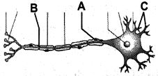
Identify $A$ and $B$ from the figure and what is the function of the part labelled as $C$?
A
$A-$ Schwann cell,$B-$ Node of Ranvier,$C-$ Dendrites,which transport impulses towards the cell body.
B
$A-$ Nucleus,$B-$ Schwann cell,$C-$ Dendrites,which transport impulses towards the cell body.
C
$A-$ Schwann cell,$B-$ Axon,$C-$ Dendrites,which transport impulses away from the cell body.
D
$A-$ Node of Ranvier,$B-$ Schwann cell,$C-$ Axon,which transports impulses away from the cell body.
Solution
(A) In the provided diagram of a neuron:
$A$ points to the Schwann cell,which forms the myelin sheath around the axon.
$B$ points to the gap between two adjacent myelin sheaths,known as the Node of Ranvier.
$C$ points to the dendrites,which are short,branched processes that receive and transport nerve impulses towards the cell body (cyton).
0 likes
View Solution113
MediumMCQ
Which of the following is not found in or a part of myelinated nerve fibres?
A
Schwann cell
B
Node of Ranvier
C
Nissl's granules
D
Synaptic knob
Solution
(C) Myelinated nerve fibres are covered by Schwann cells,which produce the myelin sheath around the axon.
Nodes of Ranvier are the gaps between two adjacent myelin sheaths.
Synaptic knobs are present at the terminal ends of the axon.
Nissl's granules are granular bodies found in the cell body (cyton) and dendrites of a neuron,but they are characteristically absent in the axon and its terminal branches.
Therefore,Nissl's granules are not found in the nerve fibre (axon).
0 likes
View Solution114
MediumMCQ
Neurons are excitable cells because...
A
They lack myelin sheath
B
They possess myelin sheath
C
Their membranes are in a polarized state
D
All of the above
Solution
(C) Neurons are considered excitable cells because their membranes are in a polarized state.
In the resting state,the axonal membrane is more permeable to $K^+$ ions and nearly impermeable to $Na^+$ ions.
This differential permeability,maintained by the $Na^+-K^+$ pump,creates an electrical potential difference across the membrane,known as the resting membrane potential.
This state of polarization allows the neuron to respond to stimuli by generating an action potential.
0 likes
View Solution115
EasyMCQ
The gaps between two adjacent myelin sheaths are called........
A
Nodes of Ranvier
B
Synapse
C
Synaptic cleft
D
Amygdala
Solution
(A) The myelin sheath is an insulating layer that forms around nerves,including those in the brain and spinal cord.
It is composed of protein and fatty substances.
This sheath allows electrical impulses to transmit quickly and efficiently along the nerve cells.
The gaps or spaces between two adjacent myelin sheaths are known as the $Nodes$ $of$ $Ranvier$.
These nodes are essential for saltatory conduction,where the nerve impulse 'jumps' from one node to the next,significantly increasing the speed of transmission.
0 likes
View Solution116
MediumMCQ
Choose the incorrect statement.
A
Myelinated nerve fibres are found in spinal and cranial nerves.
B
Unmyelinated nerve fibre is commonly found in autonomous and the somatic neural systems.
C
$A$ neuron is a microscopic structure composed of cyton only.
D
The afferent nerve fibres transmit impulses from tissues/organs to $CNS$.
Solution
(C) The correct answer is $C$.
$A$ neuron is a microscopic structure composed of three major parts: cell body (cyton),dendrites,and axon.
Statement $C$ is incorrect because it claims a neuron is composed of cyton only,ignoring the presence of dendrites and axons.
Statement $A$ is correct as myelinated nerve fibres are typically found in spinal and cranial nerves.
Statement $B$ is correct as unmyelinated nerve fibres are commonly found in autonomous and somatic neural systems.
Statement $D$ is correct as afferent (sensory) nerve fibres transmit impulses from tissues/organs to the $CNS$.
0 likes
View Solution117
EasyMCQ
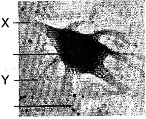
Identify $X$ and $Y$ in the given figure.
A
$X$ - axon,$Y$ - cell body
B
$X$ - cell body,$Y$ - dendrite
C
$X$ - axon,$Y$ - dendrite
D
$X$ - cell body,$Y$ - neuroglia
Solution
(C) The provided image shows a neuron (nerve cell).
In the figure,$X$ points to the long,slender projection extending from the cell body,which is the axon.
$Y$ points to the short,branched projections extending from the cell body,which are the dendrites.
Therefore,$X$ is the axon and $Y$ is the dendrite.
In the figure,$X$ points to the long,slender projection extending from the cell body,which is the axon.
$Y$ points to the short,branched projections extending from the cell body,which are the dendrites.
Therefore,$X$ is the axon and $Y$ is the dendrite.
0 likes
View Solution118
EasyMCQ
Nissl's granules in a neuron are composed of ........
A
Ribosomes
B
Proteins
C
$DNA$
D
$RNA$
Solution
(A) Nissl's granules (also known as Nissl bodies) are large granular bodies found in neurons.
They are composed of rough endoplasmic reticulum $(RER)$ with clusters of free ribosomes and are the site of protein synthesis.
Since they contain ribosomes,they are rich in $RNA$ and are responsible for the synthesis of proteins required for the maintenance and repair of the neuron.
They are composed of rough endoplasmic reticulum $(RER)$ with clusters of free ribosomes and are the site of protein synthesis.
Since they contain ribosomes,they are rich in $RNA$ and are responsible for the synthesis of proteins required for the maintenance and repair of the neuron.
0 likes
View Solution119
MediumMCQ
Which of the following is considered the structural and functional unit of neural tissue?
A
Myelin sheath
B
Axon
C
Dendrite
D
Neuron
Solution
(D) The neural tissue is composed of neurons and neuroglial cells.
Neurons are the structural and functional units of the nervous system.
They are specialized cells that receive,process,and transmit information through electrical and chemical signals.
While axons,dendrites,and myelin sheaths are parts of a neuron,the entire cell itself is the fundamental unit.
Neurons are the structural and functional units of the nervous system.
They are specialized cells that receive,process,and transmit information through electrical and chemical signals.
While axons,dendrites,and myelin sheaths are parts of a neuron,the entire cell itself is the fundamental unit.
0 likes
View Solution120
MediumMCQ
Which of the following statements is true regarding the Nodes of Ranvier?
A
The neurilemma is discontinuous.
B
The myelin sheath is discontinuous.
C
Both the neurilemma and the myelin sheath are discontinuous.
D
It is covered by the myelin sheath.
Solution
(B) The Nodes of Ranvier are periodic gaps in the myelin sheath of axons.
These gaps occur where the myelin sheath is absent,exposing the axonal membrane to the extracellular fluid.
This allows for saltatory conduction,where the nerve impulse 'jumps' from one node to the next,significantly increasing the speed of signal transmission.
The neurilemma (sheath of Schwann) remains continuous over the nodes.
0 likes
View Solution121
EasyMCQ
In a neuron,the components known as Nissl's granules are now identified as:
A
Ribosomes
B
Mitochondria
C
Cell metabolites
D
Fat granules
Solution
(A) Nissl's granules are irregular masses of rough endoplasmic reticulum $(RER)$ with numerous ribosomes attached to them,found in the cell body $(cyton)$ and dendrites of neurons. These structures are primarily sites of protein synthesis. Modern cell biology identifies these granules as clusters of ribosomes and rough endoplasmic reticulum.
0 likes
View Solution122
MediumMCQ
Which of the following pairs of structures is distinct from the other types of cells found in neurons?
A
Vacuoles and filaments
B
Flagella and myelin sheath
C
Nucleus and mitochondria
D
Perikaryon and dendrites
Solution
(B) Neurons are specialized cells that contain typical cell organelles like the $Nucleus$ and $Mitochondria$ (found in most cells). $Perikaryon$ (cell body) and $Dendrites$ are characteristic structures of neurons. $Vacuoles$ and $Filaments$ are also present in various cell types. However,the $Myelin$ $sheath$ is a specialized insulating layer formed by glial cells (Schwann cells or oligodendrocytes) around the axon,and $Flagella$ are typically absent in mature neurons. Among the given options,the pair $Flagella$ and $Myelin$ $sheath$ is the most distinct because $Myelin$ $sheath$ is a unique feature of nerve fibers,and $Flagella$ are not standard components of neuronal structure.
0 likes
View Solution123
EasyMCQ
Myelin sheath is produced by .......... .
A
Schwann cells and oligodendrocytes
B
Astrocytes and Schwann cells
C
Oligodendrocytes and osteoclasts
D
Osteoclasts and astrocytes
Solution
(A) The myelin sheath is an insulating layer that forms around nerves,including those in the brain and spinal cord.
In the Central Nervous System $(CNS)$,the myelin sheath is produced by oligodendrocytes.
In the Peripheral Nervous System $(PNS)$,the myelin sheath is produced by Schwann cells.
Therefore,the correct answer is Schwann cells and oligodendrocytes.
In the Central Nervous System $(CNS)$,the myelin sheath is produced by oligodendrocytes.
In the Peripheral Nervous System $(PNS)$,the myelin sheath is produced by Schwann cells.
Therefore,the correct answer is Schwann cells and oligodendrocytes.
0 likes
View Solution124
EasyMCQ
Nissl bodies are mainly composed of
A
Nucleic acids and $SER$
B
Proteins and lipids
C
Free ribosomes and $RER$
D
$DNA$ and $RNA$
Solution
(C) Nissl bodies (also known as Nissl substance or chromatophilic substance) are large granular bodies found in neurons.
They are primarily composed of rough endoplasmic reticulum $(RER)$ and free ribosomes.
These structures are the sites of protein synthesis within the nerve cell,which is essential for the maintenance and repair of the neuron.
They are primarily composed of rough endoplasmic reticulum $(RER)$ and free ribosomes.
These structures are the sites of protein synthesis within the nerve cell,which is essential for the maintenance and repair of the neuron.
0 likes
View Solution125
MediumMCQ
Bipolar nerve cells are present in
A
Skin tactile corpuscles
B
Spinal cord
C
Retina of eye
D
All the above
Solution
(C) Bipolar neurons are nerve cells that have two extensions: one axon and one dendrite.
These cells are specialized sensory neurons for the transmission of special senses.
They are found in the retina of the eye,the olfactory epithelium of the nose,and the ganglia of the vestibulocochlear nerve ($VIII$ cranial nerve).
These cells are specialized sensory neurons for the transmission of special senses.
They are found in the retina of the eye,the olfactory epithelium of the nose,and the ganglia of the vestibulocochlear nerve ($VIII$ cranial nerve).
0 likes
View Solution126
MediumMCQ
Multipolar nerve cells are present in
A
Cochlea
B
Dorsal root ganglia of spinal cord
C
Retina of eye
D
Brain
Solution
(D) Multipolar neurons are characterized by having one axon and two or more dendrites. They are the most common type of neurons in the vertebrate nervous system and are found in the cerebral cortex of the brain and the spinal cord. In contrast,bipolar neurons are found in the retina of the eye,and pseudounipolar neurons are found in the dorsal root ganglia of the spinal cord.
0 likes
View Solution127
MediumMCQ
Neurons receive signals through their . . . . . . and send signals to other neurons through their . . . . . . .
A
dendrites ... receptors
B
end feet ... cell bodies and dendrites
C
cell bodies and dendrites ... axons
D
transmitter vesicles ... axons
Solution
(C) Neurons are the structural and functional units of the neural system.
$1$. Dendrites are short,branched processes that receive signals from other neurons and conduct them toward the cell body $(cyton)$.
$2$. The axon is a long fiber that conducts nerve impulses away from the cell body to the synapse,where they are transmitted to other neurons or effector cells.
Therefore,the correct answer is $C$.
0 likes
View Solution128
Easy
What are axons and where are they found in the animal body?
Solution
(N/A) Axons are long,slender,cylindrical projections of neurons (nerve cells) that transmit electrical impulses away from the neuron's cell body toward other neurons,muscles,or glands.
In the animal body,axons are found as part of the nervous tissue. They aggregate in bundles to form nerves,which are distributed throughout the body to facilitate communication between different organs and the central nervous system.
In the animal body,axons are found as part of the nervous tissue. They aggregate in bundles to form nerves,which are distributed throughout the body to facilitate communication between different organs and the central nervous system.
0 likes
View Solution129
Medium
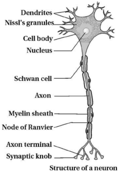
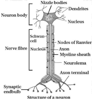
Describe the structure of a neuron with a diagram.
Solution
(N/A) Neurons are the functional units of the neural system.
In the microscopic structure of a neuron,three main parts are observed:
$(1)$ Cell body,$(2)$ Dendrites,$(3)$ Axon.
$(1)$ Cell body: The body of the neuron is called the cyton or soma. The cyton may be oval,round,or stellate-shaped. The cytoplasm of the neuron is called neuroplasm. $A$ big round nucleus is present in the center of the cyton.
Neuroplasm consists of cellular organelles and small basophilic granules called Nissl's granules. Nissl's granules are mostly found near the nucleus.
$(2)$ Dendrites: These are short fibers that branch repeatedly and project out of the cyton. They also contain Nissl's granules. Dendrites transmit impulses towards the cell body.
$(3)$ Axon: It is a long fiber; the distal end of it is branched. Each branch terminates into a bulb-like structure called a synaptic knob.
Synaptic knob: It has synaptic vesicles containing chemicals called neurotransmitters.
The axon transmits nerve impulses away from the cyton to a synapse or to a neuro-muscular junction.
Types of neurons: Based on the number of axons and dendrites,neurons are divided into three types:
$(1)$ Multipolar: One axon and two or more dendrites (e.g.,cerebral cortex).
$(2)$ Bipolar: One axon and one dendrite (e.g.,retina of the eye).
$(3)$ Unipolar: Cell body with one axon only (e.g.,present in the embryonic stage).
Based on the presence or absence of a myelin sheath,neurons are classified as:
$(i)$ Myelinated neuron $(ii)$ Non-myelinated neuron.
$(i)$ Myelinated neuron: The nerve fibers are enveloped with Schwann cells,which form a myelin sheath around the axon. The gaps between two adjacent myelin sheaths are called nodes of Ranvier. Myelinated nerve fibers are found in spinal and cranial nerves.
$(ii)$ Non-myelinated neuron: They are enveloped by Schwann cells but a myelin layer is not formed around the axon. It is found in the somatic and autonomous neural systems.
0 likes
View Solution130
Easy
Describe the structure of a neuron,its types based on processes,and their locations with examples.
Solution
(N/A) Neurons are the structural and functional units of the neural system.
The microscopic structure of a neuron consists of three main parts:
$(1)$ Cell body (Cyton),$(2)$ Dendrites,and $(3)$ Axon.
$(1)$ Cell body (Cyton): The main body of the neuron,which may be oval,round,or stellate in shape. It contains a central,large,round nucleus and cytoplasm called neuroplasm. The neuroplasm contains cellular organelles and characteristic basophilic granules called Nissl's granules,which are typically found near the nucleus.
$(2)$ Dendrites: These are short,branched processes that project out of the cyton. They also contain Nissl's granules and transmit nerve impulses towards the cell body.
$(3)$ Axon: $A$ long,single fibre that conducts nerve impulses away from the cell body to a synapse or a neuro-muscular junction. The distal end of the axon is branched,and each branch terminates into a bulb-like structure called a synaptic knob. These knobs contain synaptic vesicles filled with chemicals known as neurotransmitters.
Types of neurons based on the number of axons and dendrites:
$(1)$ Multipolar: One axon and two or more dendrites. Found in the cerebral cortex.
$(2)$ Bipolar: One axon and one dendrite. Found in the retina of the eye.
$(3)$ Unipolar: Cell body with only one axon. Found usually in the embryonic stage.
Based on the presence of a myelin sheath,neurons are classified as:
$(i)$ Myelinated neurons: The axon is enveloped by Schwann cells that form a myelin sheath. The gaps between adjacent myelin sheaths are called nodes of Ranvier. These are found in cranial and spinal nerves.
$(ii)$ Non-myelinated neurons: The axon is enclosed by Schwann cells but does not form a myelin sheath. These are found in the somatic and autonomous neural systems.
0 likes
View Solution131
EasyMCQ
Draw a labeled diagram of a neuron.
A
Cell body (Soma)
B
Dendrites
C
Axon
D
Synaptic knob
Solution
(A) neuron is the structural and functional unit of the neural system. It consists of three major parts:
$1$. Cell body (Soma): Contains cytoplasm,cell organelles,and Nissl's granules.
$2$. Dendrites: Short fibers which branch repeatedly and project out of the cell body. They transmit impulses towards the cell body.
$3$. Axon: $A$ long fiber,the distal end of which is branched. Each branch terminates as a bulb-like structure called a synaptic knob,which contains neurotransmitters.
0 likes
View Solution132
Easy
Differentiate between medullated nerve fibers and non-medullated nerve fibers.
Solution
(N/A)
| Medullated nerve fibers | Non-medullated nerve fibers |
|---|---|
| $1$. Axon is enveloped by a thick myelin sheath and Schwann cells on the outer side. | $1$. Axon is enveloped by Schwann cells,but the myelin sheath is absent or negligible. |
| $2$. Nodes of Ranvier are present at regular intervals. | $2$. Nodes of Ranvier are absent. |
| $3$. Cranial and spinal nerves are medullated. | $3$. The autonomic neural system contains non-myelinated nerves. |
0 likes
View Solution133
Medium
Define the location and function of the following:
$(1)$ Nerve net
$(2)$ Dendrites
$(1)$ Nerve net
$(2)$ Dendrites
Solution
(N/A) $(1)$ Location: Invertebrate animals like $Hydra$ possess a nervous system in the form of a nerve net.
Function: It provides coordination throughout the body.
$(2)$ Location: These are cytoplasmic processes arising from the anterior region of the cyton (cell body) of nerve cells.
Function: They conduct nerve impulses towards the cyton.
Function: It provides coordination throughout the body.
$(2)$ Location: These are cytoplasmic processes arising from the anterior region of the cyton (cell body) of nerve cells.
Function: They conduct nerve impulses towards the cyton.
0 likes
View Solution134
Easy
Rearrange the following in the correct order of involvement in electrical impulse movement:
$(i)$ Synaptic knob
$(ii)$ Dendrites
$(iii)$ Cell body
$(iv)$ Axon terminal
$(v)$ Axon
$(i)$ Synaptic knob
$(ii)$ Dendrites
$(iii)$ Cell body
$(iv)$ Axon terminal
$(v)$ Axon
Solution
(A) The electrical impulse travels through a neuron in the following sequence:
$1$. Dendrites $(ii)$: These receive the signal.
$2$. Cell body $(iii)$: The signal is processed here.
$3$. Axon $(v)$: The impulse travels along the axon.
$4$. Axon terminal $(iv)$: The signal reaches the end of the axon.
$5$. Synaptic knob $(i)$: The signal reaches the terminal bulb to trigger neurotransmitter release.
Therefore,the correct order is $(ii) \rightarrow (iii) \rightarrow (v) \rightarrow (iv) \rightarrow (i)$.
0 likes
View Solution135
Medium
Analogy type question:
$(1)$ Myelinated nerve fiber is covered by Schwann cells : The gap between two adjacent myelin sheaths : .......
$(2)$ Multipolar neuron : One axon and two or more dendrites :: Bipolar neuron : ..........
$(1)$ Myelinated nerve fiber is covered by Schwann cells : The gap between two adjacent myelin sheaths : .......
$(2)$ Multipolar neuron : One axon and two or more dendrites :: Bipolar neuron : ..........
Solution
(A) $(1)$ The gap between two adjacent myelin sheaths is known as the Node of Ranvier.
$(2)$ $A$ bipolar neuron consists of one axon and one dendrite.
$(2)$ $A$ bipolar neuron consists of one axon and one dendrite.
0 likes
View Solution136
MediumMCQ
What is a neuron?
A
$A$ type of muscle cell
B
The structural and functional unit of the nervous system
C
$A$ gland that secretes hormones
D
$A$ specialized blood cell
Solution
(B) neuron is defined as the structural and functional unit of the nervous system.
The neural system provides an organized network for quick coordination throughout the body.
In contrast,the endocrine system provides chemical integration through the secretion of hormones.
The neural system provides an organized network for quick coordination throughout the body.
In contrast,the endocrine system provides chemical integration through the secretion of hormones.
0 likes
View Solution137
EasyMCQ
Where are Nissl's granules present?
A
In the axon terminal
B
In the cell body and dendrites
C
Only in the nucleus
D
In the myelin sheath
Solution
(B) Nissl's granules are irregular masses of rough endoplasmic reticulum $(RER)$ with attached ribosomes. They are present in the cell body (cyton) and the dendrites of a neuron,but they are notably absent in the axon and the axon hillock.
0 likes
View Solution138
Easy
Explain the node of Ranvier.
Solution
(N/A) The gaps between two adjacent myelin sheaths are known as nodes of Ranvier.
These nodes are essential for the saltatory conduction of nerve impulses,where the action potential jumps from one node to the next,significantly increasing the speed of transmission.
Myelinated nerve fibres are typically found in the cranial nerves and spinal nerves.
These nodes are essential for the saltatory conduction of nerve impulses,where the action potential jumps from one node to the next,significantly increasing the speed of transmission.
Myelinated nerve fibres are typically found in the cranial nerves and spinal nerves.
0 likes
View Solution139
Medium
Why are neurons called excitable cells? Mention special features of the membrane of the neuron.
Solution
(N/A) Neurons are called excitable cells because their membranes are in a polarized state. The neuronal membrane contains various types of ion channels,making it selectively permeable to different ions.
When a stimulus reaches the neuron,it causes a change in the resting membrane potential,leading to depolarization. This disturbance travels along the axon as an action potential.
Special features of the neuronal membrane (neurilemma/axolemma) include:
$(a)$ Excitability: Neurons can respond to stimuli by undergoing a change in their membrane potential,transitioning from a resting state to an active state.
$(b)$ Conductivity: Once an action potential is generated,it is conducted along the entire length of the axon.
$(c)$ Connectivity: Neurons are interconnected to form complex networks,allowing for the transmission of signals to other neurons or effector cells.
$(d)$ Responsiveness: Neurons can process incoming signals and generate appropriate responses to stimuli.
0 likes
View Solution140
MediumMCQ
Four healthy people in their twenties faced injuries resulting in damage and death of a few cells given below. Which of the cells are least likely to be replaced by new cells?
A
Liver cells
B
Neurons
C
Malpighian layer of the skin
D
Osteocytes
Solution
(B) Neurons are the basic structural and functional units of the nervous system.
Neurons are the least likely to be replaced by new cells because they lack the ability to divide (centrioles are absent) and have negligible regenerative capacity compared to other cell types like liver cells or skin cells.
Neurons are the least likely to be replaced by new cells because they lack the ability to divide (centrioles are absent) and have negligible regenerative capacity compared to other cell types like liver cells or skin cells.
0 likes
View Solution141
EasyMCQ
Nervous tissue cells that play several supporting roles but do not transmit impulses are called
A
glial cells
B
dendrites
C
nerve cells
D
neurons
Solution
(A) Neuroglial cells (or glial cells) are specialized cells in the nervous tissue that provide structural and functional support to neurons. Unlike neurons,they are non-excitable and do not transmit nerve impulses. They perform various roles such as insulation,nutrient supply,and protection of neurons.
0 likes
View Solution142
EasyMCQ
Three essential components of most neurons are
A
simple epithelium,extracellular matrix and nerves.
B
axon,dendrites and cell body.
C
nerve cells,synapse and neuroglia.
D
myelin sheath,node of Ranvier and Schwann cells.
Solution
(B) neuron (also known as a nerve cell) is an electrically excitable cell that processes and transmits information through electrical and chemical signals.
These signals between neurons occur via synapses,which are specialized connections with other cells.
$A$ typical neuron consists of three main parts:
$1$. Cell body (soma): Contains the nucleus and cytoplasm.
$2$. Dendrites: Branch-like structures that receive signals.
$3$. Axon: $A$ long projection that transmits signals away from the cell body.
0 likes
View Solution143
EasyMCQ
Neuroglia are
A
excitable cells of neural tissue.
B
supporting and non-excitable cells of neural tissue.
C
two to three times in volume of neural tissue.
D
protective and excitable cells of neural tissue.
Solution
(B) Neurons form the structural and functional unit of nervous tissue and are excitable cells.
Neuroglial cells constitute the rest of the neural system.
They provide support and protection to the neurons.
Unlike neurons,neuroglial cells are non-excitable.
Neuroglial cells constitute the rest of the neural system.
They provide support and protection to the neurons.
Unlike neurons,neuroglial cells are non-excitable.
0 likes
View Solution144
MediumMCQ
Which one of the following pairs of structures distinguishes a nerve cell from other types of cells?
A
Vacuoles and Fibres
B
Flagellum and Medullary sheath
C
Nucleus and Mitochondria
D
Cell body and Dendrites
Solution
(D) Each neuron (nerve cell) is characterized by a distinct structure consisting of a cell body (also known as the perikaryon or soma) and specialized cytoplasmic extensions called dendrites.
While other cells contain nuclei and mitochondria,the presence of a cell body with specific branching processes like dendrites and axons is unique to the morphology of nerve cells,allowing them to receive and transmit electrical impulses.
While other cells contain nuclei and mitochondria,the presence of a cell body with specific branching processes like dendrites and axons is unique to the morphology of nerve cells,allowing them to receive and transmit electrical impulses.
0 likes
View Solution145
MediumMCQ
The nerve cells do not possess
A
Axon
B
Dendrites
C
Nerve endings
D
Adhering junctions
Solution
(D) Nerve cells do not lie in direct contact with each other. Signals are transmitted from one nerve cell to another via synapses,which are specialized junctions. Unlike epithelial tissues,nerve cells do not possess adhering junctions (or desmosomes) to bind them together,as they rely on chemical or electrical signaling across synaptic clefts.
0 likes
View Solution146
EasyMCQ
Nissl's granules are present in which part of a neuron?
A
Cyton
B
Synaptic knobs
C
Axon
D
Nerve endings
Solution
(A) Nissl granules or Nissl bodies are present in the cyton or cell body of a neuron.
Nissl bodies are basophilic structures of various shapes.
They consist of pieces of granular endoplasmic reticulum with or without free polyribosomes.
They aid in the rapid synthesis of proteins and enzymes required by neurons for their metabolic activities and maintenance.
Nissl bodies are basophilic structures of various shapes.
They consist of pieces of granular endoplasmic reticulum with or without free polyribosomes.
They aid in the rapid synthesis of proteins and enzymes required by neurons for their metabolic activities and maintenance.
0 likes
View Solution147
MediumMCQ
What are the functions and characteristics of neuroglia?
A
Protect neurons
B
Support neurons
C
Make up more than one-half the volume of neural tissue
D
All of these
Solution
(D) Neuroglial cells are specialized cells found in the brain and spinal cord that support neurons.
About more than $50\%$ of all brain cells are neuroglial cells.
These cells provide structural support,protection,and nourishment to neurons.
Therefore,all the given statements are correct.
About more than $50\%$ of all brain cells are neuroglial cells.
These cells provide structural support,protection,and nourishment to neurons.
Therefore,all the given statements are correct.
0 likes
View Solution148
MediumMCQ
Unipolar neurons with an axon and no dendrite are present in
A
Embryos
B
Dorsal root ganglia of spinal cord
C
Brain
D
Retina
Solution
(A) Unipolar neurons are characterized by having a single process (an axon) extending from the cell body,with no dendrites. These types of neurons are typically found in the early embryonic stage of development.
0 likes
View Solution149
EasyMCQ
Unipolar neurons can be seen in the
A
Embryonic stage
B
Cerebellum
C
Cerebral cortex
D
Retina of eye
Solution
(A) Unipolar neurons are neurons that possess a cell body with only a single axon. These types of neurons are primarily observed during the embryonic stage of development.
0 likes
View Solution150
EasyMCQ
Schwann cells are found around:
A
Axon
B
Cyton
C
Dendrite
D
Dendron
Solution
(A) All multicellular animals contain elongated nerve cells,called neurons.
Each neuron consists of a cell body (cyton),an axon,and smaller processes called dendrites.
An axon is the long process of a nerve cell that carries impulses away from the cell body.
Axons are surrounded along their entire length by a series of Schwann cells.
These Schwann cells are responsible for forming the myelin sheath around the axons in the peripheral nervous system.
0 likes
View SolutionNeural Control and Coordination — Neuron as Structural and Functional Unit of Neural System · Frequently Asked Questions
1Are these Neural Control and Coordination questions useful for JEE and NEET?
Yes. All questions in this section are mapped to JEE Main and NEET exam patterns. Previous year questions from JEE Main, NEET, GUJCET and state-level exams are included with full solutions.
2Can I switch to Hindi or Gujarati for these questions?
Yes. Use the language tabs in the hero section or the sidebar to view the same questions and solutions in English, Hindi or Gujarati.
3How do I generate a question paper from this subtopic?
Use the Vedclass Exam Paper Generator — select the chapter and subtopic, set difficulty, and generate Sets A, B, C, D automatically. First 3 chapters of every subject are free.
Vedclass Products
For Students
Vedclass Test Series
Mock tests in real JEE/NEET style with performance analysis. 5-day free trial.
Start Free TrialFor Teachers
Exam Paper Generator
Generate Set A/B/C/D papers from this chapter in 2 minutes. 3 chapters free.
Try FreeFor Institutes
Online Exam Module
Live online exams with unlimited students, 360° analytics & white-label branding.
See DemoFor Teachers & Institutes
Generate a Neural Control and Coordination Exam Paper in 2 Minutes
Select subtopic & difficulty — Sets A, B, C, D auto-generated with No Repeat logic.
First 3 chapters of every subject are free — no payment required.