
A English
Eye Questions in English
Class 11 Biology · Neural Control and Coordination · Eye
315+
Questions
English
Language
100%
With Solutions
Showing 50 of 315 questions in English
251
EasyMCQ
Match the following columns and select the correct option.
| Column $I$ | Column $II$ |
|---|---|
| $(a)$ Rods and Cones | $(i)$ Absence of photoreceptor cells |
| $(b)$ Blind Spot | $(ii)$ Cones are densely packed |
| $(c)$ Fovea | $(iii)$ Photoreceptor cells |
| $(d)$ Iris | $(iv)$ Visible coloured portion of the eye |
A
$(a)-(ii), (b)-(iv), (c)-(iii), (d)-(i)$
B
$(a)-(iii), (b)-(i), (c)-(ii), (d)-(iv)$
C
$(a)-(ii), (b)-(iii), (c)-(i), (d)-(iv)$
D
$(a)-(iii), (b)-(iv), (c)-(ii), (d)-(i)$
Solution
(B) The correct matching is as follows:
$(a)$ Rods and Cones are photoreceptor cells $(iii)$.
$(b)$ Blind Spot is the region where photoreceptor cells are absent $(i)$.
$(c)$ Fovea is the thinned-out portion of the retina where cones are densely packed $(ii)$.
$(d)$ Iris is the visible coloured portion of the eye $(iv)$.
Therefore,the correct sequence is $(a)-(iii), (b)-(i), (c)-(ii), (d)-(iv)$.
0 likes
View Solution252
EasyMCQ
The function of the iris in the eyes of a frog is to
A
Alter the size of the pupil
B
Move the nictitating membrane
C
Refract and regulate light rays
D
Move the lens forward and backward
Solution
(A) The iris is a contractile muscular diaphragm located in front of the lens. The smooth muscles found in the iris regulate the amount of light entering the eyeball by varying the size of the pupil.
0 likes
View Solution253
EasyMCQ
The anterior portion of the sclera is called:
A
Iris
B
Cornea
C
Ciliary body
D
Pupil
Solution
(B) The wall of the human eye is composed of three layers.
$1$. The external layer is composed of a dense connective tissue and is called the sclera.
$2$. The anterior portion of this layer is called the cornea.
$3$. The cornea is transparent and allows light to enter the eye.
Therefore,the correct option is $B$.
$1$. The external layer is composed of a dense connective tissue and is called the sclera.
$2$. The anterior portion of this layer is called the cornea.
$3$. The cornea is transparent and allows light to enter the eye.
Therefore,the correct option is $B$.
0 likes
View Solution254
EasyMCQ
Rods and cones are present in
A
Iris
B
Cornea
C
Sclerotic
D
Retina
Solution
(D) The retina is composed of several layers of cells:
$(i)$ Pigmented epithelium: Contains melanin pigment granules in the cytoplasm.
$(ii)$ Layer of photoreceptors: Consists of rods and cones,which are responsible for vision in dim and bright light,respectively.
$(iii)$ Layer of bipolar neurons: These act as both sensory and conducting neurons.
$(iv)$ Retinal ganglion cells: Their axons converge to form the optic nerve,which transmits visual information to the brain.
0 likes
View Solution255
EasyMCQ
The chemical used by doctors to dilate the pupil for examination is
A
Pilocarpine
B
Atropine
C
Actinomycin-$D$
D
Acetylcholine
Solution
(B) Atropine is an alkaloid obtained from $Atropa$ $belladonna$ and $Datura$ $stramonium$.
It acts as a competitive antagonist of muscarinic acetylcholine receptors.
By blocking these receptors in the iris sphincter muscle,it prevents the pupil from constricting,thereby causing mydriasis (dilation of the pupil),which allows doctors to examine the retina and other internal structures of the eye.
0 likes
View Solution256
EasyMCQ
The size of the pupil is controlled by the
A
Ciliary muscles
B
Suspensory ligaments
C
Cornea
D
Iris muscles
Solution
(D) The pupil is the central aperture of the iris. Its size is regulated by the contraction of radial muscles (which dilate the pupil) and circular muscles (which constrict the pupil) of the iris in response to dim and bright light,respectively.
Both of these muscle types are controlled by the autonomic nervous system.
Both of these muscle types are controlled by the autonomic nervous system.
0 likes
View Solution257
EasyMCQ
At the posterior pole of the eye lateral to the blind spot, there is a yellowish pigmented spot called
A
Corpus luteum
B
Fovea
C
Macula quadrigenina
D
Macula lutea
Solution
(D) The retina contains a yellowish pigmented spot called the $Macula \text{ } lutea$ located at the posterior pole of the eye, lateral to the blind spot.
At the center of the $Macula \text{ } lutea$, there is a shallow depression called the $Fovea \text{ } centralis$.
The $Fovea \text{ } centralis$ contains only cone cells and is the point of greatest visual acuity.
Therefore, the correct answer is $Macula \text{ } lutea$.
At the center of the $Macula \text{ } lutea$, there is a shallow depression called the $Fovea \text{ } centralis$.
The $Fovea \text{ } centralis$ contains only cone cells and is the point of greatest visual acuity.
Therefore, the correct answer is $Macula \text{ } lutea$.
0 likes
View Solution258
EasyMCQ
The retina of nocturnal birds contains:
A
Cones only
B
Rods only
C
Both $(a)$ and $(b)$
D
None of these
Solution
(B) The retina of the eye contains two types of photoreceptor cells: rods and cones.
$1$. Rods are sensitive to dim light (scotopic vision) and are responsible for vision in low-light conditions.
$2$. Cones are sensitive to bright light (photopic vision) and are responsible for color vision and high-resolution visual acuity.
$3$. Nocturnal birds,such as owls,are active during the night and require high sensitivity to dim light to hunt.
$4$. Therefore,their retina is predominantly composed of rods,which allow them to see in the dark. Thus,the retina of nocturnal birds contains only rods.
0 likes
View Solution259
EasyMCQ
In eye donation,which one of the following parts of the donor's eye is utilized?
A
Retina
B
Cornea
C
Lens
D
Iris
Solution
(B) The cornea is the anterior,smaller,transparent,thicker,and outward-bulging part of the eye. It is non-vascular and refracts incident light rays to focus them onto the retina. Because it is transparent and lacks blood vessels,it is the primary part utilized in eye donation for corneal transplantation.
0 likes
View Solution260
EasyMCQ
The blind spot is so called because of:
A
The presence of photoreceptor cells
B
The presence of optic nerves
C
The absence of photoreceptor cells
D
None of the above
Solution
(C) The optic nerves leave the eye and the retinal blood vessels enter it at a point medial to and slightly above the posterior pole of the eyeball.
Photoreceptor cells (rods and cones) are not present in this specific region,and therefore,no image is formed here,which is why it is called the blind spot.
In contrast,at the posterior pole of the eye,lateral to the blind spot,there is a yellowish pigmented spot called the macula lutea with a central pit called the fovea.
The fovea is a thinned-out portion of the retina where only the cones are densely packed,providing the greatest visual acuity.
0 likes
View Solution261
EasyMCQ
Which part of the retina consists of only cones?
A
Fovea centralis
B
Optic nerve
C
Blind spot
D
Chiasmata
Solution
(A) The retina contains a thinned-out portion called the fovea centralis,which is located lateral to the blind spot.
This specific area consists exclusively of cone cells and lacks rod cells.
Because of the high density of cones,the fovea centralis provides the highest visual acuity and color resolution.
This specific area consists exclusively of cone cells and lacks rod cells.
Because of the high density of cones,the fovea centralis provides the highest visual acuity and color resolution.
0 likes
View Solution262
EasyMCQ
The aqueous chamber,which is filled with aqueous humour,is the space:
A
Behind the lens
B
Between sclera and retina
C
Between cornea and lens
D
Between choroid and sclera
Solution
(C) The aqueous humour is a transparent,watery fluid similar to plasma,but with a low protein concentration.
It is secreted by the ciliary body.
The aqueous chamber is the space located between the cornea and the lens.
This fluid helps in maintaining the intraocular pressure and provides nutrients to the cornea and lens.
It is secreted by the ciliary body.
The aqueous chamber is the space located between the cornea and the lens.
This fluid helps in maintaining the intraocular pressure and provides nutrients to the cornea and lens.
0 likes
View Solution263
MediumMCQ
When different cones of the human eye are stimulated equally,a sensation of ...... light is produced.
A
Red
B
White
C
Green
D
Blue
Solution
(B) When the three types of cones in the human eye (which are sensitive to red,green,and blue light) are stimulated equally,the brain perceives the sensation of white light. This is based on the principle of additive color mixing in human vision.
0 likes
View Solution264
MediumMCQ
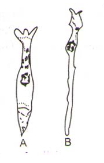
Examine the diagram of the two cell types $A$ and $B$ given below and select the correct option.
A
Cell $-A$ is the rod cell found evenly all over retina.
B
Cell $-A$ is the cone cell more concentrated in the fovea centralis.
C
Cell $-B$ is concerned with colour vision in bright light.
D
Cell $-A$ is sensitive to low light intensities.
Solution
(B) Cell $-A$ is the cone cell,which is more concentrated in the fovea centralis,the region of keenest vision. It is located in the centre of the retina,in direct line with the centre of the lens and cornea. The acuity of an animal's eye depends on the density of cones in the fovea.
Cell $-B$ is the rod cell found at the peripheral parts of the retina. Rods are high sensitivity receptors for dim light.
Cell $-B$ is the rod cell found at the peripheral parts of the retina. Rods are high sensitivity receptors for dim light.
0 likes
View Solution265
EasyMCQ
In human eyes,colour perception is done by
A
Choroid layer cells
B
Rod cells only
C
Cone cells only
D
Both $(A)$ and $(B)$
Solution
(C) The retina is the innermost neurosensory layer of the eye where images are formed. It contains two types of photoreceptor cells: rods and cones.
$1$. Rod cells are sensitive to low light intensity and are responsible for scotopic (dim light) vision,providing black and white images.
$2$. Cone cells are sensitive to high light intensity and are responsible for photopic (bright light) vision and colour perception.
Therefore,cone cells are specifically responsible for the perception and differentiation of colours in human eyes.
0 likes
View Solution266
MediumMCQ
Which of the following statements is correct regarding the pupil of the human eye?
$(I)$. It is the aperture surrounded by the iris.
$(II)$. The diameter of the pupil is regulated by the muscle fibres of the iris.
$(III)$. It is a transparent crystalline structure attached to the ciliary body.
The correct option is:
$(I)$. It is the aperture surrounded by the iris.
$(II)$. The diameter of the pupil is regulated by the muscle fibres of the iris.
$(III)$. It is a transparent crystalline structure attached to the ciliary body.
The correct option is:
A
Only $(I)$
B
Only $(III)$
C
$(I)$ and $(II)$
D
$(I, II)$ and $(III)$
Solution
(C) The middle layer of the eye is the choroid,which becomes thick on the anterior side to form the ciliary body.
The ciliary body continues forward to form the iris.
In front of the lens,the aperture surrounded by the iris is called the pupil.
Statement $(I)$ is correct as the pupil is the aperture surrounded by the iris.
Statement $(II)$ is correct because the iris contains smooth muscle fibres (sphincter pupillae and dilator pupillae) that regulate the diameter of the pupil.
Statement $(III)$ is incorrect because the transparent crystalline structure attached to the ciliary body is the lens,not the pupil.
Therefore,statements $(I)$ and $(II)$ are correct.
0 likes
View Solution267
MediumMCQ
Which of the following is not correct for rods?
$(I)$. Twilight vision is the function of the rods
$(II)$. It is responsible for daylight vision sometimes
$(III)$. The rods contain a protein called rhodopsin
$(IV)$. Rods are photoreceptor cells
Choose the correct option
$(I)$. Twilight vision is the function of the rods
$(II)$. It is responsible for daylight vision sometimes
$(III)$. The rods contain a protein called rhodopsin
$(IV)$. Rods are photoreceptor cells
Choose the correct option
A
Only $(I)$
B
Only $(II)$
C
$(I)$ and $(III)$
D
$(II)$ and $(III)$
Solution
(B) Rod cells are specialized photoreceptor cells in the retina that function in low-light conditions,providing twilight or scotopic vision.
Statement $(I)$ is correct as rods are responsible for twilight vision.
Statement $(II)$ is incorrect because rods are not responsible for daylight vision; daylight vision (photopic vision) and color vision are the functions of cone cells.
Statement $(III)$ is correct because rods contain the photopigment rhodopsin (visual purple),which is sensitive to low light.
Statement $(IV)$ is correct as rods are indeed photoreceptor cells.
Since the question asks for the statement that is not correct,only statement $(II)$ is incorrect.
0 likes
View Solution268
MediumMCQ
Which of the following statements are correct for the iris?
$(I)$. The ciliary body extends forward to form the iris.
$(II)$. It is a pigmented and opaque structure.
$(III)$. It is the visible coloured portion of the eye.
Choose the correct option.
$(I)$. The ciliary body extends forward to form the iris.
$(II)$. It is a pigmented and opaque structure.
$(III)$. It is the visible coloured portion of the eye.
Choose the correct option.
A
$(I)$ and $(II)$
B
$(I)$ and $(III)$
C
$(II)$ and $(III)$
D
$(I), (II)$ and $(III)$
Solution
(D) The correct answer is $(I), (II)$ and $(III)$.
$(I)$ The ciliary body is a forward extension of the choroid layer,which further continues to form the iris.
$(II)$ The iris is a pigmented,opaque,and circular diaphragm that controls the size of the pupil.
$(III)$ The iris is the visible coloured part of the eye that gives the eye its characteristic colour.
0 likes
View Solution269
MediumMCQ
Which of the following is correct for lens focusing while seeing distant objects?
A
Tightly stretched suspensory ligament and rounded lens
B
Contracted ciliary muscles and rounded lens
C
Relaxed ciliary muscles and tightly stretched suspensory ligament
D
Contracted ciliary muscles and relaxed suspensory ligaments
Solution
(C) Human eyes have a remarkable power of accommodation by changing the convexity of the lens.
Due to the action of the muscles of the ciliary body and the suspensory ligaments,the focal length of the lens can be changed.
This allows objects to be focused at different distances.
For the accommodation of distant objects,the ciliary muscles relax,which causes the suspensory ligaments to become tightly stretched,resulting in a flatter lens.
0 likes
View Solution270
EasyMCQ
Which layer of the wall of the eyeball looks bluish in color?
A
Sclera
B
Cornea
C
Choroid
D
Retina
Solution
(C) The $Choroid$ is the middle layer of the eyeball wall,located between the $Retina$ and the $Sclera$.
It is highly vascular and appears bluish in color due to the presence of a large amount of pigment (melanin).
The primary function of the $Choroid$ is to prevent internal reflection of light within the eye,which ensures that the image formed on the $Retina$ is sharp and not blurred.
It is highly vascular and appears bluish in color due to the presence of a large amount of pigment (melanin).
The primary function of the $Choroid$ is to prevent internal reflection of light within the eye,which ensures that the image formed on the $Retina$ is sharp and not blurred.
0 likes
View Solution271
EasyMCQ
The eye lens is held in place by:
A
Muscle fibres of the iris
B
Ligaments attached to the ciliary body
C
$A$ transparent gel called vitreous humor
D
Thin watery fluid called aqueous humor
Solution
(B) The eye lens is held in its position by the suspensory ligaments,which are attached to the ciliary body. These ligaments adjust the shape of the lens to focus light on the retina.
0 likes
View Solution272
EasyMCQ
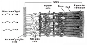
The order of three layers of cells in the retina from inside to outside is
A
Bipolar cells $\rightarrow$ Ganglion cells $\rightarrow$ Photoreceptor cells
B
Photoreceptor cells $\rightarrow$ Bipolar cells $\rightarrow$ Ganglion cells
C
Ganglion cells $\rightarrow$ Bipolar cells $\rightarrow$ Photoreceptor cells
D
Photoreceptor cells $\rightarrow$ Ganglion cells $\rightarrow$ Bipolar cells
Solution
(C) The retina consists of three layers of neural cells from the inside (towards the vitreous humor) to the outside (towards the choroid/pigmented epithelium).
These layers are:
$1$. Ganglion cells (innermost layer)
$2$. Bipolar cells (middle layer)
$3$. Photoreceptor cells (outermost layer,consisting of rods and cones).
Therefore,the correct order from inside to outside is Ganglion cells $\rightarrow$ Bipolar cells $\rightarrow$ Photoreceptor cells.
These layers are:
$1$. Ganglion cells (innermost layer)
$2$. Bipolar cells (middle layer)
$3$. Photoreceptor cells (outermost layer,consisting of rods and cones).
Therefore,the correct order from inside to outside is Ganglion cells $\rightarrow$ Bipolar cells $\rightarrow$ Photoreceptor cells.
0 likes
View Solution273
EasyMCQ
The retinal blood vessels enter the eye at
A
Fovea
B
Blind spot
C
Macula lutea
D
Crista
Solution
(B) The retinal blood vessels enter the eye and the optic nerve leaves the eye at the blind spot.
Photoreceptor cells are absent at the blind spot,hence this region is known as the blind spot.
Photoreceptor cells are absent at the blind spot,hence this region is known as the blind spot.
0 likes
View Solution274
EasyMCQ
The visual acuity is the greatest at
A
Fovea
B
Blind spot
C
Pupil
D
Ciliary body
Solution
(A) The fovea is a thinned-out portion of the retina where only cones are densely packed.
As a result,the fovea is the point where the visual acuity (resolution) is the greatest and the vision is the sharpest.
As a result,the fovea is the point where the visual acuity (resolution) is the greatest and the vision is the sharpest.
0 likes
View Solution275
MediumMCQ
Which of the following results in the generation of potential differences in the photoreceptor cells of eyes?
A
Conversion of retinal into opsin
B
Conversion of opsin into retinal
C
Dissociation of the retinal from opsin
D
Dissociation of the opsin from retinol
Solution
(C) The generation of potential differences in photoreceptor cells is triggered by the dissociation of retinal from opsin.
When light strikes the rhodopsin pigment,it causes a conformational change that leads to the splitting of retinal from the protein opsin.
This structural change results in the bleaching of the photopigment and simultaneously triggers the generation of nerve impulses in the associated ganglion cells.
0 likes
View Solution276
MediumMCQ
When the object is at a distance of more than $6 \text{ metres}$,at that time
A
Ciliary muscles are fully contracted
B
Convexity of lens is maximum
C
Eyes are fully relaxed
D
All of these
Solution
(C) When an object is at a distance of more than $6 \text{ metres}$,i.e.,when we have to see a distant object,the ciliary muscles are relaxed.
As a result,the suspensory ligaments are pulled tight,which leads to a decrease in the convexity of the lens.
In this state,the eyes are considered to be fully relaxed as there is no active accommodation required.
As a result,the suspensory ligaments are pulled tight,which leads to a decrease in the convexity of the lens.
In this state,the eyes are considered to be fully relaxed as there is no active accommodation required.
0 likes
View Solution277
EasyMCQ
In old age,the vision of the eye becomes dim. It is due to
A
Myopia
B
Hypermetropia
C
Cataract
D
Astigmatism
Solution
(C) Cataract is a condition,more prevalent in old age,which is due to an increase in the opacity of the lens.
$Myopia$: The eyeball is anterio-posteriorly elongated (short-sightedness).
$Hypermetropia$: The eyeball is anterio-posteriorly shortened (farsightedness).
$Astigmatism$: This disorder is due to the irregular curvature of the cornea or lens.
Cataract is the most common cause of vision loss in people over age $40$ and is a principal cause of blindness in the world.
0 likes
View Solution278
MediumMCQ
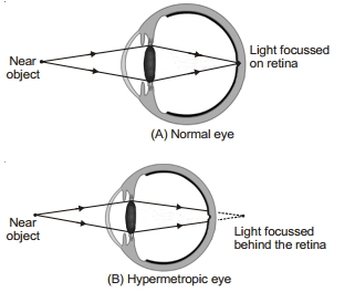
In hypermetropia,the image is formed
A
Before retina and is corrected by convex lens
B
Behind retina and is corrected by convex lens
C
Before retina and is corrected by concave lens
D
Behind retina and is corrected by concave lens
Solution
(B) In hypermetropia (farsightedness),the eyeball is anterio-posteriorly shortened,causing the image of nearby objects to be formed behind the retina. This condition is corrected by using a convex lens (converging lens),which helps to focus the light rays onto the retina.
0 likes
View Solution279
MediumMCQ
In $presbyopia$:
A
The eye ball becomes short
B
The lens becomes opaque
C
The retina gets damaged
D
Diminution of accommodation of lens due to loss of elasticity
Solution
(D) $Presbyopia$ is a vision defect commonly occurring in old age due to the loss of elasticity of the lens and reduced power of accommodation.
As a person ages,structural changes occur within the proteins of the lens,making it harder and less flexible over time.
Additionally,the ciliary muscles surrounding the lens weaken,further reducing the eye's ability to focus on near objects.
As a person ages,structural changes occur within the proteins of the lens,making it harder and less flexible over time.
Additionally,the ciliary muscles surrounding the lens weaken,further reducing the eye's ability to focus on near objects.
0 likes
View Solution280
MediumMCQ
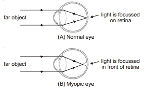
In myopia,light rays from far-off objects converge:
A
Behind the retina
B
In front of the retina
C
On the retina
D
In the retina
Solution
(B) Myopia (nearsightedness) is a vision defect in which a person can clearly see nearby objects but cannot see distant objects clearly.
In a myopic eye,the light rays from distant objects converge in front of the retina instead of on the retina.
This occurs due to the elongation of the eyeball (increase in the antero-posterior axis) or excessive curvature of the cornea.
It is corrected by using a concave lens (diverging lens),which helps to shift the image back onto the retina.
0 likes
View Solution281
EasyMCQ
Overproduction of aqueous humour results in
A
Astigmatism
B
Fovea centralis
C
Macula lutea or yellow spot
D
Glaucoma
Solution
(D) Glaucoma is a condition caused by increased intraocular pressure $(IOP)$.
This increase in pressure occurs either due to the overproduction of aqueous humour or the blockage of its drainage canal.
This elevated pressure leads to damage of the optic nerve,which can result in vision loss.
This increase in pressure occurs either due to the overproduction of aqueous humour or the blockage of its drainage canal.
This elevated pressure leads to damage of the optic nerve,which can result in vision loss.
0 likes
View Solution282
EasyMCQ
Short-sightedness or myopic vision is corrected by wearing:
A
Convex lenses
B
Concave lenses
C
Convex mirrors
D
Concave mirrors
Solution
(B) Short-sightedness or myopia is a condition in which the image of a distant object is formed in front of the retina instead of on the retina.
This defect is corrected by using a diverging lens,which is a concave lens,to shift the image back onto the retina.
This defect is corrected by using a diverging lens,which is a concave lens,to shift the image back onto the retina.
0 likes
View Solution283
MediumMCQ
$A$: Sharpest vision is in fovea centralis.
$R$: The relationship of receptor to bipolar cells to ganglion cells is $1:1:1$ within fovea centralis.
$R$: The relationship of receptor to bipolar cells to ganglion cells is $1:1:1$ within fovea centralis.
A
Assertion and Reason both are correct and Reason is the correct explanation of Assertion.
B
Assertion and Reason both are correct but Reason is not the correct explanation of Assertion.
C
Assertion is correct,but Reason is incorrect.
D
Both Assertion and Reason are incorrect.
Solution
(A) Fovea centralis is the area of the most acute vision on the retina.
In this region,the ratio of photoreceptor cells (cones) to bipolar cells to ganglion cells is $1:1:1$.
This specific arrangement allows for high visual acuity because each cone cell has a dedicated pathway to the brain,minimizing signal convergence.
Therefore,the Assertion is correct and the Reason provides the correct explanation for it.
0 likes
View Solution284
MediumMCQ
$A$: Retina is arranged anatomically in reverse order from what might be expected.
$R$: The receptor cells are towards the outside and ganglionic cells towards the inside,and light must pass through the nerve cells to reach them.
$R$: The receptor cells are towards the outside and ganglionic cells towards the inside,and light must pass through the nerve cells to reach them.
A
Assertion and Reason both are correct and Reason is the correct explanation of Assertion.
B
Assertion and Reason both are correct but Reason is not the correct explanation of Assertion.
C
Assertion is correct,but Reason is incorrect.
D
Both Assertion and Reason are incorrect.
Solution
(A) The retina is anatomically arranged in a reverse order.
In the vertebrate retina,the photoreceptor cells (rods and cones) are located at the back of the retina,furthest from the incoming light.
Light must pass through the layers of ganglion cells,bipolar cells,and nerve fibers before reaching the photoreceptor cells.
Therefore,both the Assertion and the Reason are correct,and the Reason provides the correct explanation for the Assertion.
0 likes
View Solution285
EasyMCQ
Ishihara chart is used to detect:
A
Tuberculosis
B
Eye sight
C
Colour blindness
D
Diabetes
Solution
(C) The Ishihara chart is a color perception test for red-green color deficiencies. It consists of a number of colored plates,called Ishihara plates,each of which contains a circle of dots appearing randomized in color and size. Within the pattern are dots which form a number or shape clearly visible to those with normal color vision,and invisible,or difficult to see,to those with a red-green color vision defect. Therefore,it is used to detect colour blindness.
0 likes
View Solution286
EasyMCQ
In which part of the sclera blood capillaries are absent?
A
Cornea
B
Choroid
C
Retina
D
Iris
Solution
(A) The sclera is the opaque,fibrous outer layer of the eye. The anterior portion of the sclera is modified into a transparent structure called the cornea. The cornea is avascular,meaning it lacks blood capillaries,which allows light to pass through it without obstruction.
0 likes
View Solution287
EasyMCQ
What is the ciliary body?
A
$A$ part of the retina that detects light.
B
$A$ thickened anterior part of the choroid layer.
C
$A$ structure that controls the size of the pupil.
D
The transparent layer covering the front of the eye.
Solution
(B) The choroid layer is thin over the posterior two-thirds of the eyeball,but it becomes thick in the anterior part to form the ciliary body. The ciliary body holds the lens in place and controls its shape for focusing.
0 likes
View Solution288
EasyMCQ
What is the function of cone cells?
A
Daylight vision and colour vision
B
Twilight vision and colour vision
C
Daylight vision and black-and-white vision
D
Twilight vision and black-and-white vision
Solution
(A) Cone cells contain light-sensitive proteins known as photopigments.
Daylight (photopic) vision and colour vision are the primary functions of cone cells.
In contrast,twilight (scotopic) vision is the function of rod cells.
Daylight (photopic) vision and colour vision are the primary functions of cone cells.
In contrast,twilight (scotopic) vision is the function of rod cells.
0 likes
View Solution289
EasyMCQ
Cone cells are sensitive to which main colors?
A
Red,green,and blue
B
Red,yellow,and blue
C
Green,yellow,and blue
D
Red,green,and yellow
Solution
(A) Cone cells are photoreceptor cells in the retina of the eye that are responsible for color vision.
There are three types of cone cells in the human eye,each containing a different photopigment that is sensitive to specific wavelengths of light.
These three types of cone cells are primarily sensitive to red,green,and blue light.
There are three types of cone cells in the human eye,each containing a different photopigment that is sensitive to specific wavelengths of light.
These three types of cone cells are primarily sensitive to red,green,and blue light.
0 likes
View Solution290
Medium
What are the blind spot and the fovea?
Solution
(N/A) - The $blind \ spot$ is a small area on the retina where the optic nerve exits the eye. Photoreceptor cells (rods and cones) are absent in this region,so no image is formed here.
- The $fovea$ is a central pit located within the $macula \ lutea$,a yellowish pigmented spot found at the posterior pole of the eye,lateral to the blind spot. It contains a high density of cones and is responsible for the sharpest visual acuity.
- The $fovea$ is a central pit located within the $macula \ lutea$,a yellowish pigmented spot found at the posterior pole of the eye,lateral to the blind spot. It contains a high density of cones and is responsible for the sharpest visual acuity.
0 likes
View Solution291
EasyMCQ
Photosensitive compounds are formed of which substances?
A
Opsin and Retinol
B
Opsin and Retinal
C
Rhodopsin and Retinol
D
Opsin and Vitamin $A$
Solution
(B) Photosensitive compounds (photopigments) in the human eyes are composed of opsin (a protein) and retinal (an aldehyde of vitamin $A$).
These two components combine to form the light-sensitive pigments necessary for vision.
These two components combine to form the light-sensitive pigments necessary for vision.
0 likes
View Solution292
EasyMCQ
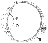
The following figure is a diagram showing parts of the human eye. Identify $P, Q$,and $R$ from the options given below.
$\quad\quad P\quad\quad\quad Q\quad\quad\quad R$
$\quad\quad P\quad\quad\quad Q\quad\quad\quad R$
A
Iris $\quad$ $\quad$ Lens $\quad$ $\quad$ Retina
B
Iris $\quad$ $\quad$ Lens $\quad$ $\quad$ Choroid
C
Lens $\quad$ $\quad$ Iris $\quad$ $\quad$ Sclera
D
Lens $\quad$ $\quad$ Iris $\quad$ $\quad$ Choroid
Solution
(D) Based on the anatomical structure of the human eye:
$P$ points to the crystalline lens,which is a transparent,biconvex structure.
$Q$ points to the iris,which is the visible colored portion of the eye that controls the size of the pupil.
$R$ points to the choroid,which is the middle vascular layer of the eye wall that contains many blood vessels.
Therefore,the correct identification is $P = \text{Lens}, Q = \text{Iris}, R = \text{Choroid}$.
This corresponds to option $D$.
0 likes
View Solution293
EasyMCQ
The anterior portion of the sclera is called the .......
A
Choroid
B
Retina
C
Cornea
D
Lens
Solution
(C) The wall of the human eye consists of three layers. The external layer is composed of a dense connective tissue and is called the sclera. The anterior portion of this layer is called the cornea. The cornea is transparent and allows light to enter the eye.
0 likes
View Solution294
EasyMCQ
The choroid layer is thin over the $.......$ of the eyeball.
A
Posterior $2/3$ of the eyeball
B
Anterior $2/3$ of the eyeball
C
Posterior $1/3$ of the eyeball
D
Anterior $1/3$ of the eyeball
Solution
(A) The choroid is the middle layer of the eyeball,which is bluish in color.
It contains many blood vessels and is thin over the posterior two-thirds of the eyeball.
However,it becomes thick in the anterior part to form the ciliary body.
It contains many blood vessels and is thin over the posterior two-thirds of the eyeball.
However,it becomes thick in the anterior part to form the ciliary body.
0 likes
View Solution295
EasyMCQ
Identify the correct sequence of cells in the retina from the outside to the inside.
A
Photoreceptor cells $\rightarrow$ Ganglion cells $\rightarrow$ Bipolar cells
B
Bipolar cells $\rightarrow$ Ganglion cells $\rightarrow$ Photoreceptor cells
C
Ganglion cells $\rightarrow$ Bipolar cells $\rightarrow$ Photoreceptor cells
D
Photoreceptor cells $\rightarrow$ Bipolar cells $\rightarrow$ Ganglion cells
Solution
(D) The retina consists of three layers of neural cells from the outside to the inside:
$1$. Photoreceptor cells (rods and cones) are located at the outermost layer.
$2$. Bipolar cells are located in the middle layer.
$3$. Ganglion cells are located at the innermost layer.
Therefore,the sequence from outside to inside is: Photoreceptor cells $\rightarrow$ Bipolar cells $\rightarrow$ Ganglion cells.
0 likes
View Solution296
EasyMCQ
The colored visible part of the eye is known as:
A
Pupil
B
Iris
C
Lens
D
Cornea
Solution
(B) The eye contains a visible colored portion known as the $Iris$. The $Iris$ is a pigmented,opaque structure that surrounds the $Pupil$. It controls the amount of light entering the eye by adjusting the size of the $Pupil$.
0 likes
View Solution297
EasyMCQ
The .......... contains many blood vessels and looks bluish in color.
A
Sclera
B
Cornea
C
Choroid
D
Retina
Solution
(C) The middle layer of the eye is called the $Choroid$.
It is rich in blood vessels and looks bluish in color.
The $Choroid$ layer is thin over the posterior two-thirds of the eyeball,but it becomes thick in the anterior part to form the ciliary body.
It is rich in blood vessels and looks bluish in color.
The $Choroid$ layer is thin over the posterior two-thirds of the eyeball,but it becomes thick in the anterior part to form the ciliary body.
0 likes
View Solution298
EasyMCQ
The choroid is thin over the posterior two-thirds of the eyeball,but it becomes thick in the anterior part to form the:
A
Lens
B
Retina
C
Pupil
D
Ciliary body
Solution
(D) The choroid is the middle layer of the eye wall,which is bluish in color.
It is thin over the posterior two-thirds of the eyeball.
However,it becomes thick in the anterior part to form the ciliary body.
The ciliary body then continues forward to form a pigmented and opaque structure called the iris,which is the visible colored portion of the eye.
It is thin over the posterior two-thirds of the eyeball.
However,it becomes thick in the anterior part to form the ciliary body.
The ciliary body then continues forward to form a pigmented and opaque structure called the iris,which is the visible colored portion of the eye.
0 likes
View Solution299
EasyMCQ
Which structure is covered by the iris?
A
Lens
B
Pupil
C
Ciliary body
D
Retina
Solution
(B) The iris is the visible colored portion of the eye. It is a pigmented and opaque structure. The iris contains a central opening called the $Pupil$. The diameter of the pupil is regulated by the muscle fibers of the iris. Therefore, the iris surrounds or covers the area around the pupil, controlling the amount of light entering the eye.
0 likes
View Solution300
MediumMCQ
Which muscles are present in the structure of the iris?
A
Smooth muscles
B
Striated muscles
C
Cardiac muscles
D
All of the above
Solution
(A) The iris is the colored part of the eye that controls the diameter and size of the pupil. It contains two types of involuntary smooth muscles: the sphincter pupillae (circular muscles) and the dilator pupillae (radial muscles). These muscles are responsible for adjusting the amount of light entering the eye. Therefore,the iris is composed of smooth muscles.
0 likes
View SolutionNeural Control and Coordination — Eye · Frequently Asked Questions
1Are these Neural Control and Coordination questions useful for JEE and NEET?
Yes. All questions in this section are mapped to JEE Main and NEET exam patterns. Previous year questions from JEE Main, NEET, GUJCET and state-level exams are included with full solutions.
2Can I switch to Hindi or Gujarati for these questions?
Yes. Use the language tabs in the hero section or the sidebar to view the same questions and solutions in English, Hindi or Gujarati.
3How do I generate a question paper from this subtopic?
Use the Vedclass Exam Paper Generator — select the chapter and subtopic, set difficulty, and generate Sets A, B, C, D automatically. First 3 chapters of every subject are free.
Vedclass Products
For Students
Vedclass Test Series
Mock tests in real JEE/NEET style with performance analysis. 5-day free trial.
Start Free TrialFor Teachers
Exam Paper Generator
Generate Set A/B/C/D papers from this chapter in 2 minutes. 3 chapters free.
Try FreeFor Institutes
Online Exam Module
Live online exams with unlimited students, 360° analytics & white-label branding.
See DemoFor Teachers & Institutes
Generate a Neural Control and Coordination Exam Paper in 2 Minutes
Select subtopic & difficulty — Sets A, B, C, D auto-generated with No Repeat logic.
First 3 chapters of every subject are free — no payment required.