
A English
Eye Questions in English
Class 11 Biology · Neural Control and Coordination · Eye
315+
Questions
English
Language
100%
With Solutions
Showing 50 of 315 questions in English
201
EasyMCQ
The middle layer of the human eye,the choroid,contains $A$ and looks $B$ in color. Choose the correct option for $A$ and $B$.
A
$A -$ blood vessels,$B -$ bluish
B
$A -$ connective tissue,$B -$ reddish
C
$A -$ bipolar cells,$B -$ blackish
D
$A -$ muscle fiber,$B -$ brownish
Solution
(A) The human eye wall is composed of three layers: the outer sclera,the middle choroid,and the inner retina.
$1$. The choroid is the middle layer and is highly vascularized,meaning it contains many blood vessels $(A)$.
$2$. Due to the presence of numerous blood vessels and pigment,the choroid appears bluish $(B)$ in color.
Therefore,the correct option is $A$.
$1$. The choroid is the middle layer and is highly vascularized,meaning it contains many blood vessels $(A)$.
$2$. Due to the presence of numerous blood vessels and pigment,the choroid appears bluish $(B)$ in color.
Therefore,the correct option is $A$.
0 likes
View Solution202
EasyMCQ
Rhodopsin is also known as visual $ \text{purple} $.
A
Green
B
Yellow
C
Brown
D
Purple
Solution
(D) Rhodopsin is a biological pigment found in the rods of the retina. It is a $G$-protein-coupled receptor that is extremely sensitive to light. Because of its characteristic color, it is commonly referred to as visual $ \text{purple} $.
0 likes
View Solution203
EasyMCQ
$X -$ Rod cells' function is twilight (scotopic) vision.
$Y -$ Cone cells' function is daylight (photopic) vision.
$Y -$ Cone cells' function is daylight (photopic) vision.
A
$X$ and $Y$ are incorrect.
B
$X$ is correct,$Y$ is incorrect.
C
$X$ is incorrect,$Y$ is correct.
D
$X$ and $Y$ are correct.
Solution
(D) The human eye contains two types of photoreceptor cells: rods and cones.
$1$. Rod cells contain the pigment rhodopsin and are highly sensitive to low light intensities,making them responsible for twilight or scotopic vision.
$2$. Cone cells contain photopsins and are responsible for color vision and daylight or photopic vision.
Since both statements $X$ and $Y$ accurately describe the physiological functions of these cells,the correct option is $D$.
0 likes
View Solution204
EasyMCQ
Which of these cells are not found in the retina?
A
Bipolar cells
B
Unipolar cells
C
Photoreceptor cells
D
Ganglion cells
Solution
(B) The retina is the innermost layer of the eye and contains three layers of neural cells from inside to outside: $1.$ Ganglion cells,$2.$ Bipolar cells,and $3.$ Photoreceptor cells (rods and cones).
Unipolar cells are typically found in sensory ganglia of the peripheral nervous system,not in the retina.
Therefore,unipolar cells are not found in the retina.
Unipolar cells are typically found in sensory ganglia of the peripheral nervous system,not in the retina.
Therefore,unipolar cells are not found in the retina.
0 likes
View Solution205
EasyMCQ
The diameter of the pupils is regulated by the muscle fibres of ...........
A
Iris
B
Fovea
C
Sclera
D
All given
Solution
(A) The iris is the visible colored part of the eye that surrounds the pupil.
It contains two sets of smooth muscle fibres: the circular sphincter pupillae and the radial dilator pupillae.
These muscles contract and relax to adjust the size of the pupil,thereby regulating the amount of light entering the eye.
It contains two sets of smooth muscle fibres: the circular sphincter pupillae and the radial dilator pupillae.
These muscles contract and relax to adjust the size of the pupil,thereby regulating the amount of light entering the eye.
0 likes
View Solution206
MediumMCQ
What is correct for fovea?
A
Thinned out portion of retina
B
Only cones are densely packed here
C
Resolution is the greatest
D
All correct
Solution
(D) The fovea is a small,central pit in the retina of the eye where visual acuity is highest.
$1$. It is a thinned-out portion of the retina where the inner layers of the retina are pushed aside,allowing light to fall directly on the photoreceptors.
$2$. It contains only cones,which are densely packed,providing high color sensitivity and sharp vision.
$3$. Because of the high density of cones and the direct path of light,the visual resolution (acuity) is the greatest at the fovea.
Therefore,all the given statements are correct.
0 likes
View Solution207
EasyMCQ
Rhodopsin is a ...... coloured protein.
A
Green
B
Yellow
C
Purplish-red
D
Blue
Solution
(C) Rhodopsin,also known as visual purple,is a biological pigment found in the rod cells of the retina. It is a $G$-protein-coupled receptor that is extremely sensitive to light. Due to its specific light-absorbing properties,it appears purplish-red in colour. Therefore,the correct option is $C$.
0 likes
View Solution208
EasyMCQ
Macula lutea is present in.
A
Iris
B
Retina
C
Vitreous chamber
D
Tympanic Membrane
Solution
(B) The $Macula \text{ } lutea$ is a yellowish pigmented spot located at the posterior pole of the eye, specifically on the $Retina$.
It contains a central pit called the $fovea \text{ } centralis$, which is responsible for the highest visual acuity and color vision.
Therefore, the correct location of the $Macula \text{ } lutea$ is the $Retina$.
It contains a central pit called the $fovea \text{ } centralis$, which is responsible for the highest visual acuity and color vision.
Therefore, the correct location of the $Macula \text{ } lutea$ is the $Retina$.
0 likes
View Solution209
EasyMCQ
It is an aldehyde of Vitamin $- A$.
A
Retinal
B
Opsin
C
Rhodopsin
D
All given
Solution
(A) Vitamin $- A$ (retinol) is converted into its aldehyde form,known as retinal,in the human eye.
Retinal combines with the protein opsin to form rhodopsin (visual purple),which is the light-sensitive pigment found in the rod cells of the retina.
Therefore,retinal is the aldehyde form of Vitamin $- A$.
Retinal combines with the protein opsin to form rhodopsin (visual purple),which is the light-sensitive pigment found in the rod cells of the retina.
Therefore,retinal is the aldehyde form of Vitamin $- A$.
0 likes
View Solution210
EasyMCQ
Which of the following are functions of the cone cells of the retina?
$1.$ Photopic vision
$2.$ Colour vision
$3.$ Scotopic vision
$1.$ Photopic vision
$2.$ Colour vision
$3.$ Scotopic vision
A
Only $2$
B
$1, 3$
C
$2, 3$
D
$1, 2$
Solution
(D) The retina contains two types of photoreceptor cells: rods and cones.
$1.$ Cone cells are responsible for daylight (photopic) vision and colour vision.
$2.$ Rod cells contain the pigment rhodopsin and are responsible for twilight (scotopic) vision.
Therefore,statements $1$ and $2$ are functions of cone cells,while statement $3$ is a function of rod cells.
Thus,the correct option is $D$.
0 likes
View Solution211
EasyMCQ
Which of the following is not a part of the human eye?
A
Ciliary body
B
Blind spot
C
Scala media
D
Iris
Solution
(C) The human eye consists of several structures including the cornea,iris,pupil,lens,ciliary body,retina,and blind spot.
$A$. The ciliary body is a part of the eye that controls the shape of the lens.
$B$. The blind spot is the area on the retina where the optic nerve exits the eye.
$C$. The scala media (also known as the cochlear duct) is a fluid-filled cavity within the cochlea of the inner ear,not the eye.
$D$. The iris is the colored part of the eye that controls the size of the pupil.
Therefore,the scala media is not a part of the eye.
0 likes
View Solution212
EasyMCQ
The space between the cornea and the lens is called...
A
Vitreous chamber
B
Aqueous chamber
C
Scala vestibuli
D
Scala tympani
Solution
(B) The human eye contains two main chambers filled with fluid.
$1$. The space between the cornea and the lens is known as the $Aqueous$ $chamber$,which is filled with a thin,watery fluid called $Aqueous$ $humor$.
$2$. The space between the lens and the retina is known as the $Vitreous$ $chamber$,which is filled with a transparent gel called $Vitreous$ $humor$.
$3$. $Scala$ $vestibuli$ and $Scala$ $tympani$ are parts of the inner ear,specifically the cochlea,and are not related to the eye structure.
Therefore,the correct option is $B$.
0 likes
View Solution213
EasyMCQ
Acute vision is found in ................
A
Eagle
B
Shark
C
Bat
D
Frog
Solution
(A) Acute vision or high visual acuity is a characteristic feature of birds of prey, such as the $Eagle$.
These birds possess a high density of photoreceptor cells (cones) in their retina, specifically in the fovea, which allows them to detect small prey from great distances with high precision.
These birds possess a high density of photoreceptor cells (cones) in their retina, specifically in the fovea, which allows them to detect small prey from great distances with high precision.
0 likes
View Solution214
EasyMCQ
The sensory pigmented layer of the eye is known as:
A
Sclera
B
Retina
C
Choroid
D
Pupil
Solution
(B) The wall of the human eye is composed of three layers:
$1$. The external layer is the sclera,which is composed of dense connective tissue.
$2$. The middle layer is the choroid,which contains many blood vessels and looks bluish in color. It is the pigmented layer.
$3$. The inner layer is the retina,which contains three layers of cells: ganglion cells,bipolar cells,and photoreceptor cells (rods and cones). The retina is the sensory layer of the eye.
Therefore,the sensory layer of the eye is the retina.
0 likes
View Solution215
EasyMCQ
The pupil is a part of the ........
A
Iris
B
Choroid
C
Choroid and Retina
D
Iris and Choroid
Solution
(A) The eye consists of three layers: the outer sclera,the middle choroid,and the inner retina.
The iris is the visible colored portion of the eye,which is a continuation of the choroid.
The pupil is the aperture or opening located in the center of the iris.
Therefore,the pupil is functionally and structurally associated with the iris,which is part of the middle layer (uvea) of the eye.
The iris is the visible colored portion of the eye,which is a continuation of the choroid.
The pupil is the aperture or opening located in the center of the iris.
Therefore,the pupil is functionally and structurally associated with the iris,which is part of the middle layer (uvea) of the eye.
0 likes
View Solution216
EasyMCQ
The amount of light entering the eye is regulated by the:
A
Pupil
B
Aperture
C
Cornea
D
Lens
Solution
(A) The $Pupil$ is a circular aperture in the center of the $Iris$. The $Iris$ contains muscles that contract or relax to change the size of the $Pupil$,thereby regulating the amount of light that enters the eye. When light intensity is high,the $Pupil$ constricts,and when light intensity is low,the $Pupil$ dilates.
0 likes
View Solution217
EasyMCQ
The function of the pupil is:
A
To move the lens forward and backward.
B
To regulate the amount of light entering the eye.
C
To induce movement of the eyelids.
D
To change the size of the eyeball.
Solution
(B) The pupil is the aperture located in the center of the iris of the eye. Its primary function is to regulate the amount of light that enters the eye. In bright light,the iris muscles contract to make the pupil smaller,reducing light entry. In dim light,the iris muscles relax to make the pupil larger,allowing more light to enter.
0 likes
View Solution218
EasyMCQ
The area of the retina that is most sensitive to light is the:
A
Optic disc
B
Sclera
C
Macula lutea
D
Fovea centralis
Solution
(D) The $Fovea$ $centralis$ is a small, central pit in the $Macula$ $lutea$ of the retina.
It contains the highest density of cone cells and is responsible for sharp, detailed central vision.
Because it lacks rod cells and has the highest concentration of cones, it is the area of the retina that is most sensitive to light and provides the highest visual acuity.
It contains the highest density of cone cells and is responsible for sharp, detailed central vision.
Because it lacks rod cells and has the highest concentration of cones, it is the area of the retina that is most sensitive to light and provides the highest visual acuity.
0 likes
View Solution219
MediumMCQ
Corneal transplantation is highly successful because:
A
The cornea can be easily preserved.
B
The cornea is not connected to blood vessels and the immune system.
C
The procedure is very simple.
D
The cornea is easily available.
Solution
(B) The cornea is an avascular tissue,meaning it lacks blood vessels. Because it is not directly connected to the circulatory system,it is not easily accessed by the immune system's cells. This lack of direct immune surveillance makes the cornea an 'immune-privileged' site,significantly reducing the risk of transplant rejection and making corneal transplantation highly successful.
0 likes
View Solution220
EasyMCQ
The light-sensitive chemical substance for vision in mammals is called:
A
Sclerotin
B
Retinal
C
Rhodopsin
D
Melanin
Solution
(C) In mammals,the light-sensitive pigments in the retina are known as photopigments. These consist of an opsin (a protein) and retinal (an aldehyde of vitamin $A$). The most well-known photopigment in the rod cells of the retina is $Rhodopsin$ (also known as visual purple). When light strikes the retina,it causes the dissociation of retinal from opsin,which triggers the generation of action potentials in the ganglion cells,leading to vision. Therefore,the correct answer is $Rhodopsin$.
0 likes
View Solution221
MediumMCQ
If a person has a deficiency of the visual pigment $Rhodopsin$, they are advised to consume more of which of the following?
A
Carrots and potatoes
B
Apples and grapes
C
Carrots and ripe papayas
D
Guavas and ripe bananas
Solution
(C) $Rhodopsin$ (also known as visual purple) is a biological pigment found in the rod cells of the retina that is responsible for vision in low-light conditions.
$Rhodopsin$ is composed of a protein called $Opsin$ and a derivative of $Vitamin A$ called $Retinal$.
$A$ deficiency of $Vitamin A$ leads to a reduced synthesis of $Rhodopsin$, which causes night blindness (nyctalopia).
Carrots and ripe papayas are rich sources of $\beta$-carotene, which is a precursor to $Vitamin A$.
Therefore, consuming these foods helps in the synthesis of $Rhodopsin$ and improves vision in low light.
0 likes
View Solution222
MediumMCQ
Which of the following is a characteristic of the human cornea?
A
It is secreted by the conjunctiva and glandular tissue.
B
It is a lacrimal gland that secretes tears.
C
Blood circulation is absent in the cornea.
D
It becomes opaque and develops a white layer with age, which causes cataracts.
Solution
(C) The human cornea is the transparent anterior part of the eye.
It is unique because it is avascular, meaning it lacks blood vessels $(blood \text{ circulation is absent})$.
Oxygen is obtained directly from the atmosphere through the tear film.
Option $A$ is incorrect as the cornea is not a secretion.
Option $B$ is incorrect as the lacrimal gland is separate from the cornea.
Option $D$ describes the lens, not the cornea.
0 likes
View Solution223
MediumMCQ
Which of the following is a correct difference between the rod cells and cone cells of our retina?
A
Visual acuity: High || Low
B
Visual pigments: Iodopsin || Rhodopsin
C
Overall function: Sensitive to dim light || Color vision and bright light vision
D
Distribution: Concentrated in the fovea || Distributed throughout the retina
Solution
(C) Rod cells contain the pigment $Rhodopsin$ and are highly sensitive to dim light (scotopic vision),but they do not provide color vision. They are distributed throughout the retina. Cone cells contain the pigment $Iodopsin$ and are responsible for color vision and high visual acuity in bright light (photopic vision). They are highly concentrated in the fovea centralis of the retina. Therefore,option $C$ correctly describes the functional difference between these two photoreceptor cells.
0 likes
View Solution224
MediumMCQ
In humans,corneal transplants are rarely rejected because,
A
its cells are less susceptible to bacterial invasion.
B
it does not have a blood supply.
C
it is composed of enucleated cells.
D
it is a non-living layer.
Solution
(B) The cornea is the transparent anterior part of the eye.
It is an avascular structure,meaning it lacks a direct blood supply.
Since the immune system's cells (like lymphocytes) reach tissues primarily through the bloodstream,the lack of blood vessels in the cornea makes it an 'immune-privileged' site.
Therefore,the body's immune system does not easily recognize or attack the transplanted corneal tissue,leading to a very low rate of rejection.
0 likes
View Solution225
EasyMCQ
The purple-red protein rhodopsin,which is present in the light-sensitive rod cells of the human eye,is a derivative of:
A
Vitamin-$B$
B
Vitamin-$C$
C
Vitamin-$D$
D
Vitamin-$A$
Solution
(D) Rhodopsin (also known as visual purple) is a biological pigment found in the rod cells of the retina.
It is a $G$-protein-coupled receptor that is extremely sensitive to light.
Rhodopsin is composed of a protein called opsin and a light-absorbing chromophore called retinal.
Retinal is an aldehyde derivative of Vitamin-$A$ (retinol).
Therefore,the correct answer is Vitamin-$A$.
It is a $G$-protein-coupled receptor that is extremely sensitive to light.
Rhodopsin is composed of a protein called opsin and a light-absorbing chromophore called retinal.
Retinal is an aldehyde derivative of Vitamin-$A$ (retinol).
Therefore,the correct answer is Vitamin-$A$.
0 likes
View Solution226
MediumMCQ
The figure shows parts of the human eye labeled as $A, B, C$ and $D$. Select the option which correctly identifies the labeled part with its function and characteristics.
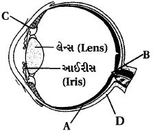
A
$D$ - Choroid - Its anterior part forms the ciliary body.
B
$A$ - Retina - Contains photoreceptor rods and cones.
C
$B$ - Blind spot - Has a very small number of rods and cones.
D
$C$ - Aqueous chamber - Reflects light that does not pass through the lens.
Solution
(B) Based on the provided diagram of the human eye:
$A$ points to the innermost layer,the retina,which contains photoreceptor cells (rods and cones).
$B$ points to the blind spot,where the optic nerve leaves the eye and photoreceptor cells are absent.
$C$ points to the ciliary body,which holds the lens in place.
$D$ points to the choroid,the middle vascular layer.
Therefore,option $B$ is the correct identification.
0 likes
View Solution227
MediumMCQ
Which of the following statements is $NOT$ correct?
A
Retinal is a light-absorbing pigment.
B
Rods in the retina contain rhodopsin pigment,while cones contain three different types of pigments.
C
Retinal is a derivative of Vitamin $C$.
D
Rhodopsin is a purplish-red protein present in rod cells.
Solution
(C) The correct answer is $C$.
Retinal is an aldehyde derivative of Vitamin $A$,not Vitamin $C$.
$A$. Retinal is indeed the light-absorbing part of the visual pigments.
$B$. Rods contain the pigment rhodopsin,and cones contain three types of photopsins (pigments) that respond to red,green,and blue light.
$D$. Rhodopsin (visual purple) is the light-sensitive pigment found in the rod cells of the retina.
0 likes
View Solution228
EasyMCQ
In the human eye,the fovea is the central point of vision,where:
A
rods are more numerous than cones.
B
cones are densely packed,but rods are absent.
C
the optic nerve exits the eye.
D
only rods are present.
Solution
(B) The fovea is a small,central pit in the retina of the eye where visual acuity is highest.
It contains a high density of cone cells,which are responsible for color vision and high-resolution detail.
Rod cells,which are responsible for vision in low light,are absent in the fovea.
Therefore,the fovea is characterized by the presence of densely packed cones and the complete absence of rods.
It contains a high density of cone cells,which are responsible for color vision and high-resolution detail.
Rod cells,which are responsible for vision in low light,are absent in the fovea.
Therefore,the fovea is characterized by the presence of densely packed cones and the complete absence of rods.
0 likes
View Solution229
MediumMCQ
Select the correct statement.
A
Photoreceptor cells in the human eye are depolarized in the dark and become hyperpolarized under the influence of light.
B
Receptor cells do not generate graded potentials.
C
Nociceptors respond to changes in pressure.
D
Meissner's corpuscles are thermoreceptors.
Solution
(A) In the human eye,photoreceptor cells (rods and cones) are in a state of depolarization in the dark,releasing neurotransmitters like glutamate. When light strikes these cells,it triggers a biochemical cascade that leads to hyperpolarization of the cell membrane,thereby reducing the release of neurotransmitters. Thus,statement $A$ is correct. Statement $B$ is incorrect because receptor cells do generate graded potentials. Statement $C$ is incorrect because nociceptors are pain receptors,not pressure receptors. Statement $D$ is incorrect because Meissner's corpuscles are mechanoreceptors that detect light touch,not thermoreceptors.
0 likes
View Solution230
EasyMCQ
What is the light-sensitive chemical present in the human eye composed of?
A
Opsin and Retinal
B
Opsin and Retinol
C
Transducin and Retinene
D
Cyanopsin and Retinol
Solution
(A) The light-sensitive pigments (photopigments) in the human eye,specifically in the rods and cones of the retina,are composed of an opsin (a protein) and retinal (an aldehyde of vitamin $A$).
When light strikes the photopigment,it causes the dissociation of retinal from opsin,resulting in changes in the structure of the opsin.
This structural change leads to the generation of action potentials in the photoreceptor cells,which are then transmitted to the brain via the optic nerve.
When light strikes the photopigment,it causes the dissociation of retinal from opsin,resulting in changes in the structure of the opsin.
This structural change leads to the generation of action potentials in the photoreceptor cells,which are then transmitted to the brain via the optic nerve.
0 likes
View Solution231
MediumMCQ
Good vision depends on adequate intake of carotene-rich food. Select the most appropriate option from the following statements.
$I.$ Vitamin $A$ derivatives are formed from carotene.
$II.$ Photopigments are embedded in the membrane discs of the outer segment.
$III.$ Retinal is a derivative of Vitamin $A$.
$IV.$ Retinal is a part of all photopigments that absorb light.
$I.$ Vitamin $A$ derivatives are formed from carotene.
$II.$ Photopigments are embedded in the membrane discs of the outer segment.
$III.$ Retinal is a derivative of Vitamin $A$.
$IV.$ Retinal is a part of all photopigments that absorb light.
A
$(I)$ and $(II)$
B
$(I), (III)$ and $(IV)$
C
$(I)$ and $(III)$
D
$(I), (II)$ and $(IV)$
Solution
(B) The correct answer is $(B)$.
$I.$ Carotene is a precursor to Vitamin $A$,which is synthesized in the body.
$II.$ Photopigments are located in the membrane discs of the outer segment of photoreceptor cells (rods and cones),not the inner segment.
$III.$ Retinal is indeed a derivative of Vitamin $A$ (specifically an aldehyde of Vitamin $A$).
$IV.$ Retinal is the light-absorbing component (chromophore) present in all photopigments (like rhodopsin and iodopsin).
Therefore,statements $(I), (III),$ and $(IV)$ are correct.
0 likes
View Solution232
EasyMCQ
The transparent lens in the human eye is held in its place by:
A
Smooth muscles attached to the ciliary body
B
Ligaments attached to the ciliary body
C
Smooth muscles attached to the iris
D
Ligaments attached to the iris
Solution
(B) The human eye contains a transparent,crystalline lens.
This lens is held in its position by suspensory ligaments.
These ligaments are attached to the ciliary body,which is an extension of the choroid.
The ciliary body contains ciliary muscles that control the shape of the lens by adjusting the tension on these ligaments,thereby facilitating accommodation for near and far vision.
0 likes
View Solution233
MediumMCQ
Which of the following statements is correct?
A
Cornea is an external,transparent and protective proteinaceous covering of the eye-ball.
B
Cornea consists of dense connective tissue of elastin and can repair itself.
C
Cornea is convex,transparent layer which is highly vascularised.
D
Cornea consists of dense matrix of collagen and is the most sensitive portion of the eye.
Solution
(D) The cornea is the anterior,transparent part of the eye-ball.
It is composed of a dense matrix of collagen fibers,which provides structural integrity and transparency.
It is avascular,meaning it lacks blood vessels,which helps maintain its transparency.
It is considered the most sensitive portion of the eye due to the presence of a high density of nerve endings.
Therefore,statement $D$ is correct.
0 likes
View Solution234
MediumMCQ
Examine the diagram of the two cell types $A$ and $B$ given below and select the correct option.
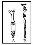
A
Cell $A$ is the rod cell found evenly all over retina.
B
Cell $A$ is the cone cell more concentrated in the fovea centralis.
C
Cell $B$ is concerned with colour vision in bright light.
D
Cell $A$ is sensitive to low light intensities.
Solution
(B) Based on the morphological structure,cell $A$ represents a cone cell,which is characterized by a conical outer segment. Cone cells are highly concentrated in the fovea centralis (yellow spot) of the retina. They are responsible for colour vision and high-resolution vision in bright light. Cell $B$ represents a rod cell,which is characterized by a cylindrical outer segment and is sensitive to low light intensities (scotopic vision) but does not provide colour vision.
0 likes
View Solution235
EasyMCQ
Which of the following is an eye disease?
A
Hepatitis
B
Measles
C
Glaucoma
D
Bronchitis
Solution
(C) Glaucoma is an eye disease characterized by increased ocular pressure within the eyeball.
It is a group of diseases of the optic nerve involving the loss of retinal ganglion cells in a characteristic pattern of optic neuropathy.
Untreated glaucoma leads to permanent damage of the optic nerve and resultant visual field loss that can progress to blindness.
$A$ (Hepatitis) is an inflammation of the liver.
$B$ (Measles) is a highly infectious viral disease that usually spreads by droplet infection.
$D$ (Bronchitis) is the inflammation of the membrane lining of the bronchial tubes.
0 likes
View Solution236
MediumMCQ
The black pigment in the eye which reduces the internal reflection is located in
A
retina
B
iris
C
cornea
D
sclerotic
Solution
(B) The middle layer of the eyeball is the choroid,which contains many blood vessels and looks bluish in color. The choroid layer is thin over the posterior two-thirds of the eyeball,but it becomes thick in the anterior part to form the ciliary body. This layer contains numerous melanocytes that produce black pigment,which absorbs light and prevents internal reflection within the eyeball.
0 likes
View Solution237
MediumMCQ
Assertion : Astigmatism is due to uneven curvature of lens.
Reason : It is treated with cylindrical lenses.
Reason : It is treated with cylindrical lenses.
A
If both Assertion and Reason are correct and the Reason is a correct explanation of the Assertion.
B
If both Assertion and Reason are correct but Reason is not a correct explanation of the Assertion.
C
If the Assertion is correct but Reason is incorrect.
D
If both the Assertion and Reason are incorrect.
Solution
(A) Astigmatism is a vision defect where the cornea or lens has an irregular curvature,causing distorted or blurred vision because light rays do not focus at a single point on the retina.
Since the curvature is uneven,the eye cannot focus light equally in all meridians.
Cylindrical lenses are specifically designed to compensate for this uneven curvature by providing different refractive powers in different meridians.
Therefore,both the Assertion and the Reason are correct,and the Reason correctly explains why cylindrical lenses are used to treat astigmatism.
0 likes
View Solution238
Medium
Answer briefly:
$(a)$ How do you perceive the colour of an object?
$(b)$ Which part of our body helps us in maintaining the body balance?
$(c)$ How does the eye regulate the amount of light that falls on the retina?
$(a)$ How do you perceive the colour of an object?
$(b)$ Which part of our body helps us in maintaining the body balance?
$(c)$ How does the eye regulate the amount of light that falls on the retina?
Solution
(N/A) The cone cells are responsible for colour vision. There are three types of cone cells that respond to green,blue,and red light. These cells are stimulated by different wavelengths of light,and the combination of signals generated by them allows us to perceive different colours.
$(b)$ The inner ear,specifically the vestibular apparatus (consisting of the semi-circular canals and the otolith organ),is responsible for maintaining body balance.
$(c)$ The pupil is a small aperture surrounded by the iris that regulates the amount of light entering the eye. The iris muscles contract or relax to change the size of the pupil,thereby controlling the amount of light reaching the retina.
0 likes
View Solution239
EasyMCQ
The region of the vertebrate eye, where the optic nerve passes out of the retina, is called the
A
fovea
B
iris
C
blind spot
D
optic chiasm
Solution
(C) The region of the vertebrate eye where the optic nerve exits the retina and blood vessels enter it is devoid of photoreceptor cells (rods and cones).
Because there are no photoreceptors in this specific area, no image is formed there, and it is therefore known as the $blind \text{ spot}$.
Because there are no photoreceptors in this specific area, no image is formed there, and it is therefore known as the $blind \text{ spot}$.
0 likes
View Solution240
Medium
Describe in detail the structure of the eye,its various parts,and their functions.
Solution
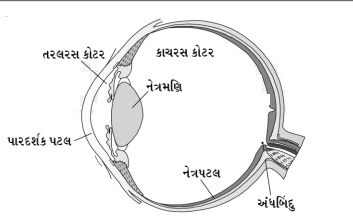
(N/A) Paired eyes are located in sockets of the skull called orbits.
Parts of an eye: Each eyeball is nearly spherical in structure.
Its diameter is approximately $2.5 \ cm$ and it weighs around $6$ to $8 \ g$.
The wall of the eyeball is composed of three layers:
$(a)$ Sclera $(b)$ Choroid $(c)$ Retina
$(a)$ Sclera: It is a fibrous,white layer. The posterior $5/6$ part is a white membrane formed of collagen fibers,whereas the anterior $1/6$ part is formed of fibrous tissue devoid of blood vessels and is called the cornea.
Conjunctiva: Conjunctiva is a thin,transparent membrane made up of stratified epithelium. It externally surrounds the transparent cornea and the exposed part of the white layer. It is the thinnest epithelium of the animal body.
$(b)$ Choroid: It is the middle layer,contains many blood vessels,and looks bluish in color.
The choroid layer is thin over the posterior two-thirds of the eyeball,but it becomes thick in the anterior part to form the ciliary body.
The ciliary body continues forward to form a pigmented and opaque structure called the iris. The iris is the visible colored portion of the eye.
The eyeball contains a transparent crystalline lens which is held in place by ligaments attached to the ciliary body.
In front of the lens,the aperture surrounded by the iris is called the pupil. The diameter of the pupil is regulated by the muscle fibers of the iris.
$(c)$ Retina: It is the innermost layer and it contains three layers of neural cells from inside to outside: Ganglion cells,Bipolar cells,and Photoreceptor cells.
Photoreceptor cells are of two types: rod cells and cone cells.
These cells contain light-sensitive proteins called photopigments.
Daylight (photopic) vision and color vision are functions of cones,and twilight (scotopic) vision is the function of the rods.
The rod cells contain a purplish-red protein called rhodopsin,which contains a derivative of Vitamin $A$.
In the human eye,there are three types of cones which possess their own photopigments that respond to red,green,and blue lights.
The sensations of different colors are produced by various combinations of these cones and their photopigments.
When these cones are stimulated equally,a sensation of white light is produced.
The optic nerves leave the eye and the retinal blood vessels enter it at a point medial to and slightly above the posterior pole of the eyeball.
Photoreceptor cells are not present in that region,and hence it is called the blind spot.
At the posterior pole of the eye,lateral to the blind spot,there is a yellowish pigmented spot called the macula lutea with a central pit called the fovea.
The fovea is a thinned-out portion of the retina where only the cones are densely packed. It is the point where the visual resolution is the greatest.
The space between the cornea and the lens is called the aqueous chamber and contains a thin,watery fluid called aqueous humor.
The space between the lens and the retina is called the vitreous chamber and is filled with a transparent gel called vitreous humor.
0 likes
View Solution241
Medium
Explain the mechanism of vision in detail.
Solution
(N/A) The light rays in the visible wavelength focused on the retina through the cornea and lens generate potentials (impulses) in rods and cones.
Photosensitive compounds (photopigments) in the human eyes are composed of opsin (a protein) and retinal (an aldehyde of vitamin $A$).
Light induces the dissociation of the retinal from the opsin,resulting in changes in the structure of opsin. This causes membrane permeability changes. As a result,potential differences are generated in the photoreceptor cells.
This produces a signal that generates action potentials in the ganglion cells through the bipolar cells.
These action potentials (impulses) are transmitted by the optic nerves to the visual cortex area of the brain.
Neural impulses are analyzed,and the image formed on the retina is recognized based on earlier memory and experience.
0 likes
View Solution242
Medium
Differentiate between rod cells and cone cells.
Solution
(N/A)
| Rod cells | Cone cells |
|---|---|
| $(1)$ These are rod-shaped photoreceptor cells. | $(1)$ These are cone-shaped photoreceptor cells. |
| $(2)$ They contain the photopigment rhodopsin (visual purple). | $(2)$ They contain photopigments known as iodopsins. |
| $(3)$ They are responsible for scotopic vision (vision in dim light). | $(3)$ They are responsible for photopic vision (daylight vision) and colour vision. |
| $(4)$ They are highly sensitive to light but do not provide colour information. | $(4)$ They are less sensitive to light but provide sharp images and colour discrimination. |
0 likes
View Solution243
Medium
Differentiate between the yellow spot and the blind spot.
Solution
(N/A)
| Yellow spot (Macula lutea) | Blind spot |
| $(1)$ It is a small, yellowish depression on the retina, specifically the fovea centralis. | $(1)$ It is the point on the retina where the optic nerve exits the eye. |
| $(2)$ It contains a high density of cone cells. | $(2)$ It lacks photoreceptor cells (both rods and cones). |
| $(3)$ It is the area of greatest visual acuity. | $(3)$ It is insensitive to light, resulting in no image formation. |
0 likes
View Solution244
Medium
Distinguish between:
$(1)$ Receptor proteins and Receptor cells.
$(2)$ Aqueous humor and Vitreous humor.
$(1)$ Receptor proteins and Receptor cells.
$(2)$ Aqueous humor and Vitreous humor.
Solution
(N/A) $(1)$ Receptor proteins: When an impulse arrives at the axon terminal,synaptic vesicles fuse with the plasma membrane and release their neurotransmitters into the synaptic cleft. The released neurotransmitter binds to specific proteins on the postsynaptic membrane called receptor proteins.
Receptor cells: These are specialized cells that detect stimuli. For example,the organ of Corti is located on the basilar membrane and contains hair cells,which act as auditory receptor cells.
$(2)$ Aqueous humor: This is a thin,watery fluid present in the space between the cornea and the lens.
Vitreous humor: This is a transparent,jelly-like substance present in the space between the lens and the retina.
0 likes
View Solution245
Medium
Define the location and function of the following structures:
$(1)$ Epithalamus
$(2)$ Rod cells
$(1)$ Epithalamus
$(2)$ Rod cells
Solution
(N/A) $(1)$ Location: It is a non-nervous part of the diencephalon that is fused with the piamater to form the anterior choroid plexus.
Function: It is involved in the secretion of cerebrospinal fluid and helps in the regulation of the endocrine system.
$(2)$ Location: These are rod-shaped photoreceptor cells found in the retina of the eye.
Function: They are specialized for vision in dim light (scotopic vision) and contain the pigment rhodopsin.
0 likes
View Solution246
EasyMCQ
Which cells of the retina enable us to see coloured objects around us?
A
Rod cells
B
Cone cells
C
Bipolar cells
D
Ganglion cells
Solution
(B) Cone cells of the retina help us to perceive colours.
There are three types of cone cells present in the retina,which contain specific photopigments.
These cells mainly respond to Red,Green,and Blue colours,allowing us to perceive a wide spectrum of colours.
There are three types of cone cells present in the retina,which contain specific photopigments.
These cells mainly respond to Red,Green,and Blue colours,allowing us to perceive a wide spectrum of colours.
0 likes
View Solution247
Easy
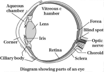
Label the following parts in the given diagram of the human eye:
$(a)$ Aqueous chamber
$(b)$ Cornea
$(c)$ Lens
$(d)$ Retina
$(e)$ Vitreous chamber
$(f)$ Blind spot.
$(a)$ Aqueous chamber
$(b)$ Cornea
$(c)$ Lens
$(d)$ Retina
$(e)$ Vitreous chamber
$(f)$ Blind spot.
Solution
(N/A) The human eye is a complex sensory organ. Based on the provided diagram,the parts are labeled as follows:
$(a)$ Aqueous chamber: The space between the cornea and the lens,filled with aqueous humor.
$(b)$ Cornea: The transparent anterior portion of the sclera.
$(c)$ Lens: $A$ transparent,biconvex crystalline structure held in place by ligaments attached to the ciliary body.
$(d)$ Retina: The innermost layer of the eye containing photoreceptor cells (rods and cones).
$(e)$ Vitreous chamber: The large space between the lens and the retina,filled with vitreous humor.
$(f)$ Blind spot: The point on the retina where the optic nerve leaves the eye; it lacks photoreceptor cells.
0 likes
View Solution248
MediumMCQ
Analogy type question:
$(1)$ Photoreceptor cells : Cones : Dim light vision : ........
$(2)$ Eyeball contains transparent crystalline : Lens : : In front of the lens : .........
$(1)$ Photoreceptor cells : Cones : Dim light vision : ........
$(2)$ Eyeball contains transparent crystalline : Lens : : In front of the lens : .........
A
$1$: Rods,$2$: Pupil
B
$1$: Rods,$2$: Iris
C
$1$: Cones,$2$: Cornea
D
$1$: Bipolar cells,$2$: Retina
Solution
(A) $(1)$ Cones are responsible for daylight (photopic) vision and color vision,whereas Rods are responsible for dim light (scotopic) vision. Therefore,the answer is Rods.
$(2)$ The eyeball contains a transparent crystalline lens. The structure located in front of the lens,which regulates the amount of light entering the eye,is the Pupil.
$(2)$ The eyeball contains a transparent crystalline lens. The structure located in front of the lens,which regulates the amount of light entering the eye,is the Pupil.
0 likes
View Solution249
MediumMCQ
Select the correct option:
$(1)$ There are no photoreceptor cells slightly above the posterior pole of the eyeball,hence it is called the blind spot.
$(2)$ Rod cells contain a purple-red protein called rhodopsin.
$(1)$ There are no photoreceptor cells slightly above the posterior pole of the eyeball,hence it is called the blind spot.
$(2)$ Rod cells contain a purple-red protein called rhodopsin.
A
Statement $(1)$ is correct,Statement $(2)$ is incorrect.
B
Statement $(1)$ is incorrect,Statement $(2)$ is correct.
C
Both statements $(1)$ and $(2)$ are correct.
D
Both statements $(1)$ and $(2)$ are incorrect.
Solution
(B) $(1)$ The statement is incorrect because the blind spot is located at the posterior pole of the eyeball,not slightly above it.
$(2)$ The statement is correct as rod cells contain the pigment rhodopsin,which is a derivative of Vitamin $A$ and is responsible for vision in dim light (scotopic vision).
$(2)$ The statement is correct as rod cells contain the pigment rhodopsin,which is a derivative of Vitamin $A$ and is responsible for vision in dim light (scotopic vision).
0 likes
View Solution250
MediumMCQ
Which cells are affected in a person suffering from color blindness?
A
Rod cells
B
Cone cells
C
Optic nerve
D
Bipolar neurons
Solution
(B) Color blindness is a sex-linked recessive disorder due to defect in either red or green cone cells of the eye resulting in failure to discriminate between red and green color.
Cone cells are responsible for color vision and detailed vision in bright light.
Rod cells are responsible for vision in dim light (scotopic vision).
Therefore,the correct option is $B$.
Cone cells are responsible for color vision and detailed vision in bright light.
Rod cells are responsible for vision in dim light (scotopic vision).
Therefore,the correct option is $B$.
0 likes
View SolutionNeural Control and Coordination — Eye · Frequently Asked Questions
1Are these Neural Control and Coordination questions useful for JEE and NEET?
Yes. All questions in this section are mapped to JEE Main and NEET exam patterns. Previous year questions from JEE Main, NEET, GUJCET and state-level exams are included with full solutions.
2Can I switch to Hindi or Gujarati for these questions?
Yes. Use the language tabs in the hero section or the sidebar to view the same questions and solutions in English, Hindi or Gujarati.
3How do I generate a question paper from this subtopic?
Use the Vedclass Exam Paper Generator — select the chapter and subtopic, set difficulty, and generate Sets A, B, C, D automatically. First 3 chapters of every subject are free.
Vedclass Products
For Students
Vedclass Test Series
Mock tests in real JEE/NEET style with performance analysis. 5-day free trial.
Start Free TrialFor Teachers
Exam Paper Generator
Generate Set A/B/C/D papers from this chapter in 2 minutes. 3 chapters free.
Try FreeFor Institutes
Online Exam Module
Live online exams with unlimited students, 360° analytics & white-label branding.
See DemoFor Teachers & Institutes
Generate a Neural Control and Coordination Exam Paper in 2 Minutes
Select subtopic & difficulty — Sets A, B, C, D auto-generated with No Repeat logic.
First 3 chapters of every subject are free — no payment required.